1. Introduction
In the range of important techniques used for studying nucleic acid-containing systems (including e.g. nuclear magnetic resonance (NMR), X-ray crystallography, electrophoresis, footprinting, calorimetry, enzymatic methods and UV-Vis absorption), fluorescence is one of the most sensitive, versatile, easily accessible, fast and straightforward ones. For this reason, it has found use in single-molecule real-time dynamics of nucleic acids as well as proteins, cell microscopy, nucleic acid detection and nucleic acid–protein interaction measurements. Thus, fluorescence studies at both the ensemble and single-molecule level are at the fore of biochemical as well as biophysical research on nucleic acids, and will increase in importance in the expanding field of DNA nanotechnology as these constructs keep decreasing in dimensions (Aldaye & Sleiman, Reference Aldaye and Sleiman2006; Seeman, Reference Seeman2003; Tumpane et al. Reference Tumpane, Kumar, Lundberg, Sandin, Gale, Nandhakumar, Albinsson, Lincoln, Wilhelmsson, Brown and Nordén2007). However, to investigate nucleic acids using fluorescence techniques, an emissive reporter group, a fluorophore, first has to be covalently or non-covalently introduced to the system since the nucleobases themselves are virtually non-fluorescent under normal conditions. This fluorophore labelling will be the focus of this review and most attention will be given to the covalent internal modifications and more specifically to fluorescent nucleobase analogues. Since most of the fluorescent modifications in nucleic acid research have been performed on DNA-containing systems, the review will primarily discuss these; however, for most of the modifications, one can equally well imagine using them for RNA systems.
2. Fluorescent labelling of nucleic acids
The lack of adequate intrinsic emission in DNA and RNA has resulted in the development of a variety of classes of molecules for fluorescent labelling of nucleic acids. One of those classes has been developed to interact non-covalently with the nucleic acid and has been used, for example, to visualize DNA in gel electrophoresis or cell microscopy. Examples of such molecules are DNA intercalators like ethidium bromide, YO (oxazole yellow) and its homodimer YOYO, as well as DNA groove binders such as 4′,6-diamidino-2-phenylindole (DAPI) and Hoechst derivatives. More importantly, molecules that enable external and internal covalent modifications of nucleic acids have also been developed and will be described in more detail below.
2.1 External modifications
Modifying nucleic acids by covalently attaching fluorophores to the backbone at the end of or within an oligonucleotide sequence, but outside the actual base stack, can be referred to as an external modification. Since there are many fluorophores commercially available for this purpose and also other ones still on the research level (Mayer et al. Reference Mayer, Valis, Wagner, Rist, Amann and Wagenknecht2004; Varghese & Wagenknecht, Reference Varghese and Wagenknecht2009), this is currently the most common way of labelling DNA. Fluorophores in the commercial class include, for example, fluorescein and rhodamine derivatives, Alexa dyes and Cy dyes. These molecules have high to very high molar absorptivities (up to several hundred thousand) combined with a significant to very high fluorescence quantum yield depending on the nature of the system under investigation (the reader could refer to commercial sources to find details about these fluorophores). As a consequence, the fluorophores are extremely bright and, thus, especially useful as probe molecules in gel electrophoresis experiments, different fluorescence microscopy techniques, or single- or few-molecule experiments. In general, they are also more photostable than the internal fluorescent modifications that will be thoroughly discussed below.
In some cases, molecules that are primarily used for non-covalent interaction with DNA such as ethidium, YO and the other ones mentioned above have also been utilized as external modifications by means of a covalently attached linker. The increase in fluorescence upon intercalation of the covalently attached cyanine dye TO (thiazole yellow) has, for example, been used to study the duplex formation of matched and mismatched sequences aiming at specific nucleic acid detection (Svanvik et al. Reference Svanvik, Westman, Wang and Kubista2000).
2.2 Internal modifications
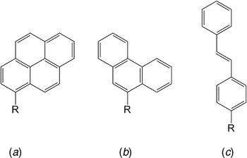
An internal modification means that the fluorophore is covalently attached aiming at replacing the nucleobase inside the base stack. In this group of molecules you find the fluorescent base analogues that are described in detail below, and also other planar aromatic compounds attached at the nucleobase position. These planar aromatics are generally different in shape and size and lack the possibility to form hydrogen bonds with the base on the opposite strand. For example, Kool et al. have replaced nucleobases with polycyclic hydrocarbons such as pyrene, phenanthrene and stilbene (Fig. 1) (Gao et al. Reference Gao, Strassler, Tahmassebi and Kool2002; Ren et al. Reference Ren, Chaudhuri, Paris, Rumney and Kool1996; Strässler et al. Reference Strässler, Davis and Kool1999; Wilson & Kool, Reference Wilson and Kool2006). Since these modifications allow for stacking interaction inside the base stack, they can be incorporated with merely minor perturbation to the DNA structure, even though they lack the hydrogen bonding capacity. There is even an example of enzymatic incorporation of this kind of molecule by DNA polymerase (Matray & Kool, Reference Matray and Kool1999). Other interesting candidates in the group of aromatic compounds for internal modification have been developed and studied by the groups of Romesberg (Berger et al. Reference Berger, Ogawa, Mcminn, Wu, Schultz and Romesberg2000; Matsuda & Romesberg, Reference Matsuda and Romesberg2004; Ogawa et al. Reference Ogawa, Wu, Berger, Schultz and Romesberg2000a, Reference Ogawa, Wu, Mcminn, Liu, Schultz and Romesbergb) and Benner (Piccirilli et al. Reference Piccirilli, Krauch, Moroney and Benner1990; Schneider & Benner, Reference Schneider and Benner1990; Switzer et al. Reference Switzer, Moroney and Benner1989, Reference Switzer, Moroney and Benner1993). These nucleobase analogues have been shown not to destabilize the DNA and are expected not to distort the structure substantially. However, so far there has been no or only little effort made on examining the potential use of these analogues as fluorescent probes.
Polycyclic hydrocarbons for incorporation into the nucleobase stack: (a) pyrene, (b) phenanthrene and (c) stilbene. R=(deoxy)ribose.

2.3 Internal versus external modifications
Either external or internal modification could be preferable when using fluorescence to investigate nucleic acid-containing systems. Therefore, it is impossible and incorrect to claim that a certain fluorophore or even group of fluorophores is, overall, best suited for fluorescently modifying DNA. Instead one needs to carefully consider each system under examination and try to optimize the choice of fluorescent probe molecule thereafter.
Generally, as mentioned above, the external commercially available fluorophores are very bright and photostable. Sometimes it is also preferable to have the probe molecule covalently attached via a long flexible linker and, thus, far from the site of interest. However, for investigations where the level of detail needs to be high, a modification in which a fluorescent base analogue is incorporated is generally preferable, since this enables the probe to be very close to the site of examination. Also, the bulkiness of the external fluorophores could give rise to problems concerning the binding of other molecules to the nucleic acid. Moreover, a modification in which a fluorescent base analogue is used normally minimizes perturbations to the native structure and behaviour of the nucleic acid. Consequently, measuring properties of the nucleic acid systems using internal modifications, especially fluorescent base analogues, normally results in an answer that is closer to the native behaviour than when using external modifications.
3. Fluorescent base analogues
It could be discussed how fluorescent ‘base analogues’ should be defined. Occasionally even molecules that (1) lack the ability to form hydrogen bonds to any of the natural bases and are considerably larger than a normal nucleobase as well as (2) natural nucleobases with externally covalently attached fluorophores are referred to as fluorescent base analogues. In this review, however, the term fluorescent base analogue will be used merely for significantly fluorescent molecules that resemble the shape of the natural nucleobases and also have some ability to form hydrogen bonds to a base in the complementary strand, i.e., they should not seriously perturb the overall structure of the nucleic acid.
3.1 Environment-sensitive fluorescent base analogues
With the exception of the family of tricyclic cytosines presented later in this review in section 3.2, all fluorescent base analogues have a quantum yield that is somewhat to highly sensitive to their immediate surroundings. Factors such as hydrogen bonding (Wilhelmsson et al. Reference Wilhelmsson, Sandin, Holmén, Albinsson, Lincoln and Nordén2003), single- or double-stranded environment, and neighbouring base are important factors governing the fluorescence quantum yield of a base analogue inside the base stack. Generally, fluorescent base analogues are highly quenched inside DNA, and normally the effect is most significant if surrounded by neighbouring purines (see e.g. Hawkins, Reference Hawkins2001; Rachofsky et al. Reference Rachofsky, Osman and Ross2001; Ward et al. Reference Ward, Reich and Stryer1969).
3.1.1 2-Aminopurine (2-AP)
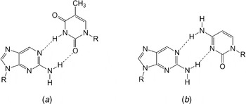
In 1969 Ward et al. discovered that 2-AP (Fig. 2) (Ward et al. Reference Ward, Reich and Stryer1969), currently the most used fluorescent base analogue, is highly fluorescent. 2-AP, which is one of the few commercially available fluorescent base analogues, is an adenine (6-AP) analogue forming stable base pairs with thymine (Fig. 2a) and uracil but also moderately stable base pairs with cytosine (Fig. 2b) (Freese, Reference Freese1959; Sowers et al. Reference Sowers, Fazakerley, Eritja, Kaplan and Goodman1986, Reference Sowers, Boulard and Fazakerley2000). The lowest energy absorption band of 2-AP is centred at 305 nm and has a molar absorptivity (ε) of 6000 M−1 cm−1 (Albert & Taguchi, Reference Albert and Taguchi1973; Holmén et al. Reference Holmén, Nordén and Albinsson1997). The position of the band is outside the absorption of the nucleic acid and, thus, 2-AP can be selectively excited in the presence of the natural nucleobases.
Fluorescent base analogue 2-AP base paired with thymine (a) and cytosine (b). R=(deoxy)ribose.

The high fluorescence quantum yield of 2-AP free in solution (0·68) is considerably reduced (~100 times but highly dependent on base sequence) when incorporated into nucleic acids (Ward et al. Reference Ward, Reich and Stryer1969). This sensitivity to the microenvironment has been utilized in a range of investigations, including nucleic acid structure and dynamics (Guest et al. Reference Guest, Hochstrasser, Sowers and Millar1991; Rachofsky et al. Reference Rachofsky, Osman and Ross2001; Stivers, Reference Stivers1998). This property of 2-AP has also been utilized to study DNA–protein interactions such as the interaction with the EcoRI DNA methyltransferase (Allan & Reich, Reference Allan and Reich1996), the Klenow fragment of DNA polymerase (Bloom et al. Reference Bloom, Otto, Beechem and Goodman1993; Hochstrasser et al. Reference Hochstrasser, Carver, Sowers and Millar1994; Joyce et al. Reference Joyce, Potapova, Delucia, Huang, Basu and Grindley2008), the EcoRI endonuclease (Lycksell et al. Reference Lycksell, Gräslund, Claesens, Mclaughlin, Larsson and Rigler1987; Nordlund et al. Reference Nordlund, Andersson, Nilsson, Rigler, Gräslund and Mclaughlin1989) and the uracil DNA glycosylate (Stivers et al. Reference Stivers, Pankiewicz and Watanabe1999). More recently, 2-AP has been used as a probe in the rapidly expanding field of small RNAs and RNA elements that control gene expression at a number of levels. For example, the groups of Lilley and Batey have investigated riboswitch folding properties and thermodynamics as well as kinetics of ligand binding to the purine riboswitch aptamer domain (Gilbert et al. Reference Gilbert, Stoddard, Wise and Batey2006; Lemay et al. Reference Lemay, Penedo, Tremblay, Lilley and Lafontaine2006). On a rather different subject, 2-AP has also found use in studies of photo-induced electron transfer through the DNA stack (Kelley & Barton, Reference Kelley and Barton1999).
As illustrated above, the sensitivity of the emission of 2-AP to the microenvironment is very useful. However, the less efficient and less specific base-pairing relative to adenine introduces a perturbation to the native structure of DNA and also gives 2-AP increased dynamics within the DNA helix. This, in combination with the sensitivity of the fluorescence quantum yield to the environment, makes 2-AP less suited as a probe for studies of molecular dynamics and DNA–protein interaction using techniques such as fluorescence anisotropy and fluorescence resonance energy transfer (FRET).
3.1.2 Pteridines
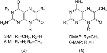
An interesting and frequently utilized family of fluorescent base analogues are the pteridines mainly developed by Hawkins and Pfleiderer (Hawkins, Reference Hawkins2001). The most promising analogues within this family are the guanine analogues 3-MI and 6-MI (Fig. 3a) and the adenine analogues 6-MAP and DMAP (Fig. 3b). Like 2-AP, these probes are very sensitive to the microenvironment and are highly quenched, with fluorescence quantum yields ranging from <0·01 to 0·3 depending on the nature of the surrounding base pairs, when incorporated into DNA (Hawkins, Reference Hawkins2001; Hawkins et al. Reference Hawkins, Pfleiderer, Mazumder, Pommier and Falls1995, Reference Hawkins, Pfleiderer, Balis, Porter and Knutson1997, Reference Hawkins, Pfleiderer, Jungmann and Balis2001). The lowest energy excitation maxima for both the adenine and guanine analogues are located between 310 and 350 nm, and are thus well resolved from the absorption of the natural nucleobases and easy to selectively excite. The molar absorptivities of the low-energy transition of the pteridine base analogues are not extensively examined; however, values between 5000 and 15 000 M−1 cm−1 have been reported, e.g. 6-MAP in methanol ε329=8500 M−1 cm−1 (Stanley et al. Reference Stanley, Hou, Yang and Hawkins2005) and 3-MI in water ε351=13 000 M−1 cm−1 (Sanabia et al. Reference Sanabia, Goldner, Lacaze and Hawkins2004). A slight drawback with this group of fluorescent base analogues is that duplex melting temperature studies have shown that, with the exception of 6-MI, they reduce the thermal stability of oligonucleotides (Hawkins et al. Reference Hawkins, Pfleiderer, Balis, Porter and Knutson1997, Reference Hawkins, Pfleiderer, Jungmann and Balis2001). For example, as an effect of the methyl group in the 3-position of 3-MI, the hydrogen bonding to cytosine is hindered, resulting in a reduced stability approximately equivalent to a single base-pair mismatch.
Fluorescent guanine analogues 3-MI and 6-MI (a) and fluorescent adenine analogues DMAP and 6-MAP (b). R=(deoxy)ribose.

The fluorescent pteridines have already been successfully utilized in a range of investigations. 3-MI has among other things, been utilized in a real-time assay for O 6-alkylguanine-DNA alkyltransferase (Moser et al. Reference Moser, Patel, Yoo, Balis and Hawkins2000), DNA binding of the non-specific histone-like protein HU (Wojtuszewski et al. Reference Wojtuszewski, Hawkins, Cole and Mukerji2001), and as a probe of hybridization specificity (Hawkins & Balis, Reference Hawkins and Balis2004). Furthermore, 3-MI has been evaluated for single-molecule detection purposes (Sanabia et al. Reference Sanabia, Goldner, Lacaze and Hawkins2004). The other guanine analogue, 6-MI, has been used in recombination to study the mechanism of the RecA-mediated DNA strand exchange (Roca & Singleton, Reference Roca and Singleton2003; Xiao et al. Reference Xiao, Lee and Singleton2006) as well as in structural studies of the RecA-DNA filament (Singleton et al. Reference Singleton, Roca, Lee and Xiao2007). The adenine analogue 6-MAP, on the other hand, has been used in premelting transition studies of DNA A-tracts (Augustyn et al. Reference Augustyn, Wojtuszewski, Hawkins, Knutson and Mukerji2006) and as a probe for base flipping by DNA photolyase (Yang et al. Reference Yang, Matsika and Stanley2007).
3.1.3 Pyrrolo-dC
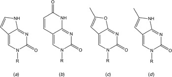
In the late 1980s, Inoue and co-workers reported on the fluorescence properties of the pyrrolo-dC derivative dF* (Fig. 4a) inside an oligonucleotide (Inoue et al. Reference Inoue, Imura and Ohtsuka1987), which was a follow-up on their studies on dF (Fig. 4b) some years earlier (Bergstrom et al. Reference Bergstrom, Inoue and Reddy1982). In those studies, it was also found that an F*⋅G base pair had a stability similar to that of a C⋅G base pair. Later a fluorescent nucleobase named furano-dT (Fig. 4c) was introduced (Woo et al. Reference Woo, Meyer and Gamper1996). However, after incorporation of this non-specific nucleobase into an oligonucleotide and the subsequent ammonia treatment at the final stage of the DNA solid-phase synthesis, it was found that the nucleobase had reacted to form the C-analogue pyrrolocytosine (pyrrolo-dC; Fig. 4d) (Berry et al. Reference Berry, Jung, Wise, Sercel, Pearson, Mackie, Randolph and Somers2004). This fluorescent base analogue is a 6-methyl derivative of the previously mentioned dF* (Fig. 4a). It has been shown in several studies that pyrrolo-dC hybridizes selectively with G (dG>>dT>dA>dC) and that duplexes containing pyrrolo-dC in place of dC are virtually as stable as the corresponding unmodified ones (Berry et al. Reference Berry, Jung, Wise, Sercel, Pearson, Mackie, Randolph and Somers2004). As for the pteridines mentioned above, this commercially available fluorescent base analogue can be selectively excited since the low-energy absorption band at 350 nm (ε350=5900 M−1 cm−1) is well separated from both nucleic acid and protein absorption (Liu & Martin, Reference Liu and Martin2001). The emission maxima for the pyrrolo-dC monomer before and after incorporation into duplex contexts are 460 and 473 nm, respectively. The fluorescence quantum yield of the monomer has been reported to be 0·20 (Liu & Martin, Reference Liu and Martin2001). However, after incorporation into a single strand the quantum yield drops significantly and subsequent hybridization with a complementary strand to form a duplex reduces the quantum yield even more (Berry et al. Reference Berry, Jung, Wise, Sercel, Pearson, Mackie, Randolph and Somers2004; Dash et al. Reference Dash, Rausch and Le Grice2004; Hardman & Thompson, Reference Hardman and Thompson2006; Liu & Martin, Reference Liu and Martin2001). Very little has been reported on the influence of neighbouring bases on the quantum yield of pyrrolo-dC; however, quantum yields of 0·03 in dsDNA (TT neighbours) and approximately 0·05 in dsRNA (CA neighbours) have been reported.
Fluorescent base analogues in the pyrrolo-dC-‘family’: (a) dF*, (b) dF, (c) furano-dT and (d) pyrrolo-dC. R=(deoxy)ribose.

Since pyrrolo-dC is commercially available, it has already been used in several biochemical applications. In the study by Liu and Martin, at the time when it was found that furano-dT modified strands actually were transformed to pyrrolo-dC during the DNA solid-phase synthesis, they used the fluorescence of the C-analogue to characterize the transcription bubble in the elongation complexes of the T7 RNA polymerase (Liu & Martin, Reference Liu and Martin2001). Later, in 2005 Zang et al. utilized pyrrolo-dC to examine the kinetics of parts of the repair of damaged DNA by a human alkyltransferase (Zang et al. Reference Zang, Fang, Pegg and Guengerich2005). Furthermore, pyrrolo-dC has been incorporated into DNA strands that subsequently have been annealed with an RNA strand to form a DNA/RNA hybrid and used to investigate the HIV-1 polypurine tract (PPT) in which abnormal base pairing has been reported (Dash et al. Reference Dash, Rausch and Le Grice2004). In the search for high-affinity and highly selective triplex-forming oligonucleotides (TFOs), the groups of Brown and Fox have developed methylated pyrrolo-dC derivatives for use in the duplex recognition strand (Ranasinghe et al. Reference Ranasinghe, Rusling, Powers, Fox and Brown2005). These derivatives might be interesting as fluorescent probes for DNA triplexes, but this has so far not been investigated. In a different approach optimizing the wavelength of excitation and emission as well as the position of the involved probes, Turro and co-workers have shown that pyrrolo-dC can be utilized in pair with 2-AP in a molecular beacon-like fashion (Marti et al. Reference Marti, Jockusch, Li, Ju and Turro2006). Recently pyrrolo-dC has also been used for investigating the structure and thermodynamics of single strands that are able to form DNA hairpins (Zhang & Wadkins, Reference Zhang and Wadkins2009), the secondary structure of RNA (Tinsley & Walter, Reference Tinsley and Walter2006) as well as the propagation of base-pair flipping in a DNA photolyase complex (Yang & Stanley, Reference Yang and Stanley2008).
3.1.4 Other environment-sensitive fluorescent base analogues
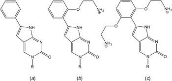
With the aim of improving both the hybridization and fluorescence properties of the above-mentioned pyrrolo-dC, the group of Hudson started to substitute the 6-methyl group with various molecular entities with a focus on phenyl derivatives (PhpC, moPhpC and boPhpC; Fig. 5) (Hudson et al. Reference Hudson, Viirre, Liu, Wojciechowski and Dambenieks2003; Wojciechowski & Hudson, Reference Wojciechowski and Hudson2008). These phenyl derivatives show increased duplex (peptide nucleic acid (PNA)/DNA hybrid) stability compared to the unmodified case, good base-pairing selectivity and a promisingly high fluorescence quantum yield. The reported quantum yield for boPhcP monomer in aqueous solution is 0·32 (approx. 50% higher than pyrrolo-dC), whereas it was found to be approximately 50 and 25% in the two investigated single and double strands, respectively (Wojciechowski & Hudson, Reference Wojciechowski and Hudson2008). The high quantum yield in the single-stranded PNAs and in the PNA/DNA hybrids in combination with the molar absorptivity of 6650 M−1 cm−1 at 365 nm makes this base analogue an interesting fluorescent probe. However, the dependence of the quantum yield on surrounding bases still remains to be studied. Recently, boPhpC was incorporated into antisense PNA, and it was demonstrated that the stability of boPhcP–PNA/RNA duplexes was increased (Hu et al. Reference Hu, Dodd, Hudson and Corey2009). In another effort to increase the stability of (hybrid) duplexes, the group of Hudson developed additional modifications of PhpC that also show fluorescence properties (Wojciechowski & Hudson, Reference Wojciechowski and Hudson2009). Other derivatives of pyrrolocytosine have also been developed and incorporated into nucleic acid, and their quantum yields have been determined to be relatively low (ϕf≈0·06) (Seela & Sirivolu, Reference Seela and Sirivolu2008).
Phenyl derivatives of pyrrolo-dC: (a) PhpC, (b) moPhpC and (c) boPhpC. R=(deoxy)ribose.

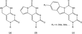
Sekine et al. have focused on another group of fluorescent cytosine analogues, where they have expanded the cytosine one-ring system to different two-, three- and four-ring systems while maintaining the hydrogen bonding possibilities with guanine (Fig. 6). The two-ring cytosine analogue, dChpp (Fig. 6a), has been investigated for its fluorescence in DNA single and double strands (Miyata et al. Reference Miyata, Tamamushi, Ohkubo, Taguchi, Seio, Santa and Sekine2006). The emission in the single-stranded case has a peak at approximately 370 nm that is considerably quenched upon hybridization with a complementary strand (G opposite). However, having a mismatch A opposite the dChpp keeps the emission virtually unchanged relative to the single-stranded case. Furthermore, it has been shown that dChpp forms not only stable base pairs with G but also fairly stable ones with A, where the melting temperature is increased by 10°C compared to the corresponding C–A mismatch. The quantum yield of dChpp inside DNA has not been reported but has been measured for the monomeric form (ϕf=0·12) (Miyata et al. Reference Miyata, Tamamushi, Ohkubo, Taguchi, Seio, Santa and Sekine2006). To improve the fluorescence properties and also red-shift the wavelength of excitation compared to dChpp (λmax=300 nm), a third ring was attached to the system to form the cytosine analogue dCppp (Fig. 6b) (Miyata et al. Reference Miyata, Mineo, Tamamushi, Mizuta, Ohkubo, Taguchi, Seio, Santa and Sekine2007). This analogue has an excitation maximum in the low-energy band at 360 nm and an emission maximum at 490 nm with a quantum yield of approximately 0·11, but has not yet been investigated inside nucleic acid sequences. In yet another study concerning cytosine analogues, Sekine et al. attached an additional ring to dCppp forming various derivatives of dCPPI (Fig. 6c) (Mizuta et al. Reference Mizuta, Seio, Miyata and Sekine2007). The unsubstituted dCPPI was shown to have a quantum yield that was reduced almost 20 times compared to the parent compound dCppp. For some of the other derivatives of dCPPI, a quantum yield comparable to dCppp as well as both excitation and emission maxima that were slightly red-shifted were found (Mizuta et al. Reference Mizuta, Seio, Miyata and Sekine2007). As for the bicyclic dChpp, the dCPPI derivatives form stable base pairs with G and fairly stable ones with A (Mizuta et al. Reference Mizuta, Seio, Ohkubo and Sekine2009). Moreover, the quantum yields on average decrease in going from single strands (ϕf≈0·06) to fully complementary duplexes (ϕf≈0·015). However, for one of the derivatives where dCPPI mismatches with a C, the quantum yield has been shown to be as high as 0·27 (Mizuta et al. Reference Mizuta, Seio, Ohkubo and Sekine2009). It should be noted that the influence of different surrounding bases on quantum yield has not been investigated in detail. The three- and four-ring systems have also been investigated in a recent study, where they were utilized as base analogues that could increase the stability of DNA triplexes (Mizuta et al. Reference Mizuta, Banba, Kanamori, Tawarada, Ohkubo, Sekine and Seio2008).
Fluorescent cytosine analogues developed by Sekine et al.: (a) dChpp, (b) dCppp and (c) dCPPI. R=(deoxy)ribose.

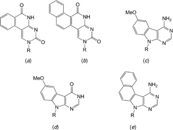
The group of Saito has developed a range of base-discriminating fluorescent (BDF) nucleoside analogues (Okamoto et al. Reference Okamoto, Saito and Saito2005). Many of these have common fluorophores covalently attached to a normal nucleobase and show promising properties (Bag et al. Reference Bag, Saito, Hanawa, Kodate, Suzuka and Saito2006; Saito et al. Reference Saito, Hanawa, Motegi, Omoto, Okamoto and Saito2005, Reference Saito, Hanawa, Kawasaki, Bag and Saito2006, Reference Saito, Bag, Kusakabe, Nagai, Matsumoto, Mizuno, Kodate, Suzuka and Saito2007, Reference Saito, Motegi, Bag and Saito2008; Tainaka et al. Reference Tainaka, Tanaka, Ikeda, Nishiza, Unzai, Fujiwara, Saito and Okamoto2007); however, as mentioned above, here the focus will be on internal fluorescent base analogues. The fluorescent base analogue BPP (Fig. 7a) has an absorption maximum around 350 nm, well separated from the nucleobases, and an emission centred at approximately 390 nm (Okamoto et al. Reference Okamoto, Tainaka and Saito2003a). It has a considerable similarity with the base analogues by Sekine et al., can form stable base pairs with both A and G, and can be utilized to discriminate between the two purines using the change in quantum yield when base paired with a G (ϕf≈0·0018) or an A (ϕf≈0·035) (Okamoto et al. Reference Okamoto, Tainaka and Saito2003a). The structurally related NPP (Fig. 7b), having an additional benzene ring compared to BPP, has also been shown to have base-discriminating properties (Okamoto et al. Reference Okamoto, Tainaka and Saito2003b). The peak of emission is found around 420 nm and the quantum yield again depends on base pairing with G (ϕf≈0·007) or A (ϕf≈0·096). Using a combination of the two fluorescent base analogues MDA and MDI (Fig. 7 c and d) has been suggested to be a powerful tool for T/C single nucleotide polymorphism (SNP) typing (Okamoto et al. Reference Okamoto, Tanaka, Fukuta and Saito2003c). In combination with C in the complementary strand, a quantum yield of 0·081 and a peak at 424 nm have been observed for MDA. For all other base-pair combinations, the quantum yield was found to be lower. The corresponding values for MDI (ϕf≈0·011; Emmax=424 nm) are found in combination with T in the complementary strand. Finally, Saito and co-workers have used another BDF nucleoside, NDA (Fig. 7e), in FRET in combination with fluorescein to distinguish C from all other bases (Okamoto et al. Reference Okamoto, Tanaka, Fukuta and Saito2004).
BDF nucleoside analogues by Saito et al.: (a) BPP, (b) NPP, (c) MDA, (d) MDI and (e) NDA. R=(deoxy)ribose.

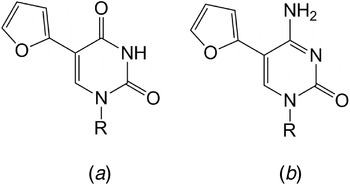
Important contributions to the field of fluorescent nucleobase analogues have also been made by the group of Tor. In an attempt to develop emissive nucleosides that have a high sensitivity to the microenvironment, they attached different five-membered heterocycles to both pyrimidines and purines (Fig. 8). Although the eight-modified purines show favourably high quantum yields in water (0·69 and 0·57 for the adenine and guanine analogue, respectively) (Greco & Tor, Reference Greco and Tor2007), the research has focused on the 5-position furan-modified T/U (Fig. 8a) due to its high responsiveness to the surrounding microenvironment (Greco & Tor, Reference Greco and Tor2005, Reference Greco and Tor2007). The fluorescence quantum yield of this thymine (or uridine) analogue in water has been determined to be 0·03 and the corresponding maxima in the lowest energy absorption band and the fluorescence have been shown to be at 316 and 431 nm, respectively. It has been successfully incorporated into DNA sequences, where it is still emissive in both single- and double-stranded systems (Greco & Tor, Reference Greco and Tor2007). Moreover, the stability of a furan-dT-modified oligonucleotide has been shown to be exactly the same as that of the corresponding unmodified sequence. It has been shown to be useful for abasic site detection (Greco & Tor, Reference Greco and Tor2005) as well as major groove polarity measurements in DNA (Sinkeldam et al. Reference Sinkeldam, Greco and Tor2008). Furthermore, the related uridine analogue has been synthesized in its triphosphate form for subsequent successful exploitation as in RNA HIV-1 TAR recognition studies (Srivatsan & Tor, Reference Srivatsan and Tor2007b) and as a substrate in in vitro transcription reactions using the T7 RNA polymerase (Srivatsan & Tor, Reference Srivatsan and Tor2007a). Additionally, the furan derivative of cytosine (Fig. 8b) shows similar photophysical properties as its thymine counterpart, works very well as a C-analogue, and has been shown to efficiently distinguish between G, 8-oxoG and T in a non-destructive method for in vitro detection of a DNA oxidative damage pathway (Greco et al. Reference Greco, Sinkeldam and Tor2009).
Fluorescent furan-modified pyrimidines: (a) thymine analogue and (b) cytosine analogue. R=(deoxy)ribose.

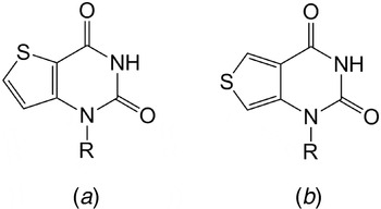
Tor et al. have also designed and developed new isomorphic fluorescent nucleobase analogues using the thieno[3,2-d]- or thieno[3,4-d]-pyrimidine core (Srivatsan et al. Reference Srivatsan, Weizman and Tor2008b; Tor et al. Reference Tor, Del Valle, Jaramillo, Srivatsan, Rios and Weizman2007). The thieno[3,2-d]pyrimidine T-analogue (Fig. 9a) has been shown to have a quantum yield in water of approximately 0·06 and the corresponding maxima in the lowest energy absorption band and the fluorescence have been shown to be situated at 292 and 351 nm, respectively. Corresponding values for the thieno[3,4-d]pyrimidine T-analogue (Fig. 9b) are ϕf≈0·50, 304 nm and 412 nm (Srivatsan et al. Reference Srivatsan, Weizman and Tor2008b). Thus, this T-analogue has a more promising quantum yield as well as a low-energy absorption band that is more well separated from the intrinsic nucleic acid absorption. It has been synthesized in its triphosphate form, and enzymatically incorporated into RNA oligonucleotides (Srivatsan & Tor, Reference Srivatsan and Tor2009) and has also been used for sensing mismatch pairing and signalling the activity of toxic ribosome-inactivating proteins (Srivatsan et al. Reference Srivatsan, Greco and Tor2008a).
Fluorescent thymine analogues developed by Tor et al.: (a) thieno[3,2-d]pyrimidine and (b) thieno[3,4-d]pyrimidine. R=(deoxy)ribose.

Recently, the group of Tor designed and synthesized the uracil base analogue, 5-methoxy-quinazoline-2,4-(1H,3H)-dione, and incorporated it into RNA (Xie et al. Reference Xie, Dix and Tor2009). This base analogue has an emission maximum at 395 nm with a quantum yield of 0·16 in its monomeric form that drops to 0·03 inside the RNA construct. With an absorption maximum at 320 nm, this U-analogue is easily selectively excited and for the sequence used in the investigation it has minimal influence on the stability of the folded RNA. In the investigation, the authors use the U-analogue as a FRET donor in pair with coumarin-labelled aminoglycosides and demonstrate the use of this pair as an analysis and discovery platform for antibiotics targeting the bacterial rRNA A-site.
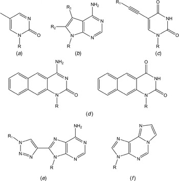
5-Methylpyrimidin-2-one (Fig. 10a) is referred to as a fluorescent T-analogue (Laland & Serckhanssen, Reference Laland and Serckhanssen1964). It has been shown to be predominantly stacked in oligonucleotide context; however, it does not base-pair well with A. It has its excitation and emission maxima at 310 and 380 nm, respectively, and the emission is efficiently quenched when the base analogue is inside the base stack. Apart from base (un)stacking determinations (Wu et al. Reference Wu, Nordlund, Gildea and Mclaughlin1990), it has been used for probing the RecA nucleoprotein filament (Singleton et al. Reference Singleton, Shan, Kanan, Mcintosh, Stearman, Helm and Webb2001). Other fluorescent base analogues worth mentioning are the 7-deazapurines (Fig. 10b) studied by Seela et al. (Seela & Zulauf, Reference Seela and Zulauf1998; Seela et al. Reference Seela, Zulauf, Sauer and Deimel2000), the 5-alkynyluridines (Fig. 10c) of Hudson & Ghorbani-Choghamarani (Reference Hudson and Ghorbani-Choghamarani2007), the benzoquinazolines (Fig. 10d) of Godde, Toulmé and Moreau that have been utilized in triplexes (Godde et al. Reference Godde, Toulme and Moreau1998, Reference Godde, Toulme and Moreau2000), the triazoleadenosines (Fig. 10e) of Wilhelmsson and Grøtli (Dyrager et al. Reference Dyrager, Börjesson, Diner, Elf, Albinsson, Wilhelmsson and Grøtli2009) and last but definitely not the least the 1,N 6-ethenoadenosine (Fig. 10f) that was developed, investigated and utilized by Secrist et al. already in the early 1970s (Secrist et al. Reference Secrist, Barrio and Leonard1972).
Fluorescent base analogues (a) 5-methylpyrimidin-2-one, (b) 7-deazapurines, (c) 5-alkynyluridines, (d) benzoquinazolines, (e) triazoleadenosines and (f) 1,N 6-ethenoadenosine. R=(deoxy)ribose.

3.2 Environment-insensitive fluorescent base analogues
The base analogues described in previous sections all have an emission that is highly sensitive to their immediate surroundings. 2-AP, for example, has a quantum yield of 0·68 as a monomer, but is quenched approximately 100 times upon incorporation into a DNA duplex (Ward et al. Reference Ward, Reich and Stryer1969). Another example of how sensitive these base analogues are to the microenvironment are the pteridines that can have quantum yields that change up to 30 times depending on the nature of the surrounding bases (Hawkins, Reference Hawkins2001; Hawkins et al. Reference Hawkins, Pfleiderer, Balis, Porter and Knutson1997, Reference Hawkins, Pfleiderer, Jungmann and Balis2001). In this section, fluorescent base analogues that have quantum yields that are essentially or fairly insensitive to the immediate surroundings of the analogue and their use will be discussed.
3.2.1 Why are environment-insensitive fluorescent base analogues important?
At first consideration, it may be easy to think that an environment-sensitive fluorescent probe is always more useful than an insensitive one. However, in experiments where the emission is used merely for detection, a high and stable quantum yield in combination with a high molar absorptivity (brightness∝ϕf⋅ε) is more important. Moreover and more importantly for the fluorescent base analogues that are environment insensitive is their use in FRET and anisotropy measurements. For these kinds of experiments, the high and virtually unaffected quantum yield and the single fluorescence lifetime in combination with the high control of orientation and position within the nucleic acid are essential properties that are unique for the base analogues of the tC family discussed below. As will be described in detail later in the text, a stable quantum yield (ϕf) and high control of position and orientation (κ2) are crucial in accurately determining the Förster distance (R 0) and, thus, also for the precision of distances (R DA) estimated using FRET. Furthermore, a single lifetime and strong emission in combination with a firm stacking within the base stack is essential for accurately determining the mobility of nucleic acids using fluorescence anisotropy.
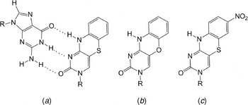
3.2.2 tC – 1,3-diaza-2-oxophenothiazine
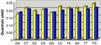
The tricyclic cytosine analogue tC (Fig. 11a), 1,3-diaza-2-oxophenothiazine, was originally developed by Matteucci and co-workers for antisense purposes (Lin et al. Reference Lin, Jones and Matteucci1995). Several years later we found that tC is highly fluorescent with an emission maximum at 505 nm and has a quantum yield that is roughly the same in the monomeric form, in a PNA single strand and in a PNA–DNA hybrid duplex (Wilhelmsson et al. Reference Wilhelmsson, Holmén, Lincoln, Nielsen and Nordén2001). Later, using a set of ten different base-pair surroundings, we could conclude that tC in fact has a fluorescence quantum yield that is virtually unaffected by its immediate surroundings (Fig. 12) (Sandin et al. Reference Sandin, Wilhelmsson, Lincoln, Powers, Brown and Albinsson2005, Reference Sandin, Lincoln, Brown and Wilhelmsson2007). The quantum yields in single- and double-stranded systems are found to be 0·17–0·24 and 0·16–0·21, respectively. Furthermore, we found that tC displays single fluorescence lifetimes in both single and double strands (τf(ss)=5·7 ns and τf(ds)=6·3 ns averaged over the ten sequences). The lowest energy absorption band centred around 395 nm in duplex DNA is very well separated from the absorption of the natural nucleobases and tC can hence be selectively excited (Wilhelmsson et al. Reference Wilhelmsson, Sandin, Holmén, Albinsson, Lincoln and Nordén2003). This band consists of a single electronic transition polarized 35° counterclockwise from the long axis of tC (Wilhelmsson et al. Reference Wilhelmsson, Sandin, Holmén, Albinsson, Lincoln and Nordén2003): parameters that are essential for further use in FRET experiments (vide infra).
Tricyclic cytosine analogues: (a) guanine base paired with tC, which displays a virtually environment-insensitive emission quantum yield, (b) highly emissive tCO and (c) base analogue FRET acceptor tCnitro. R=(deoxy)ribose.

Fluorescence quantum yield of tC in DNA single- (yellow) and double-stranded (blue) systems. Letters denote bases surrounding tC (5′- …X(tC)Y …-3′).

Earlier it was found that tC is a good cytosine analogue (Eldrup et al. Reference Eldrup, Nielsen, Haaima, Rasmussen, Kastrup, Christensen and Nielsen2001; Lin et al. Reference Lin, Jones and Matteucci1995). It base-pairs with guanine and discriminates well between guanine and adenine (Lin et al. Reference Lin, Jones and Matteucci1995). Using NMR, we have discovered that DNA adopts a normal B-form after incorporation of tC in place of cytosine and that only minor local distortions are present in the vicinity of tC (Engman et al. Reference Engman, Sandin, Osborne, Brown, Billeter, Lincoln, Nordén, Albinsson and Wilhelmsson2004). The overall bent tC-DNA NMR-structure conformation suggested in that NMR article is most likely an effect of the difficulties to draw conclusions regarding long-range features based on the short-range information provided by nuclear overhauser effect (NOEs) and torsion angle constraints. NMR data further confirm correct base pairing and a base-flipping rate of tC that is equal to the natural bases. Moreover, we found that the circular dichroism (CD) of the ten investigated tC duplexes showed features consistent with B-form DNA. We also discovered that one tC on average increases the stability of a 10-mer duplex by approximately 3°C (Engman et al. Reference Engman, Sandin, Osborne, Brown, Billeter, Lincoln, Nordén, Albinsson and Wilhelmsson2004). These findings all suggest that tC does not perturb the DNA structure significantly and that it is firmly stacked with a well-defined geometry (orientation) inside the DNA helix.
The firm stacking, low base-flipping rate, and single and comparably long fluorescence lifetime of tC in duplex is a set of truly unique properties for a fluorescent base analogue. These properties are central in studying the overall motion of nucleic acid structures without interference from any motions of the fluorescent probe itself. In a recent study, we show strong evidence that tC works excellently as a probe for monitoring motions of the overall nucleic acid structure rather than a combination of motion of the overall structure and the probe itself (Sandin et al. Reference Sandin, Börjesson, Li, Mårtensson, Brown, Wilhelmsson and Albinsson2008), as is the case for fluorescent base analogues that are not firmly stacked, or has increased base-flipping rates. Furthermore, we have utilized tC as a FRET donor in pair with rhodamine in a PNA–DNA hybrid (Wilhelmsson et al. Reference Wilhelmsson, Holmén, Lincoln, Nielsen and Nordén2001), and in pair with Alexa-555 in a study of conformational dynamics of DNA polymerase (Stengel et al. Reference Stengel, Gill, Sandin, Wilhelmsson, Albinsson, Nordén and Millar2007). In the latter study, tC was present adjacent to the primer 3′ terminus of the DNA primer/template that was bound in the active site of the polymerase. In another polymerase study, we show that the 5′-triphosphate of tC (and tCO, vide infra), which was synthesized for the first time, is efficiently incorporated into DNA by the Klenow fragment (Sandin et al. Reference Sandin, Stengel, Ljungdahl, Börjesson, Macao and Wilhelmsson2009a). This shows that the active site of the Klenow fragment is flexible enough to tolerate these size-expanded bases and constitutes the first report where the insertion of a size-expanded base is demonstrated to be more efficient than the corresponding natural base. However, we also find that tC(O) can be misincorporated opposite adenine, which suggests that there is a loss of selectivity (Sandin et al. Reference Sandin, Stengel, Ljungdahl, Börjesson, Macao and Wilhelmsson2009a). In a later comprehensive study by Stengel et al. using also the human DNA polymerase α, a similar conclusion could be drawn about the efficiency and loss of selectivity (Stengel et al. Reference Stengel, Purse, Wilhelmsson, Urban and Kuchta2009a). Furthermore, they show that tC(O) works well as templating bases and suggest that different tautomeric forms of tC(O) might explain the loss of selectivity since the imino-form could base-pair with adenine. In a related study by the same group, it was shown, however, that DNA human primase strongly discriminates against polymerizing tC opposite adenines (Urban et al. Reference Urban, Joubert, Purse, Hocek and Kuchta2010). In addition to these DNA polymerization investigations, the ribonucleotide form of tC has been synthesized and tested as a substrate for T7 RNA polymerase. Interestingly, it was also found in this study that T7 RNA polymerase does not misincorporate tC opposite adenine and it was shown that the ribonucleotide form of tC could replace normal cytosine in an ~800 nucleotide RNA, enabling straightforward fluorescent labelling of long RNAs (Stengel et al. Reference Stengel, Urban, Purse and Kuchta2010). It is also worth mentioning that for future investigations of polymerases using tC, also the dideoxy derivative has been synthesized (Porterfield & Tahmassebi, Reference Porterfield and Tahmassebi2009).
3.2.3 tCO – 1,3-diaza-2-oxophenoxazine
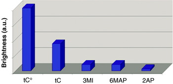
Also the oxo-homologue of tC called tCO (Fig. 11b), 1,3-diaza-2-oxophenoxazine, was originally developed by Matteucci and co-workers (Lin et al. Reference Lin, Jones and Matteucci1995) and proved to be a good cytosine analogue. Encouraged by the successful development of tC, we started to investigate the spectral properties of tCO. Like tC, tCO can easily be selectively excited. It has the lowest energy absorption band centred around 365 nm in duplex DNA as well as in ssDNA (Sandin et al. Reference Sandin, Börjesson, Li, Mårtensson, Brown, Wilhelmsson and Albinsson2008). This band consists of a single electronic transition polarized 33° counterclockwise from the long axis of tCO. Furthermore, the emission maximum of tCO is situated at 455 nm; its quantum yield in duplex DNA was found to be high (ϕf =0·22 on average) and the decay mono-exponential (Sandin et al. Reference Sandin, Börjesson, Li, Mårtensson, Brown, Wilhelmsson and Albinsson2008). In combination with a high molar absorptivity (ε=9000 M−1 cm−1) for a base analogue, this makes tCO on average the brightest fluorescent base analogue in duplex context currently available. It is more than two times brighter on average than tC and up to 10–50 times brighter than fluorescent base analogues like 3-MI, 6-MAP and 2-AP (Fig. 13). It should be pointed out that some of these base analogues even have higher brightness values than tCO for certain sequences, but on average they are considerably less bright. In contrast to tC and to the case for tCO in duplex, the quantum yield of tCO is sensitive to its surrounding bases in the single-stranded case (ϕf =0·14–0·41); in addition, the fluorescence decays in general need bi-exponential expressions to be adequately fitted (Sandin et al. Reference Sandin, Börjesson, Li, Mårtensson, Brown, Wilhelmsson and Albinsson2008). This difference distinguishes the photophysical properties of tCO in the single- and double-stranded case and, thus, it is possible to use tCO as a probe of nucleic acid secondary (and tertiary) structure (vide infra).
Average brightness (∝ϕf⋅ε) of common fluorescent base analogues in dsDNA calculated using values in Sandin et al., Ward et al., Sanabia et al., Driscoll et al., Hawkins et al. and Hawkins (average taken over various surrounding base pairs to the base analogue) (Driscoll et al. Reference Driscoll, Hawkins, Balis, Pfleiderer and Laws1997; Hawkins, Reference Hawkins2001; Hawkins et al. Reference Hawkins, Pfleiderer, Mazumder, Pommier and Falls1995; Sanabia et al. Reference Sanabia, Goldner, Lacaze and Hawkins2004; Sandin et al. Reference Sandin, Wilhelmsson, Lincoln, Powers, Brown and Albinsson2005, Reference Sandin, Börjesson, Li, Mårtensson, Brown, Wilhelmsson and Albinsson2008; Ward et al. Reference Ward, Reich and Stryer1969).

We also found that tCO, like tC, stabilizes a DNA duplex by approximately 3°C (Sandin et al. Reference Sandin, Börjesson, Li, Mårtensson, Brown, Wilhelmsson and Albinsson2008). Furthermore, we demonstrated that with an intelligent choice of neighbouring bases (GT, AA and AC) the change in duplex stability can be reduced to zero. Using CD, we established that duplexes containing a tCO in place of a cytosine still exist in the natural B-form (Sandin et al. Reference Sandin, Börjesson, Li, Mårtensson, Brown, Wilhelmsson and Albinsson2008). Further evidence of tCO being a non-perturbing base analogue that is nicely base paired and firmly stacked in the duplex comes from fluorescence anisotropy measurements. In these experiments, it was shown that the emission of tCO reports on the overall mobility of the DNA duplex without any interference from intrinsic mobility of the base analogue itself (Sandin et al. Reference Sandin, Börjesson, Li, Mårtensson, Brown, Wilhelmsson and Albinsson2008).
Apart from the study mentioned above where tCO reports on the mobility of a nucleic acid system, we have recently used it in two studies for monitoring nucleic acid sequence and structure. In the first study, we show that tCO can be used as an excellent probe for the detection of individual melting processes of complex nucleic acid structures containing a large number of separate secondary structure motifs (Börjesson et al. Reference Börjesson, Sandin and Wilhelmsson2009b). Since conventional UV-melting investigations only monitor the global melting process of the whole nucleic acid structure, e.g. multi-hairpin systems in RNA/DNA, and thus are incapable of estimating individual melting transitions of such systems, tCO represents a new method of characterization. Furthermore, we found in that study that tCO may be used to detect bulges and loops in nucleic acids as well as to distinguish a matched base pair from several of the mismatched. In the other study, we used the fluorescence properties of tCO to successfully probe the individual melting of all six sides in a DNA nanohexagon after repeated failures using commercially available covalently attached FRET probes (Sandin et al. Reference Sandin, Tumpane, Börjesson, Wilhelmsson, Brown, Nordén, Albinsson and Lincoln2009b). The commercial probes proved to disturb the efficiency of cyclization of this DNA nanoconstruct. In addition to these applications, derivatives of tCO, including the G-clamp (Lin & Matteucci, Reference Lin and Matteucci1998; Nakagawa et al. Reference Nakagawa, Ono, Li, Tsujimoto and Sasaki2007; Ortega et al. Reference Ortega, Blas, Orozco, Grandas, Pedroso and Robles2007; Rajeev et al. Reference Rajeev, Maier, Lesnik and Manoharan2001) that increases base-pair stability as well as the rigid nitroxid spin label, Ç (Barhate et al. Reference Barhate, Cekan, Massey and Sigurdsson2007; Cekan & Sigurdsson, Reference Cekan and Sigurdsson2008), have also been developed and used. Finally, it should be mentioned again that tCO has been used in studies concerning DNA polymerases (vide supra) (Sandin et al. Reference Sandin, Stengel, Ljungdahl, Börjesson, Macao and Wilhelmsson2009a; Stengel et al. Reference Stengel, Purse, Wilhelmsson, Urban and Kuchta2009a) as well as for high density labelling of polymerase chain reaction (PCR) products using the Deep Vent DNA polymerase (Stengel et al. Reference Stengel, Urban, Purse and Kuchta2009b).
3.2.4 Development of the first base analogue FRET pair
As discussed above, tC/tCO have several properties that make them ideally suited as FRET donors in nucleic acid systems: (1) a high and virtually unaffected quantum yield (ϕD), (2) a high control of orientation and position within the nucleic acid (κ2), (3) minimal perturbation to the structure of the nucleic acid, and (4) the possibility of having the probe close to the site of examination. The first two properties are important in order to have a high control of the Förster distance (R 0; Eq. 1), which is essential to know in order to be able to determine distances (R DA, distance between the donor and acceptor) using FRET efficiency (E; Eq. 2).


The geometric factor (κ2) takes the direction of the donor and acceptor transition dipoles into consideration and is described by Eq. (3):

where e1 and e2 are unit vectors of the donor and acceptor transition dipoles and e12 is the unit vector between their centres. The value of κ2 can range from 0 to 4. Thus, to be able to extract detailed structural information from the measured FRET efficiency, an accurate estimate of κ2 is required. The most frequently used (both correctly and incorrectly) κ2 is 2/3, which corresponds to freely rotating donor and acceptor transition dipoles and is therefore generally used in the case of donors and acceptors that are covalently attached using flexible linkers (e.g. rhodamines, fluoresceins, Cy dyes and Alexa dyes). However, several of these donor/acceptor chromophores interact with the nucleic acid structure (Dolghih et al. Reference Dolghih, Roitberg and Krause2007; Iqbal et al. Reference Iqbal, Wang, Thompson, Lilley and Norman2008b; Norman et al. Reference Norman, Grainger, Uhrin and Lilley2000), and thus the use of a κ2 of 2/3 may be an inaccurate assumption that results in considerable errors in structural interpretations. Various excellent investigations, such as those by Lewis et al., Iqbal et al. and Hurley and Tor have achieved an improved control of the FRET orientation factor in nucleic acid systems (Hurley & Tor, Reference Hurley and Tor2002; Iqbal et al. Reference Iqbal, Arslan, Okumus, Wilson, Giraud, Norman, Ha and Lilley2008a; Lewis et al. Reference Lewis, Zhang and Zuo2005).
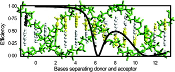
In an attempt to achieve the highest possible control of donor/acceptor orientation in nucleic acid systems, we have therefore developed a novel FRET pair composed of tCO as the donor and tCnitro (Fig. 11c) as the acceptor (Börjesson et al. Reference Börjesson, Preus, El-Sagheer, Brown, Albinsson and Wilhelmsson2009a). As for the other two members of the tricyclic cytosine family, tCnitro was shown to give minimal perturbation to both the structure and stability of DNA. The lowest energy absorption of tCnitro has a peak at approximately 440 nm, oriented ~27° clockwise from the molecular long axis towards the nitro group, resulting in a good overlap with the emission of tCO (and tC) (Preus et al. Reference Preus, Börjesson, Kilså, Albinsson and Wilhelmsson2010). In the investigations, we used a set of oligonucleotides that gave us the possibility of monitoring the FRET efficiency between tCO and tCnitro separated by 2–13 bases (approx. 10–50 Å). The results in Fig. 14 show a FRET efficiency that is highly dependent on both distance and orientation (oscillates with cos2 of the angle between the transition dipole moments of the donor and acceptor), which is expected for two chromophores that are firmly stacked in their positions during the lifetime of donor fluorescence. The data suggest that we have successfully designed an excellent nucleic acid base analogue FRET pair covering up to more than one turn of B-form DNA (Börjesson et al. Reference Börjesson, Preus, El-Sagheer, Brown, Albinsson and Wilhelmsson2009a).
Efficiency of energy transfer for the base analogue FRET-pair tCO–tCnitro estimated using decreases in tCO emission (light grey circles) and tCO average lifetimes (dark grey squares) as the two analogues are separated by 2–13 bases in a DNA duplex.

In conclusion, tCO and tCnitro constitute the first nucleic acid base analogue FRET pair. As a consequence of the unique property of both the analogues being rigidly located within the base stack and the donor quantum yield being stable, this system enables very high control of the Förster distance. A set of strands containing the FRET pair at strategically chosen positions, that is, where the slopes are steep in Fig. 14, will, hence, make it possible to accurately distinguish distance from orientation changes using FRET. In combination with the favourable base-pairing properties, this will facilitate detailed studies of the inherent dynamics of nucleic acid structures. Moreover, the placement of FRET-pair chromophores inside the base stack will be a great advantage in studies where other (biomacro)molecules interact with the nucleic acid.
4. Conclusions
When selecting the best or at least an appropriate fluorescent probe for the nucleic acid-containing system under investigation, there are several fluorophores to choose among ranging from large covalently attached external fluorophores like fluoresceins, rhodamines, Cy dyes or Alexa dyes to fluorescent base analogues with normal base-pairing properties. Either external or internal modification could be preferable and, therefore, it is impossible and incorrect to claim that a certain fluorophore or even group of fluorophores is, overall, best suited for fluorescently modifying DNA. Instead one needs to carefully consider each system under examination and try to optimize the choice of fluorescent probe molecule thereafter. However, with the increasing number of promising fluorescent base analogues developed in recent years as well as an enhanced sensitivity of fluorescence equipments, the odds of finding a base analogue that is appropriate for the required investigation has increased dramatically. There is no doubt that several of the current fluorescent base analogues have enabled some previously impossible investigations and in combination with the fact that there is still a large need for novel fluorescent base analogues of all the natural bases with improved as well as different spectral and photophysical properties, the development of this kind of fluorophore will be of great importance for a long time to come.
This review strives to cover the current state of the art in fluorescent nucleic acid base analogues. However, as with any review it is by no means fully comprehensive. To get a more complete picture of the whole literature of this field, readers could also refer to the excellent reviews by Millar, Rist and Marino, Asseline, Wilson and Kool as well as other reviews mentioned above (Asseline, Reference Asseline2006; Millar, Reference Millar1996; Rist & Marino, Reference Rist and Marino2002; Wilson & Kool, Reference Wilson and Kool2006).
Acknowledgements
This work was supported by the Swedish Research Council. I wish to thank all the co-authors of my articles concerning the tricyclic cytosine analogues and special thanks go to Dr Peter Sandin, Karl Börjesson, Dr Gudrun Stengel, Søren Preus as well as Professor Tom Brown and his group at University of Southampton, UK.