Case report
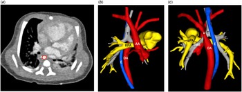
An 11-day-old full-term neonate with birth weight of 3.5 kg was admitted to our centre due to tachypnoea and decreased feeding. Echocardiography at presentation revealed left aortic arch, coarctation of the aorta with arch hypoplasia, a large perimembranous ventricular septal defect, a patent foramen ovale, and moderate degree of left ventricular systolic and diastolic dysfunction. Patent arterial duct was not observed. High-flow oxygen therapy and infusion of dopamine and prostaglandin E1 were initiated. CT confirmed the findings of echocardiography and additionally revealed the descending aorta that coursed from left to right traversing the midline (Fig. 1). After a few days of medical treatment, the left ventricular function has improved, but the arterial duct remained closed despite the use of prostaglandin E1. In our centre, we usually use end-to-side repair techniqueReference Lee, Brink and Galati1 for coarctation of the aorta with arch hypoplasia. However, there is a concern that such conventional aortic arch repair in the setting of malalignment of the aortic arch and descending aorta may cause airway compression by the abnormally coursing descending aorta.Reference Miwa, Iwai and Nagashima2 For this reason, we planned descending aortic translocation to prevent postoperative left main bronchial stenosis.
(a) Preoperative axial CT image showing the descending aorta (red circle) located on the right side of the vertebra. Preoperative 3D digital models reconstructed from CT images viewed from front (b) and back (c). The descending aorta courses from left to right traversing the midline. The asterisk represents the coarctation segment. AA = ascending aorta; DA = descending aorta; E = oesophagus; LMB = left main bronchus; LPA = left pulmonary artery; RPA = right pulmonary artery; T = trachea.

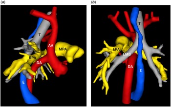
Surgery was performed at 18 days of age. Innominate artery and both caval veins were cannulated for cardiopulmonary bypass. Left-sided arterial duct was identified and divided. During cooling to 22°C, extensive mobilisation of the descending aorta was performed. After cardioplegic arrest, the ventricular septal defect and the patent foramen ovale were closed through a right atrial incision. Ligation of the aortic isthmus and clamping of the descending aorta were performed, and the ductal tissue was completely excised from the descending aorta. The descending aorta was then mobilised through the transverse sinus inferior to the tracheal carina and right pulmonary artery. Selective cerebral perfusion was initiated, and the aortic cross-clamp was removed. An incision was made on the posterior aspect of the proximal ascending aorta, and an anastomosis of the descending aorta to the ascending aorta was accomplished using a 7–0 polypropylene running suture. After deairing, whole-body perfusion was restored. The duration of selective cerebral perfusion time was 21 minutes. Post-operative course was uncomplicated, and the patient was discharged on the twenty-fifth post-operative day. CT before discharge revealed widely patent pathway to the descending aorta and both main bronchi (Fig. 2).
Postoperative 3D digital models reconstructed from CT images viewed from right (a) and back (b), revealing widely patent pathway to the descending aorta and both main bronchi. AA = ascending aorta; DA = descending aorta; E = oesophagus; LMB = left main bronchus; LPA = left pulmonary artery; MPA = main pulmonary artery; RPA = right pulmonary artery; T = trachea.

Discussion
Circumflex retroesophageal aortic arch is a rare form of aortic arch anomaly in which the aortic arch and the descending aorta are placed on opposite sides of the spine.Reference Wernovsky, Anderson and Kumar3 In the presence of an ipsilateral arterial duct with the descending aorta, as is often the case, it forms a complete vascular ring. Left aortic arch with right descending aorta associated with coarctation of the aorta is an even rarer congenital cardiac anomaly.Reference Miwa, Iwai and Nagashima2,Reference Sheth, Varghese and Sivakumar4 In our patient, the aortic arch and the descending aorta were on opposite sides of the midline, although the aorta did not course behind the oesophagus and the arterial duct was left-sided (contralateral to the descending aorta).
Left main bronchial stenosis is a well-known complication that may occur after repair of coarctation of the aorta. Usually, the mechanism is an external compression of the left main bronchus by the anteriorly relocated descending aorta. In patients with a circumflex aortic arch, this complication may occur more frequently because the descending aorta should be brought up towards the aortic arch after crossing the midline. Miwa et al reported three patients with left aortic arch, right descending aorta, and coarctation who were treated with conventional resection and direct anastomosis.Reference Miwa, Iwai and Nagashima2 In two patients, left main bronchial stenosis developed by gradual traction of the descending aorta towards the left side, thereby narrowing the space between the descending aorta and the right pulmonary artery. They suggested that alternative surgical techniques, such as arch reconstruction with an autologous pulmonary artery patch, should be considered in this situation to prevent postoperative left main bronchial stenosis.
Descending aortic translocation, first reported by McKenzie et al, is a procedure originally designed to relieve the airway compression by the midline-crossing descending aorta without associated coarctation of the aorta.Reference McKenzie, Roeser and Thompson5 In this procedure, the proximal descending aorta is transected, brought up through the transverse sinus caudad to the tracheal carina and pulmonary artery, and anastomosed in an end-to-side fashion to the proximal ascending aorta. In this way, the descending aorta is relocated away from the airway. Sheth et al reported successful use of this technique in a 5-month-old infant with left aortic arch, right descending aorta, coarctation of the aorta, and severe preoperative left main bronchial stenosis.Reference Sheth, Varghese and Sivakumar4 In our patient, there was no airway stenosis preoperatively. However, we were concerned about potential postoperative left main bronchial stenosis, given that we favour end-to-side repair techniqueReference Lee, Brink and Galati1 for coarctation of the aorta with associated arch hypoplasia. Descending aortopexy to address this issue may be ineffective in cases with midline-crossing descending aorta. Therefore, we decided to perform descending aortic translocation to prevent postoperative left main bronchial stenosis. Although a little more dissection in the posterior mediastinum was required compared with conventional repair, this simple manoeuver relocated the descending aorta well away from the airway (Fig. 2). One of the potential complications of this procedure may be chylothorax due to extensive dissection of the descending aorta. We believe that descending aortic translocation may be a useful surgical option to prevent airway stenosis in patients with coarctation of the aorta and midline-crossing descending aorta. However, long-term effect of the resultant non-anatomical aortic arch geometry remains to be elucidated.Reference Ou, Celermajer and Raisky6
Acknowledgements
None.
Financial support
This research received no specific grant from any funding agency, commercial, or not-for-profit sectors.
Competing interests
None.
Ethical standard
This study was reviewed and approved by the Institutional Review Board of Seoul St. Mary’s Hospital.