Introduction
Bidirectional communication between oocytes and cumulus cells is essential for proper maturation of oocytes, fertilization and further embryonic development (Buccione et al., Reference Buccione, Schroeder and Eppig1990; Eppig, Reference Eppig1991; Matzuk et al., Reference Matzuk, Burns, Viveiros and Eppig2002). During oocyte maturation, the cumulus cells provide a network of gap junction transmembrane channels that allows the transport of nutrient and paracrine factor between oocytes and cumulus cells (Eppig, Reference Eppig1991; Grazul-Bilska et al., Reference Grazul-Bilska, Reynolds and Redmer1997; Tanghe et al., Reference Tanghe, Soom, Nauwynck, Coryn and Kruif2002; Gilchrist et al., Reference Gilchrist, Ritter and Armstrong2004). This finding has been demonstrated by the removal of cumulus cells before maturation whereby oocyte maturation was suppressed (Mori et al., Reference Mori, Amano and Shimizu2000; Vozzi et al., Reference Vozzi, Formenton, Chanson, Senn, Sahli, Shaw, Nicod, Germond and Haefliger2001; Fatehi et al., Reference Fatehi, Zeinstra, Kooij, Colenbrander and Bevers2002; Wongsrikeao et al., Reference Wongsrikeao, Kaneshige, Ooki, Taniguchi, Agung, Nii and Otoi2005; Assidi et al., Reference Assidi, Dufort, Ali, Hamel, Algriany, Dielemann and Sirard2008). In contrast, oocyte-secreted factors (OSFs) are believed to regulate key cumulus cell functions, which in turn enhance positive regulatory factors that are required during subsequent development of the oocytes (Sugiura et al., Reference Sugiura, Pendola and Eppig2005).
Oocytes are known to have high transcriptional activity in the germinal vesicle (GV) stage leading to metaphase II (MII). The stored mRNAs and proteins in the oocyte support early embryogenesis (Memili & First, Reference Memili and First1999; Dalbies-Tran & Mermillod, Reference Dalbies-Tran and Mermillod2003; Vallee et al., Reference Vallee, Gravel, Palin, Reghenas, Stothard, Wishart and Sirard2005). However, as development proceeds and maternally inherited informational molecules decay, early embryogenesis becomes dependent on the expression of genetic information from the embryo genome (Telford et al., Reference Telford, Watson and Schultz1990). This dynamic change in transcript abundance before and after in vitro maturation of bovine oocytes can suggest the presence of temporal control of gene expression during oocyte maturation, fertilization and early embryo development (Dalbies-Tran & Mermillod, Reference Dalbies-Tran and Mermillod2003; Fair et al., Reference Fair, Carter, Park, Evans and Lonergan2007).
Similar to mRNA, microRNA (miRNA; a class of small non-coding RNAs of 18–25 nucleotides in length) are also believed to change their expression during oocyte maturation and embryonic development. For instance, we have shown previously dynamic changes in miRNA expression during oocyte maturation in bovine (Tesfaye et al., Reference Tesfaye, Worku, Rings, Phatsara, Tholen, Schellander and Hoelker2009), comparable with that reported in mouse (Tang et al., Reference Tang, Kaneda, O'Carroll, Hajkova, Barton, Sun, Lee, Tarakhovsky, Lao and Surani2007). Furthermore, the presence of regulatory miRNAs has been reported in oocytes and in numerous reproductive tissues (Murchison et al., Reference Murchison, Stein, Xuan, Pan, Zhang, Schultz and Hannon2007; Hong et al., Reference Hong, Luense, McGinnis, Nothnick and Christenson2008; Nagaraja et al., Reference Nagaraja, Andreu-Vieyra, Franco, Ma, Chen, Han, Zhu, Agno, Gunaratne, DeMayo and Matzuk2008; Carletti & Christenson, Reference Carletti and Christenson2009; Creighton et al., Reference Creighton, Benham, Zhu, Khan, Reid, Nagaraja, Fountain, Dziadek, Han, Ma, Kim, Hawkins, Anderson, Matzuk and Gunaratne2010). In addition, disruption of Dicer (a polymerase II enzyme responsible for miRNA processing and a central enzyme in miRNA processing) in mouse oocytes was found to result in defects in meiotic spindle organization and chromosome congression, reduced maturation rate and reduced the transcripts of Dicer miRNAs (Murchison et al., Reference Murchison, Stein, Xuan, Pan, Zhang, Schultz and Hannon2007; Liu et al., Reference Liu, Tang, He and Rosenwaks2010). Apart from oocytes, the role of miRNAs in maternal–zygotic transition has been reported in mouse (Tang et al., Reference Tang, Kaneda, O'Carroll, Hajkova, Barton, Sun, Lee, Tarakhovsky, Lao and Surani2007; Lykke-Andersen et al., Reference Lykke-Andersen, Gilchrist, Grabarek, Das, Miska and Zernicka-Goetz2008) and zebrafish (Giraldez et al., Reference Giraldez, Mishima, Rihel, Grocock, Van Dongen, Inoue, Enright and Schier2006).
The accumulating evidence supports the potential role of these non-coding miRNAs in post-transcriptional regulation of genes involved in various aspects of mammalian reproduction. However, so far, little information is known about the abundance of miRNAs in bovine oocytes and their surrounding cumulus cells before and after oocyte maturation, which can elucidate bidirectional communication between the two cell types. Therefore, here we report the comparative expression analysis of a set of miRNAs in bovine oocytes and in the surrounding cumulus cells at the GV and MII stages. Moreover, the expression profile of six miRNAs enriched in oocytes was investigated at different time points during oocyte maturation and preimplantation embryo development. The spatio-temporal expression of two miRNAs was investigated in sections at different follicular stages, in COCs and in bovine preimplantation stage embryos.
Materials and methods
Cumulus–oocyte complex (COC) collection and in vitro maturation
Bovine ovaries were obtained from a local slaughterhouse and transported to the laboratory in a thermoflask that contained a 0.9% saline solution and within 2 h after slaughter. COCs were aspirated from follicles of 2–8 mm, only morphologically uniform and good quality COCs (oocytes with multiple layers of cumulus cells and with a translucent and homogeneous ooplasm) were used for this study. COCs were washed in maturation medium supplemented with 15% estrous cow serum (ECS), 0.5 mM l-glutamine, 0.2 mM pyruvate, 50 mg/ml gentamycin sulphate and 10 μl/ml follicle-stimulating hormone (FSH; Follitropin®) before set into culture to obtain matured oocytes or directly freezed as immature oocytes. The COCs were cultured in groups of 50 in 400 μl of maturation medium under mineral oil in four-well dishes (Nunc). Maturation was performed for 24 h at 39°C under a humidified atmosphere of 5% CO2 in air. Both immature and matured COCs were collected and subjected to mechanical separation of the oocytes and the surrounding cumulus cells using gentle vortexing. Denuded oocytes in group of 200s and the corresponding cumulus cells were snap frozen in cryo-tubes that contained lysis buffer (0.8% Igepal; Sigma), 40 U RNasin and 5 mM dithiothreitol (DTT; Promega) and stored at –80°C until further use. A total of three replicate pools that each contained 1600 oocytes and their corresponding cumulus cells from both immature and matured COCs were utilized for the analysis of miRNA using a PCR-based miRNA array. In addition, three replicate pools of COC (n = 400) were collected at different maturation times (0, 8, 16 and 22 h) to analyse the temporal expression of selected miRNAs.
In vitro embryo production
Independent samples of COCs were collected and used for in vitro maturation, in vitro fertilization and in vitro culture to generate embryos of different stages of development (matured oocytes, zygote, 2-cell, 4-cell, 8-cell, morula and blastocyst). In vitro matured oocytes were collected after COCs were left to mature for 24 h. A total of six independent in vitro maturation runs were conducted to collect three independent pools of 450 matured oocytes. To generate different stages of preimplantation embryo, in vitro matured COCs were fertilized in vitro in Fert-TALP medium using proven bull spermatozoa at the concentration of 1 × 10⁶ cells/ml. After 20 h of co-incubation with spermatozoa, cumulus cells were removed and cumulus-free zygotes were washed and transferred to CR1aa culture media (Rosenkrans & First, Reference Rosenkrans and First1994) supplemented with essential and non-essential amino acids. Following this step, zygotes, 2-cell, 4-cell, 8-cell, morula and blastocyst stage embryos were collected from the culture at 20, 30, 42, 70, 120 and 168 h after fertilization, respectively. From six independent IVF runs, three independent pools that each consisted of 150 zygotes, 100 2-cell, 80 4-cell, 60 8-cell, 50 morula and 30 blastocyst stage embryos were collected and snap frozen in cryo-tubes that contained lysis buffer. These embryos and the matured oocytes were utilized for the expression analysis of selected miRNAs and their predicted target mRNAs.
Preparation and culture of oocyte-ectomized cumulus cells and denuded oocytes
To investigate the effect of presence or absence of cumulus cells on expression of miRNA in oocytes and vice versa, oocytes or cumulus cells were matured in vitro in the presence or absence of the other. For this step, mechanical removal of oocyte cytoplasm from COCs was performed using a micromanipulator fitted to an inverted microscope (Leica DM-IRB). In full, a group of 50 COCs was placed into manipulation medium [HEPES-buffered tissue culture medium 199 (H-TCM199)] under mineral oil to remove all contents of the oocyte cytoplasm, including the nucleus; the COCs were held with a holding pipette and the cytoplasm contents sucked out using an enucleating pipette attached to the micromanipulator. To collect denuded oocytes for maturation, immature COCs were subjected to gentle vortexing in phosphate-buffered saline (PBS) for mechanical removal of the cumulus cells. The complete removal of the cumulus cells was checked; then denuded oocytes and oocyte-ectomized cumulus cells were washed twice in drops of fresh maturation medium and placed in culture for in vitro maturation. Each time, morphologically good quality COCs were categorized into three groups: group 1 included COCs matured in vitro as intact; group 2 included those oocytes matured without the surrounding cumulus cells (denuded oocytes); and group 3 included cumulus cells matured without the oocyte cytoplasm (oocyte-ectomized). The experiment was conducted four times with the same handling and culture conditions. Each time, 250 COCs from each of the three mentioned groups were snap frozen in cryo-tubes that contained lysis buffer. Collected samples were used to analyse the expression of selected miRNAs.
RNA isolation and first strand synthesis
In all experiments total RNA enriched with miRNAs was isolated from the experimental samples using an miRNeasy® Mini kit (QIAGEN). Furthermore, the small RNAs were separated from the large RNA using RT2 qPCR-Grade miRNA Isolation Kit (SABiosciences). In all experiments the resulting small RNAs and fractionated large RNAs were checked for their concentration and integrity (based on 260/280 nm measurement) using a NanoDrop 8000 spectrophotometer (peqLab Biotechnologies GmbH). Small RNAs from each treatment groups were used for first strand cDNA synthesis using the RT2 miRNA First Strand Kit (SABiosciences) following manufacturer's protocols. Briefly, 50 ng of small RNA samples were incubated with a mixture that contained 2 μl of 5 × miRNA RT buffer, 1 μl miRNA RT primer, 1 μl DTT (100 mM) and 1 μl miRNA RT enzyme mix in a 10 μl reaction volume. Reaction incubation was performed at 37°C for 2 h followed by heating at 95°C for 5 min to inactivate the reverse transcriptase. Depending on the type of experiment and the amount of small RNA used for RT-PCR, the resulting cDNA samples were diluted before use as template for the miRNA qPCR assay. The large RNA fractions from the same samples of preimplantation embryos were recovered and utilized for RT-PCR after removal of contaminating DNA using the TURBO DNA-free kit (Ambion) according to manufacturer's instructions. All DNA-free RNA samples were reverse transcribed using the Superscript II reverse transcriptase kit (Invitrogen). Briefly, maximum volume of 10 μl RNA (with 100 ng of input RNA) from each treatment group were co-incubated with 1 μl of random hexamer (Invitrogen) and 1 μl of oligo(dT)23 at 70°C for 3 min followed by incubation on ice for 2 min. Reverse transcription was performed in a 20 μl reaction volume that contained 4 μl 5 × first strand buffer, 2 μl DTT, 1 μl dNTP, 0.3 μl RNasin and 0.7 μl RT enzyme. The reaction was incubated for 90 min at 42°C, and terminated by heating at 75°C for 15 min.
RT profiler PCR array and data analysis
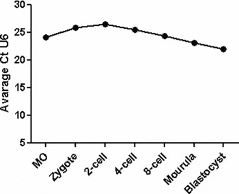
RT profiler PCR array were performed using sequence-specific miRNA qPCR primer sets and the RT2 Real-Time™ PCR master mix (SABiosciences) in ABI Prism 7000 instrument (Applied Biosystems). Expression profiling of miRNAs between oocytes and cumulus cells before and after maturation was performed using an miRNA PCR array related to human cell development and differentiation (SA Biosciences) that contained 88 well known human homologue miRNAs, four housekeeping small RNAs (SNORD44, SNORD47, SNORD48 and U6) and RT negative controls in a 96-well format. Prior to real-time PCR profiling, 90 μl DNase/RNase-free water was added to each of the 10 μl first strand cDNA product from each biological replicates of immature and matured oocytes and their corresponding cumulus cells. A PCR master mix was prepared using the 100 μl diluted cDNA template, 1275 μl 2× RT2 SYBR Green PCR master mix (SABiosciences) and 1175 μl DNase-/RNase-free water. Twenty five μl of this mixture was distributed to each well of the 96-well plate that contained sequence-specific miRNA primer sets and the respective controls. Following brief centrifugation, the plate was loaded onto ABI Prism 7000 real-time PCR apparatus and run with a thermal programme of initial heating at 95°C for 10 min followed by 40 cycles of 95°C for 15 s and 60°C for 1 min. The specificity of amplification was controlled using a melting curve generated at the end of the PCR protocol. The data were analysed by the comparative threshold (Ct) method and normalization was done using the geometric mean of four endogenous controls (SNORD44, SNORD47, SNORD48 and U6) through SABiosciences's PCR array data analysis on-line web portal provided with t-test (http://www.sabiosciences.com/pcr/arrayanalysis.php). Selection of miRNAs for further characterization was performed based on the differential expression intensity between oocytes before and after maturation. Similarly, quantitative real-time PCR analysis of selected miRNAs was performed in oocyte at different maturation time and preimplantation stages embryos using RT2 miRNA qPCR assay primer set and U6 as endogenous controls. The stability of the U6 expression was checked (Appendix Figure 1) before it was utilized for normalizing the relative abundance of specific miRNAs. Relative quantification of miRNA expression was calculated using the Ct method as described in our previous study (Tesfaye et al., Reference Tesfaye, Worku, Rings, Phatsara, Tholen, Schellander and Hoelker2009).
miRNA target prediction and in silico functional analysis
The target mRNAs of the differentially expressed miRNAs were predicted using four different search algorithms, TARGETSCAN 5.1 (http://www.targetscan.org/), PICTAR (http://www.pictar.org), miRDB (http://www.miRDB.org/), and MicroRNA.org-Target and Expression (http://www.microrna.org/). The targets that were predicted by two or more target search tools were accepted as a target gene for the miRNA under consideration. The biological functions of these predicted targets and the molecular pathways in which they could be involved were analysed through the use of IPA (Ingenuity® Systems, www.ingenuity.com)
Quantitative real-time PCR analysis of target mRNAs
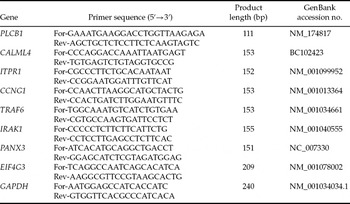
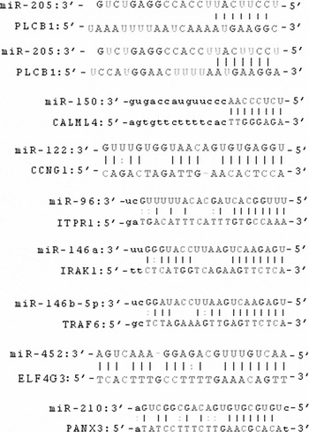
Quantitative RT-PCR was performed for one target gene per miRNA using iTaq™ SYBR® Green Supermix with ROX (BioRad) in an ABI Prism 7000 instrument (Applied Biosystems). Predicted target transcripts that were quantified in preimplantation stages embryos included phospholipase C, beta 1 (PLCB1) for miR-205, calmodulin-like 4 (CALML4) for miR-150, cyclin G (CCNG1) for miR-122, 1,4,5-triphosphate receptor 1 (ITPR1) for miR-96, interleukin-1 receptor-associated kinase 1 (IRAK1) for miR-146a, TNF-receptor-associated factor 6 (TRAF6) for miR-146b-5p, eukaryotic translation initiation factor 4 gamma, 3 (EIF4G3) for miR-452 and pannexin 3 (PANX3) for miR-210. Sequence complementarities between miRNAs and their target mRNA 3′UTR is indicated in Appendix Figure 2. Quantification of all mRNA transcripts of interest was performed using GAPDH as endogenous control (El-Sayed et al., Reference El-Sayed, Hoelker, Rings, Salilew, Jennen, Tholen, Sirard, Schellander and Tesfaye2006) after evaluating its stability across the different samples. The list of primers and corresponding sequences are indicated in Table 1. Final quantification analysis was preformed using a standard curve method (user bulletin #2 ABI PRISM 7700 SDS, http://docs.appliedbiosystems.com/pebiodocs/04303859.pdf) and results were reported as the fold change difference to the calibrator cDNA after normalization of the transcript amount to the endogenous control.
Sequence-specific primers used for quantitative PCR

In situ detection of miR-205 and miR-210
Bovine ovaries at 21 days of estrous cycle were fixed in 4% paraformaldehyde (PFA) overnight at 4°C followed by overnight incubation in PBS with 30% sucrose at 4°C and frozen in Tissue-Tek OCT reagent (Sakura Finetek). Cryo-section (10 μm) preparation, post-fixation, acetylation and proteinase K treatment were done as described previously (Obernosterer et al., Reference Obernosterer, Martinez and Alenius2007). Two hours of pre-hybridization was performed at 61°C in hybridization solution (50% formamide, 5 × sodium chloride/sodium citrate [SSC; pH 6.0], 0.1% Tween-20, 50 μg/ml heparin, and 500 mg/ml yeast tRNA). Then, ovarian sections were incubated overnight at 61°C with 3′-digoxigenin (DIG)-labelled, LNA-modified oligonucleotide ISH probes (1 pM) for miR-205, miR-210, U6 RNA and scrambled RNA sequence (Exiqon) in hybridization buffer under humidified condition. Blocking, incubation with anti-DIG-AP antibody, washing and colour development using Fast Red reaction was performed as described previously (Obernosterer et al., Reference Obernosterer, Martinez and Alenius2007). The slides were mounted with VectaShield containing DAPI (Vector Laboratories) and analysed under a confocal laser scanning microscope (CLSM LSM-510, Carl Zeiss).
Whole mount in situ hybridization of immature COC and in vitro-produced bovine preimplantation embryos were performed in 4-well culture dishes according to the high-resolution whole mount in situ hybridization protocol from Exiqon, with some modification. Signal detection was performed as described for cryo-sections. COC (n = 5) and embryos (n = 7 embryos per stage) were handled and managed with handmade glass pipettes (opening 250 μm) under a stereo-microscope. In all cases, embryos were briefly washed once before each step to avoid cross-contamination of solutions. Images for the different layers or the position of COC and embryos were taken by confocal laser scanning microscope (CLSM LSM-510, Carl Zeiss) using Z-Stack with 1 μm interval. Three-dimensional images from all the layers or Z positions of single embryo were constructed using the software ZEN 2008 Light Edition (Carl Zeiss).
Results
Expression profiling of miRNAs in immatured oocytes vs. their surrounding cumulus cells
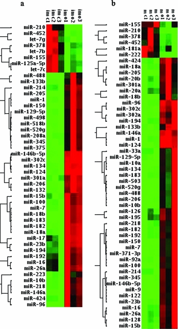
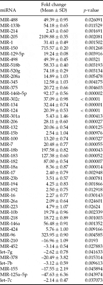
Expression profiling of 88 miRNAs between immatured oocytes and their surrounding cumulus cells revealed 47 differentially expressed miRNAs (p < 0.05, fold change > 2; Fig. 1a) between the two cell types. Among these examples, the expression level of eight miRNAs, including miR-125a-5p, miR-155, miR-210, miR-378 and miR-452, were enriched in cumulus cells. Conversely, the expression levels of 39 differentially expressed miRNAs were enriched in immatured oocytes compared with their surrounding cumulus cells. Among these, miR-146a, miR-146b-5, miR-183, miR-18b, miR-96, miR-150 and miR-205 exhibited a 56- to 2109-fold change increase in the oocytes. The list of all differentially expressed miRNAs between immatured oocytes and their surrounding cumulus cells is described in Appendix Table 1.
Hierarchical clustering of differentially expressed miRNAs between immature oocytes (imo) vs. cumulus cells (imc) (a) and matured oocytes (mo) vs. cumulus cells (mc) (b). Columns represent the similarities or differences in the expression pattern within or between biological replicates of oocyte or cumulus cells and the horizontal dendrogram represent the clustering of miRNA according to their expression similarities. This figure is available in colour in the online version of this paper. Red and green colours indicate the higher and lower expression level, respectively.

Expression profiling of miRNAs in matured oocytes vs. their surrounding cumulus cells
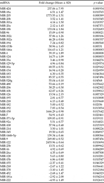
The differentially expressed miRNA between matured oocytes vs. their surrounding cumulus cells were also analysed using miRNA PCR array that consisted of 88 miRNAs. The result revealed that 51 miRNAs were differentially expressed (p < 0.05 and fold change > 2) between these two groups of cells. From those, six miRNAs (miR-155, miR-210, miR-378, miR-452, miR-18a and miR-222) were enriched in cumulus cells and 45 miRNAs were enriched in matured oocytes (Fig. 1b). Several miRNAs including miR-317–3P, miR146a, miR-18b, miR-146b-5p, miR-424, miR-9, miR-150, miR-205 and miR-122 were among the top miRNAs (109- to 2433-fold changes) enriched in matured oocytes compared with their surrounding cumulus cells. The detail list of all differentially expressed miRNAs is described in Appendix Table 2.
Commonly differentially expressed miRNA in immatured or matured oocyte vs. their surrounding cumulus cells
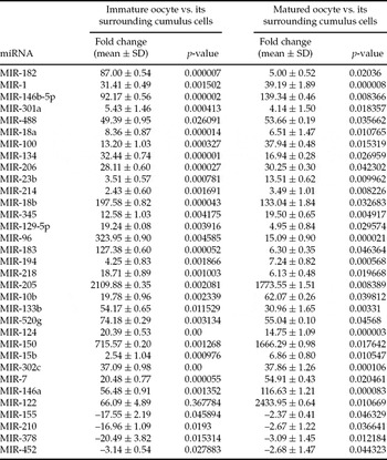
We merged the two data sets described above to identify the miRNA that are constantly expressed in oocytes or cumulus during oocytes maturation. From this analysis, we have identified 33 miRNAs commonly differentially expressed between oocytes and surrounding cells before and after maturation. From those miRNAs, miR-155, miR-210, miR-378, miR-452 were enriched in cumulus cells compared with the oocytes before and after maturation (Table 2). In addition, 29 miRNAs (including miR-205, miR-150, miR-96, miR-122, miR-146a and miR-146b-5p) were enriched in immatured and matured oocytes compared with their surrounding cumulus cells (Table 2).
List of commonly differentially expressed miRNA between oocytes and their corresponding cumulus cells after and before maturation

Positive fold change means up-regulated in oocytes and negative fold change means down-regulated in oocytes compared with the corresponding cumulus cells.
Temporal expression profile of selected miRNAs in oocytes and preimplantation embryo
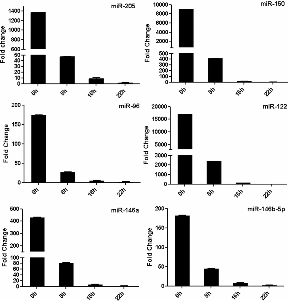
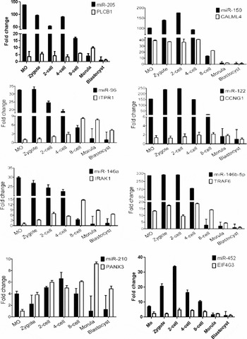
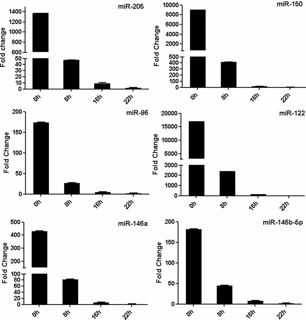
Following identification of commonly differentially expressed miRNA in immatured or matured oocyte vs. surrounding cumulus cells, six miRNAs enriched in oocyte (miR-205, miR-150, miR-96, miR-122, miR-146a and miR-146b-5p) were selected based on their expression patterns. The expression patterns of these miRNAs were then analysed at different time points in oocyte maturation (0 h, 8 h, 16 h, and 22 h). The results showed that the expression of these miRNAs was higher relatively at 0 and 8 h followed by reduction at 16 until 22 h of maturation in vitro (Fig. 2). Following this step, we evaluated whether the expression of these miRNAs would be retained or degraded during early embryo formation. For this evaluation, the expression pattern of these miRNAs was examined in different stages of preimplantation embryos and these miRNAs were found to be highly abundant in early stages of development up to the 4-cell embryo stage, followed by a dramatic reduction in expression at the 8-cell stage up to the blastocyst stage. Similarly, two miRNAs (miR-452 and miR-210) found to be enriched in cumulus cells were selected based on their expression patterns, and their expression profiles were analysed at different stages of preimplantation embryo development. Unlike those miRNAs enriched in oocytes, these miRNAs showed no defined pattern of expression throughout the developmental stages, and could be detected at relatively lower levels in all stages of development (Fig. 3).
Relative abundance of miR-205, 150, 96, 122, 146a and 146b-5p in oocyte at different maturation times.
Relative abundance of miRNAs enriched in oocytes (miR-205, 150, 96, 122, 146a and 146b-5p), miRNAs enriched in cumulus cells (miR-210 and miR-452) and their predicted genes in bovine mature oocyte (MO) and preimplantation stage embryos.

Expression profiling of genes targeted by differentially expressed miRNAs
Targets genes were selected using four different target search tools. The expression profiles of one target gene for each selected miRNA were analysed at different stages of preimplantation embryos. The results showed that three genes, namely PLCB1, ITPR1 and IRAK1, had a ‘reversed’ expression profile compared with the profile observed by their targeting miRNAs (miR-205, miR-96 and miR-146a), respectively (Fig. 3). However, CALML4 and TRAF6 transcripts showed a similar expression profile to their targeting miRNAs, namely miR-150 and miR-146b-5p. In contrast, the relative abundance of CCNG1 (the target of miR-122) was measurable up to the later stages of development, irrespective of the expression of the targeting miRNA. As it is the case for targeting miRNAs (miR-210 and miR-452), the target genes PANX3 and EIF4G3 showed no clear trend in expression profile throughout the preimplantation period (Fig. 3).
Effect of oocyte and cumulus cell factors for the expression of miRNAs
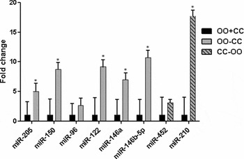
In order to determine whether the oocyte (as source of OSFs) is required for the expression of miRNAs in the surrounding cumulus cells, and vice versa, comparative analyses of oocytes matured with or without cumulus cells and cumulus cells matured with or without the oocyte cytoplasm were performed using the eight miRNAs. As shown in Fig. 4, the relative abundance of the majority of miRNAs was found to be higher (p < 0.05) in oocytes matured without the surrounding cumulus cells, except for miR-96. In contrast, a 16-fold increase in the relative abundance of miR-210 was observed in cumulus cells matured without the oocyte cytoplasm compared with those cells matured in the presence of oocyte cytoplasm. However, no significant differences were found in the relative abundance of miR-452 in these experimental groups.
Relative expression level of miRNAs in oocytes matured with (OO+CC) or without (OO-CC) the surrounding cumulus cells and in cumulus cells matured with (CC+OO) or without (CC-OO) the oocyte cytoplasm. Samples with the highest normalized value were used as a calibrator. Statistical significance (p < 0.05) between oocytes or cumulus cells is indicated by a star (*).

Spatio-temporal distribution of miR-205 and miR-210 in ovarian follicle sections
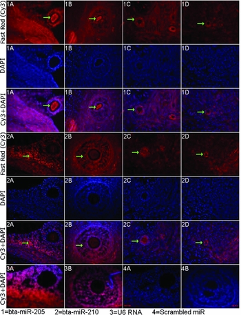
In situ hybridization was performed to examine the cell type distribution of miR-205 and miR-210 enriched in oocyte and cumulus cells, respectively in sections of bovine follicles (primary, secondary, early antral and tertiary follicles). A strong fluorescence signal of miR-205 was detected in the oocyte cytoplasm compared with the surrounding cumulus cells and other ovarian cells during the later stages of follicular development namely: early antral and tertiary follicles (Fig. 5). However, the expression level of miR-205 was not detectable in the oocyte cytoplasm at primary and secondary follicles (Fig. 51C, 1D). In contrast, miR-210 was found to be highly abundant in cumulus and granulosa cells compared with the oocyte cytoplasm in all stages of follicular development. Differences in miR-210 signal intensity between oocyte and surrounding cells was clear at the later stages of follicular development (early antral and tertiary follicles). Moreover, miR-210 was found to be localized in a single layer of cumulus cells in the primary follicles (Fig. 52D) and in multiple layers of cumulus cells in secondary, early antral and tertiary follicles (Fig. 52A–2C). While the oocyte cytoplasm in the primary follicle showed no miR-210 expression (Fig. 52D), slight signals were detected in oocyte cytoplasm of the secondary, early antral and tertiary follicle stages (Fig. 52A–2C). The nuclear U6 RNA, which was used as the positive control in this study, was detected in all follicular sections as it was localized in the nucleus (Fig. 53A, 3B).
In situ detection of miR-205 and 210 in bovine follicular cryo-sections. Ovarian follicular cryo-sections were in situ hybridized with 3′-digoxigenin labelled locked nucleic acid (LNA) microRNA probes of miR-205 (1), miR-210 (2), U6 RNA (3) and scrambled miRNA (4). Cells with higher expression are marked by arrows. A, B, C and D represent tertiary, early antral, secondary and primary follicle, respectively. This figure is available in colour in the online version of this paper.

In situ localization of miR-205 and miR-210 in in vitro produced oocytes and preimplantation stage embryos
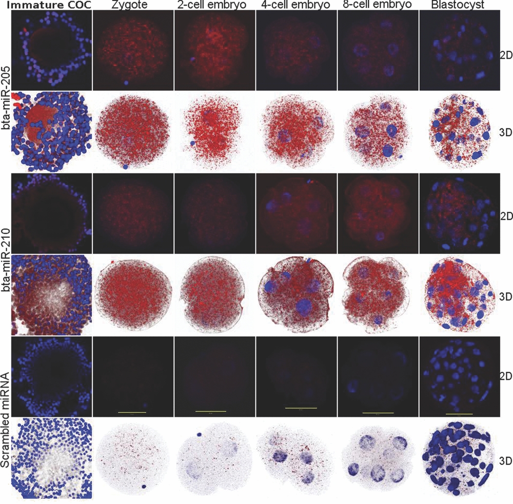
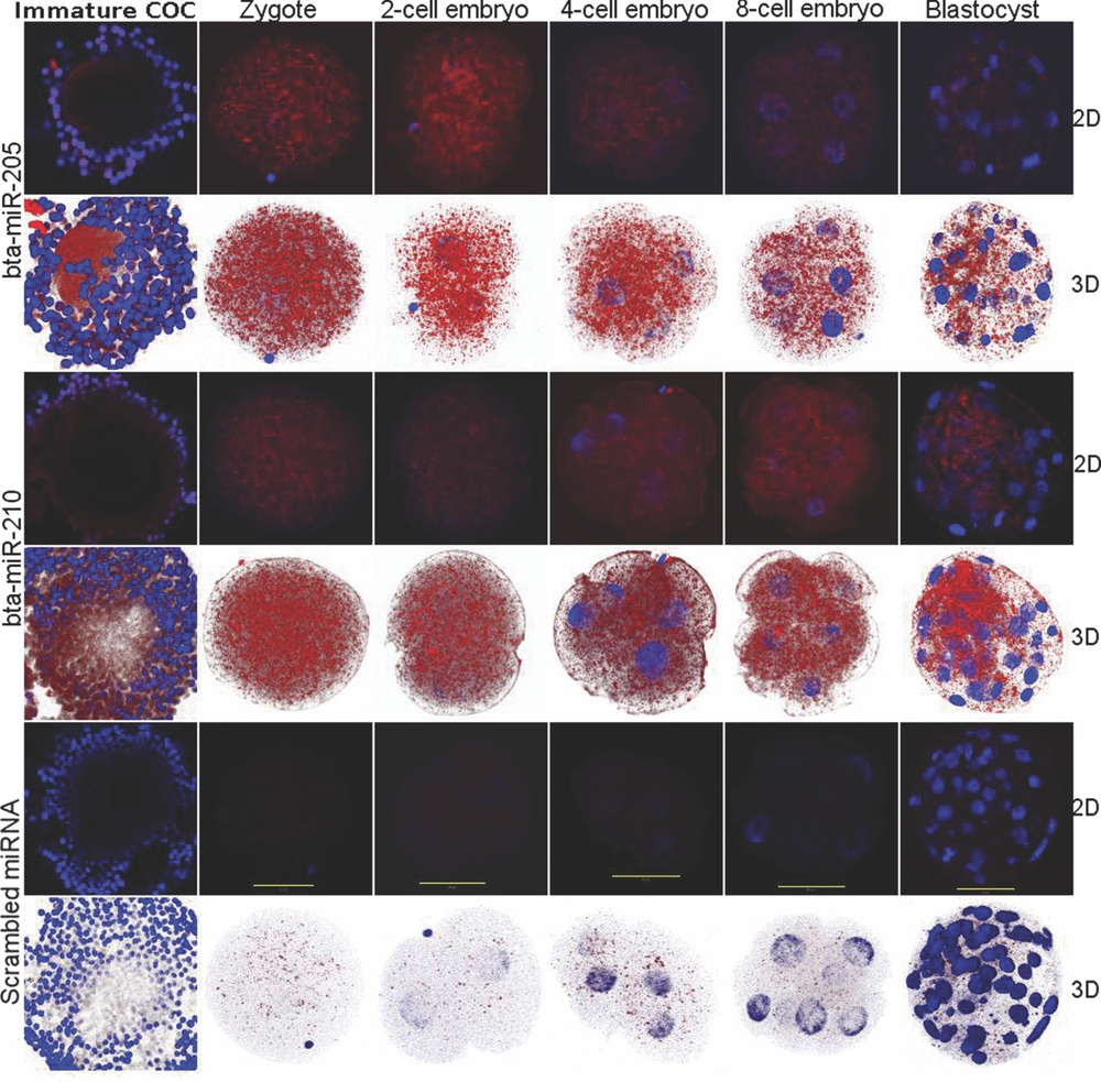
Cumulus–oocyte complex and in vitro produced bovine post-fertilization developmental stages (zygote, 2-cells, 4-cells, 8-cells and blastocyst) were used to investigate the cellular distribution of these miRNAs in early stages of embryo development. Both two- and three-dimensional presentations showed that miR-205 was detected in oocytes and in all stages of development with a reduced signal between the 8-cell and blastocyst stages compared with the signal detected at the oocyte, zygote, 2-cell and 4-cell stages (Fig. 6). However, miR-210 is detected in cumulus cells in the COC and at all stages of development, but without a noticeable difference in signal intensity between the developmental stages.
In situ detection of miR-205 and 210 in whole mount cumulus oocyte complex and post-fertilization stage bovine embryos. Whole mount bovine preimplantation embryos were in situ hybridized with 3′-digoxigenin labelled locked nucleic acid (LNA) microRNA probes of miR-205, miR-210 and scrambled miRNA. Hybridization signal and nucleus of the embryos were visualized by Fast Red (Cy-3) and DAPI, respectively. Images from at least 20 different layers or positions (Z positions) of single embryos were used to construct three-dimensional images and two-dimensional images, represented here, from only one position. Scale bar denotes 50 μm. This figure is available in colour in the online version of this paper.
Functional classification of genes targeted by differentially expressed miRNAs
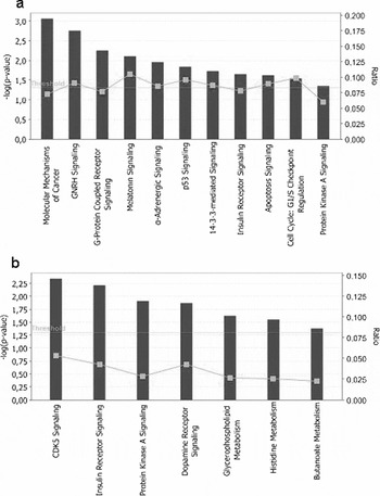
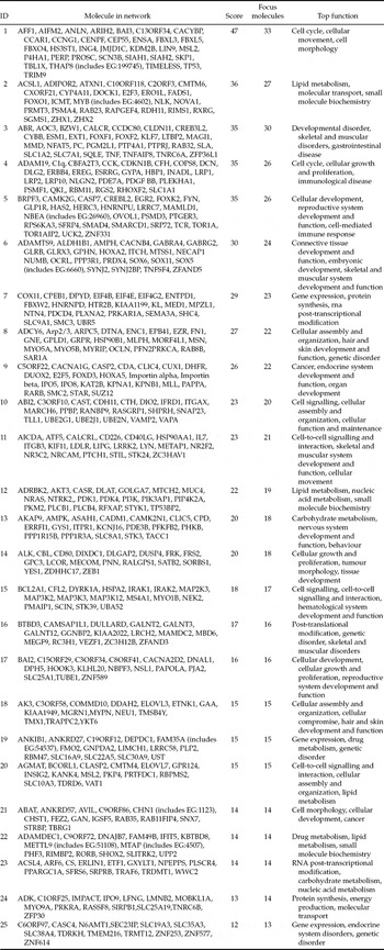
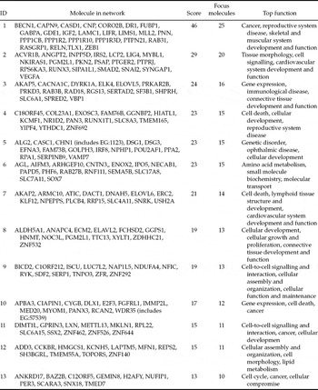
To obtain an overview on the functional role of differentially expressed miRNAs, the target genes of those miRNAs was identified using four on-line tools, TARGETSCAN, PICTAR, miRDB, and MicroRNA.org-Target and Expression. Accordingly, 854 and 234 genes were found to be targeted by miRNAs enriched in oocytes and cumulus cells, respectively. The predicted targets genes were then uploaded into the IPA system to identify relevant molecular networks (biological functions) and pathways. The analysis identified 25 and 13 molecular networks that contained the miRNA target genes enriched in oocytes and cumulus cells, respectively (Appendix Tables 3 and 4). Among these results, cellular assembly and organization, cell cycle, carbohydrate metabolism, molecular transport, cellular development, cell signalling, cell death, cellular growth and proliferation, reproductive system development and function, embryonic development, amino acid metabolism, small molecule biochemistry, gene expression, cell-to-cell signalling and interaction, and lipid metabolism were the most representative molecular networks identified by this analysis. Moreover, genes targeted by miRNAs enriched in oocytes were found to be involved in 11 significant pathways including the GnRH signalling pathway, cell cycle, G-protein coupled receptor signalling pathway, and apoptosis signalling pathway (Fig. 7a). Similarly, the genes targeted by miRNA enriched in cumulus cells were found to be involved in seven different key molecular pathways including the CDK5 signalling pathway, insulin receptor signalling and protein kinase A signalling pathway (Fig. 7b).
The most significant canonical pathway involved by genes targeted by miRNA enriched in oocytes (a) and miRNA enriched in cumulus cells (b). The bars represent the p-value for each pathway. The line with □ indicates the ratio of genes targeted by miRNAs to the total number of genes involved in the pathway.

Discussion
Mammalian oocytes, cumulus cells and other follicular cells are known to have distinct transcriptional profiles; efforts are being made to correlate these profiles or the expression of specific marker genes with oocyte developmental competence (Robert et al., Reference Robert, Gagne, Bousquet, Barnes and Sirard2001; Hamel et al., Reference Hamel, Dufort, Robert, Gravel, Leveille, Leader and Sirard2008). More recently, distinct set of proteins have been found by comparative analysis of bovine GV oocytes and their surrounding cumulus cells (Peddinti et al., Reference Peddinti, Memili and Burgess2010; Memili et al., Reference Memili, Peddinti, Shack, Nanduri, McCarthy, Sagirkaya and Burgess2007). However, the identities and the role of post-transcriptional regulator miRNAs in bovine COC are poorly understood. As most microRNA sequences are conserved across related animal species (Griffiths-Jones et al., Reference Griffiths-Jones, Grocock, van Dongen, Bateman and Enright2006; Niwa & Slack, Reference Niwa and Slack2007), in the present study we have used a human cell development and differentiation miRNA PCR array to compare the miRNA expression profile of oocytes and cumulus cells at the GV and MII stages and distinct sets of miRNAs were identified as being enriched either in oocytes or cumulus cells. Our results show that oocytes at both GV and MII stages are enriched with large numbers of miRNAs under investigation compared with their surrounding cumulus cells. In contrast, recent studies by other groups have shown a higher level of protein expression in cumulus cell when compared with GV oocytes (Memili et al., Reference Memili, Peddinti, Shack, Nanduri, McCarthy, Sagirkaya and Burgess2007; Peddinti et al., Reference Peddinti, Memili and Burgess2010).
As miRNAs are known to be expressed in a developmental time and tissue-specific manner (Plasterk, Reference Plasterk2006), our previous study (Tesfaye et al., Reference Tesfaye, Worku, Rings, Phatsara, Tholen, Schellander and Hoelker2009) and also the present data showed that bovine oocyte maturation involves a wave of expression of miRNAs in oocytes and cumulus cells, which suggests their potential role during oocyte maturation. The expression analysis of the oocyte-enriched miRNAs during oocyte maturation follows a similar trend of gene transcript abundance during oocyte maturation (De La Fuente, Reference De La Fuente2006). This involves the timely synthesis and expression of factors that are essential for oocyte growth and differentiation, as well as the synthesis and storage of transcripts required for meiotic maturation and the first cleavage divisions. Similarly, a massive destruction of transcripts during oocyte maturation has been reported in mouse (Su et al., Reference Su, Sugiura, Woo, Wigglesworth, Kamdar, Affourtit and Eppig2007) and bovine oocytes (Fair et al., Reference Fair, Carter, Park, Evans and Lonergan2007). Considering the reported protein synthesis during bovine oocyte maturation (Tomek et al., Reference Tomek, Torner and Kanitz2002), sequential expression of miRNAs in the present study follow an apposite trend. This finding may give a clue to the potential role of miRNAs in regulation of genes post transcriptionally and contribution to germinal vesicle breakdown (GVDB). Despite the fact that miRNA abundance showed reduction at 22 h post maturation, this level is still higher than the subsequent cleavage stage embryos after fertilization, as shown in Fig. 3. However, the reduction in miRNA abundance after maturation is not a global phenomenon as there are still miRNAs that show increased expression after maturation, as we have reported previously for miR-206, miR-145, miR-292–3p and miR-496 (Tesfaye et al., Reference Tesfaye, Worku, Rings, Phatsara, Tholen, Schellander and Hoelker2009).
Early mammalian embryo development is dependent on the maternal stores of mRNA and protein before the activation embryonic genome. The transition from maternal to zygotic regulation is the first major change that occurs after fertilization and comprises a dramatic reprogramming of gene expression that is essential for the development of most organisms. The involvement of regulatory maternal miRNAs in maternal–zygotic transition is evidenced in mouse (Murchison et al., Reference Murchison, Stein, Xuan, Pan, Zhang, Schultz and Hannon2007; Tang et al., Reference Tang, Kaneda, O'Carroll, Hajkova, Barton, Sun, Lee, Tarakhovsky, Lao and Surani2007) and zebrafish (Giraldez et al., Reference Giraldez, Mishima, Rihel, Grocock, Van Dongen, Inoue, Enright and Schier2006). In the present study, expression profiling of selected miRNAs and one of each target mRNA in early stages of bovine preimplantation embryos showed various patterns of expression. Especially, miRNAs enriched in oocytes (miR-205, miR-150, miR-96, miR-122, miR-146a and miR-146b-5p) showed a typical maternal expression profile in which expression was higher at earlier stages of development and reduced drastically between 8-cell and blastocyst stages (Fig. 3). This finding is additional evidence for dependence of early bovine embryo development on maternally inherited miRNAs, which are known to regulate the degradation of maternal mRNA or protein as observed in mouse embryos (Lykke-Andersen et al., Reference Lykke-Andersen, Gilchrist, Grabarek, Das, Miska and Zernicka-Goetz2008), up to the major activation bovine embryonic genome at the 8–16-cell stage (Memili & First, Reference Memili and First1999). Comparative analysis of differentially expressed miRNAs and their predicted target genes during preimplantation embryo development revealed a reciprocal expression pattern for miR-205 and the PLCB1 transcript, miR-96 and ITPR1 transcript, and miR-146a and IRAK1 transcripts (Fig. 3). This may show the possible mRNA degrading mode-of-action of the miRNAs in the post-transcriptional regulation of the corresponding target genes. The PLCB1 gene is an important element of the phosphoinositide pathway; this pathway acts principally by generating calcium release. The nuclear phosphoinositide cycle in mouse oocytes seems to be regulated essentially by the PLCB1 isoform; microinjected into the GV, the anti-PLCB1 induced inhibition of both nuclear and cytoplasmic Ca2+ oscillations and meiosis resumption (Avazeri & Lefevre, Reference Avazeri and Lefevre1998; Avazeri et al., Reference Avazeri, Courtot, Pesty, Duquenne and Lefevre2000). The functional role of ITPR1 (target of miR-96) in operating Ca2+ release in oocytes at fertilization has been demonstrated in the golden hamster, mouse and in frogs (Miyazaki et al., Reference Miyazaki, Yuzaki, Nakada, Shirakawa, Nakanishi, Nakade and Mikoshiba1992; Xu et al., Reference Xu, Kopf and Schultz1994; Runft et al., Reference Runft, Watras and Jaffe1999). In human oocytes, the type I InsP3R is expressed prominently in oocytes, zygotes and embryos and is redistributed dynamically through maturation, fertilization and early embryogenesis (Goud et al., Reference Goud, Goud, Oostveldt and Dhont1999). In addition, functional analysis using IPA revealed that GnRH signalling and G-protein-coupled receptor signalling are dominant pathways for oocyte-enriched miRNA targets (Fig. 7a). GnRH signalling controls the biosynthesis and release of luteinizing hormone (LH) and follicle-stimulating hormone (FSH) that in turn regulate development and activity of the ovaries (Jeong & Kaiser, Reference Jeong, Kaiser and Neill2006). GnRH acts via G-protein-coupled receptors (Gq/G11) and leads to activation of phospholipase C (PLC, apparently PLCβ1), which hydrolyzes phosphatidylinositol 1,4,5-bisphosphate (PIP2) to inositol-1,4,5 triphosphate (IP3) and diacylglycerol (DAG). IP3 binds to receptors in the endoplasmic reticulum to release Ca2+ transiently from these intracellular stores, which in turn elicits a rapid spike of LH release. Also, DAG activates protein kinase Cs (PKCs) that phosphorylate protein involved in the more sustained release of LH and in gonadotrophin biogenesis (Kraus et al., Reference Kraus, Naor and Seger2001; McArdle et al., Reference McArdle, Franklin, Green and Hislop2002; Miller, Reference Miller and Kastin2006). The expression of the CCNG1 gene, which may play a role as a positive modulator of cell cycle in ovarian follicular development and oocyte maturation (Liu et al., Reference Liu, Zhang, He, Zhang and Yue2006), remained constant throughout the developmental stages irrespective of the expression profile of its targeting miR-122.
Similarly, miRNAs enriched in cumulus cells (miR-452 and miR-210) and their corresponding predicted target mRNA transcripts showed a similar expression pattern in which all are detected in a relatively lower level in all stages of development, without any defined profile. This different relationship in expression profile of miRNA and target mRNA reflects the existing challenge in target identification because of varying outcome of binding of miRNAs to their target genes, which might not result in alternation of target gene expression (Brennecke et al., Reference Brennecke, Stark, Russell and Cohen2005; Grimson et al., Reference Grimson, Farh, Johnston, Garrett-Engele, Lim and Bartel2007). This interaction rather results in translational repression, which appears to be cap and polyA tail dependent during initiation steps (Humphreys et al., Reference Humphreys, Westman, Martin and Preiss2005; Pillai et al., Reference Pillai, Bhattacharyya, Artus, Zoller, Cougot, Basyuk, Bertrand and Filipowicz2005).
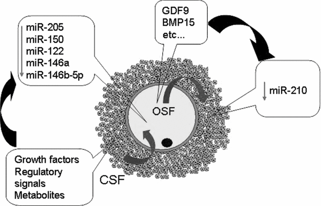
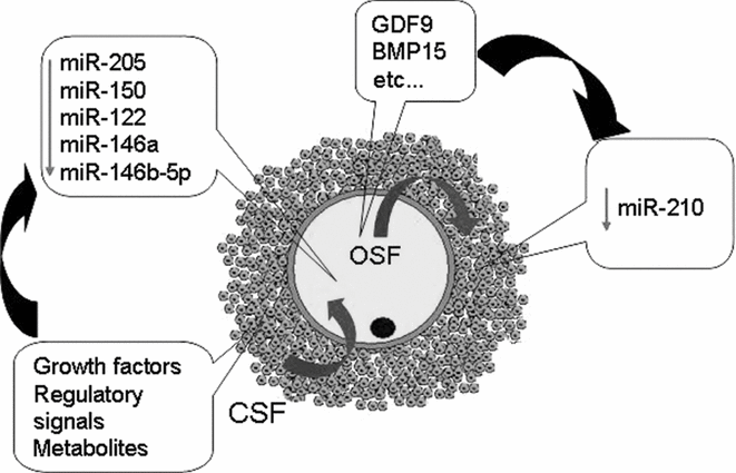
While paracrine growth factors secreted by oocytes are involved in expansion of cumulus cell number and function, regulation of follicular cell function and preovulatory and post-ovulatory events (Gilchrist et al., Reference Gilchrist, Ritter and Armstrong2001), the cumulus cell supports oocyte metabolic processes such as glycolysis and amino acid uptake (Buccione et al., Reference Buccione, Schroeder and Eppig1990). So far, a limited number of oocyte-secreted factors (OSFs) is known to regulate cumulus cell and granulosa cell function. These factors include GDF9 and BMP15, which have a profound effect on the expression of genes in cumulus cells and enhance oocyte developmental competence (Hussein et al., Reference Hussein, Thompson and Gilchrist2006). In the present study the presence of oocyte or cumulus cells was found to be important in the regulation of miRNA expression in either of the two cell types. Except for cumulus-enriched miRNAs, all oocyte-enriched miRNAs were found to be upregulated in the absence of the surrounding cumulus cells during maturation. The role of increased relative abundance of our candidate miRNAs in oocytes matured without cumulus cells needs further investigation, despite the fact that removal of cumulus cells at the beginning of in vitro oocyte maturation or shortly before fertilization resulted in a detrimental effect on oocyte maturation, fertilization and subsequent development (Zhang et al., Reference Zhang, Jiang, Wozniak, Yang and Godke1995; Fatehi et al., Reference Fatehi, Zeinstra, Kooij, Colenbrander and Bevers2002). From these results, we hypothesis that these miRNAs may be involved in the regulation of genes associated with impaired nuclear maturation of oocytes due to the absence of cumulus cell factors. In contrast, interestingly, the absence of oocyte factors increased the expression of miR-210 in cumulus cells compared with those miRNAs matured with oocyte cytoplasm. The summarized hypothesis in Fig. 8 on dependency of the two cell types to each other for miRNA expression needs further investigation in order to identify the specific oocyte or cumulus cells factors and the mode of transcriptional regulation of the miRNAs.
Hypothetical illustration depicting the dependency of bovine oocyte and cumulus cells in the relative abundance of miRNAs transcribed from their genome on the presence or absence of cumulus cell factor (CSF) and oocyte secreting factors (OSF), respectively. Green arrows indicate reduced level of miRNA expression. The inward and outward arrows indicate the effect of CSF and OSF on oocyte and cumulus cell development, respectively.
Mammalian folliculogenesis is a complex process through which primordial follicles develop into preovulatory follicles. During this time, oocytes increase in size accompanied by the proliferation and differentiation of their surrounding granulosa and theca cells. For this spatio-temporal expression, hundreds of genes have been shown to be essential (Matzuk et al., Reference Matzuk, Burns, Viveiros and Eppig2002; Roy & Matzuk, Reference Roy and Matzuk2006). The role of miRNAs as potential regulators of these genes, have been evidenced in the ovaries through impaired function of the corpus luteum and by infertility in Dicer knockout mice (Otsuka et al., Reference Otsuka, Zheng, Hayashi, Lee, Yoshino, Lin and Han2008). As observed by the genes, the results in the present study have illustrated the spatio-temporal expression of miRNAs during bovine follicular development.
Differences in the signal intensity in localization of miR-205 and 210 were found between oocytes and their surrounding cumulus cells as early as the primary follicle stage and persisted until the matured follicle stage. Despite the fact that loss-of-function studies on miRNA processing key proteins (DICER and AGO/EIF2C2) in mouse revealed a negative effect on female fertility linked to abnormal oogenesis (Tam et al., Reference Tam, Aravin, Stein, Girard, Murchison, Cheloufi, Hodges, Anger, Sachidanandam, Schultz and Hannon2008; Watanabe et al., Reference Watanabe, Totoki, Toyoda, Kaneda, Kuramochi-Miyagawa, Obata, Chiba, Kohara, Kono, Nakano, Surani, Sakaki and Sasaki2008), the exact role of these non-coding RNAs during bovine follicular development needs to be the focus of future research.
In conclusion, to the best of our knowledge, here we reported for the first time the classes of miRNA enriched in bovine oocytes or in the surrounding cumulus cells during oocyte maturation, which is a step forward in identifying miRNAs required for oocyte–cumulus communication. Moreover, we have also demonstrated the dynamic degradation of miRNAs during oocyte maturation and interdependency of oocyte and cumulus cells on the activity of the miRNAs. Furthermore, the potential role of maternal miRNAs in early bovine embryo development could be depicted from their expression dynamics during the preimplantation stage period. However, further functional studies on those differentially expressed miRNAs are required to understand the key role of miRNAs during oocyte maturation and its subsequent effect on further embryonic development.
Acknowledgements
We are grateful to Dr Kirfel from the Institute of Cell Biology the University of Bonn for laser microscopic imaging of stained ovarian sections and preimplantation embryos.
The average threshold cycle (Ct) of the endogenous control U6 RNA through out the preimplantation stages.

Sequence complementarities between selected miRNAs and their target mRNA 3′ UTR.

List of differentially expressed miRNA between immature oocytes and their surrounding cumulus cells

List of significant expressed miRNA between mature oocytes and their surrounding cumulus cells

Molecular networks involved by target genes of miRNA enriched in oocyte function

Molecular networks involved by target genes of miRNAs enriched in cumulus cells