Introduction
Cicatricial pemphigoid, also known as mucous membrane pemphigoid, is a rare, chronic autoimmune vesiculobullous disease characterised by subepidermal bullae primarily involving the oral and ocular mucous membrane.Reference Hanson, Olsen and Rogers1 Nasal, oropharyngeal and laryngeal involvements are rarely observed, and hypopharyngeal involvement is even less frequent.Reference Alexandre, Brette, Pascal, Tsianakas, Fraitag and Doan2 As the blisters tend to heal with scarring, significant complications including blindness, and stenosis of the pharynx, larynx or nasal cavity can occur. We present a rare case of cicatricial pemphigoid with hypopharyngeal involvement, and demonstrate findings from videofluorography with barium swallow and direct laryngoscopy.
Case report
A 69-year-old male had been treated in the department of dermatology at our hospital with 10 mg of prednisolone daily for 5 years under a diagnosis of cicatricial pemphigoid. He was referred to our department because he began to complain of difficulty swallowing.
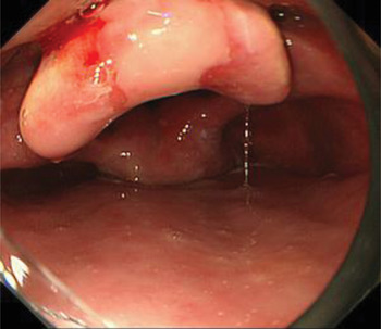
Fibre-optic laryngoscopy revealed saliva residue in the pyriform sinuses and vallecula. After removing the saliva, the pyriform sinus mucosa appeared normal (Figure 1). Mobility of the bilateral vocal folds was normal. Supraglottic stenosis was not observed, but the tip of the epiglottis was deformed due to scarring (Figure 1).
Fibre-optic laryngoscopy showed that the pyriform sinus mucosa appeared normal. The tip of the epiglottis was deformed due to scarring.

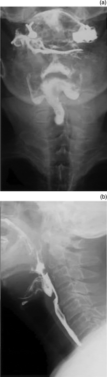
Videofluorography with barium swallow demonstrated relatively narrow flow through the postcricoid hypopharynx. However, there was no flow through the right and left pyriform sinuses, although apparent residue of barium was observed there (Figure 2a). The lateral view demonstrated a postcricoid filling defect (Figure 2b). There were no signs of oesophageal stenosis, web or intramural pseudodiverticulum. Although there was slight laryngeal but not tracheal penetration, there was no apparent nasopharyngeal reflux.
(a) Videofluorography (anterior view) demonstrated narrow flow through the medial hypopharynx, but no flow through the lateral hypopharynx. (b) The lateral view showed a postcricoid filling defect.

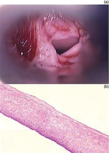
Direct laryngoscopic observation was performed under general anaesthesia. Both the right and left pyriform sinuses terminated abruptly due to dense scarring, although the mucosa appeared normal. The postcricoid hypopharyngeal lumen demonstrated narrowing due to circumferential scar formation (Figure 3a). Interestingly, thin membranous webs were observed beyond the circumferential scar. Although these thin webs spontaneously disappeared, there was no attempt at treatment such as balloon dilatation because the dense scar tissue was hard and considered resistant to conservative procedures.
(a) Direct laryngoscopy revealed narrowing of the post-cricoid hypopharyngeal lumen due to circumferential scar formation. (b) Histologically, the thin webs that were removed were identified as layers of squamous cell epithelia (H&E; ×200).

Histologically, the thin webs that were removed were identified as layers of squamous cell epithelia (Figure 3b). However, because the specimens lacked subepithelial tissues, neither subepithelial cleavage between the epithelia and connective tissues, nor blister formation associated with mucous membrane pemphigoid, were informative.
The patient felt slight but not dramatic improvement of swallowing after surgery. No further treatment was scheduled because the patient was suffering from dementia with Lewy bodies.
Discussion
Diagnosis of cicatricial pemphigoid is based on clinical presentation, evidence of subepithelial vesicles or bullae on routine histologic analysis, and direct and indirect immunofluorescence studies.Reference Bruch-Gerharz, Hertl and Ruzicka3 Cicatricial pemphigoid is characterised by linear deposition of immunoreactants, principally immunoglobulin G and complement factor 3, along epithelial basement membranes.Reference Bruch-Gerharz, Hertl and Ruzicka3, Reference Anhalt and Morrison4 In the present case, diagnosis of cicatricial pemphigoid had already been established by immunofluorescence study.
In most cases, cicatricial pemphigoid is a chronic and progressive disease that rarely goes into spontaneous remission. Currently, there is no gold standard for medical treatment. Treatment regimens usually depend on the site and severity of involvement. For patients with ocular or laryngeal disease, systemic corticosteroids alone or in combination with immunosuppressants such as azathioprine or cyclophosphamide are indicated.Reference Bruch-Gerharz, Hertl and Ruzicka3
A review of recent literature describing lesion patterns and frequencies of cicatricial pemphigoid in various sites revealed one meta-analysis that included several case reports, one retrospective study and one prospective study.Reference Hanson, Olsen and Rogers1, Reference Alexandre, Brette, Pascal, Tsianakas, Fraitag and Doan2, Reference Ahmed and Hombal5 Ahmed and Hombal summarised the clinical features of 457 patients with cicatricial pemphigoid reported in 16 papers published between 1965 and 1982.Reference Ahmed and Hombal5 In that meta-analysis, the incidences of nasal, pharyngeal and laryngeal involvement in cases of cicatricial pemphigoid were 15 per cent, 19 per cent and 8 per cent, respectively. However, the specific details with regard to the subsite and severity of the cicatricial pemphigoid lesions were not described. Similarly, Hanson et al. reported their experiences in treating 142 patients with cicatricial pemphigoid at the Mayo Clinic (Minnesota, USA).Reference Hanson, Olsen and Rogers1 In that report, the incidence of nasal, pharyngeal and laryngeal involvement in cases of cicatricial pemphigoid were 23 per cent, 8 per cent and 9 per cent, respectively. The typical finding was multiple ulcers on the posterior or lateral pharyngeal walls. One case demonstrated postcricoid ulcers; however, none of the patients had pyriform sinus disease. A prospective study by Alexandre et al., which examined 110 cases of mucous membrane pemphigoid (another term for cicatricial pemphigoid), demonstrated that at least 27 per cent, 5 per cent and 17 per cent of patients had clinical lesions in the nose, pharynx and larynx, respectively.Reference Alexandre, Brette, Pascal, Tsianakas, Fraitag and Doan2 Most of the pharyngeal lesions were erosion or erythema of the oropharynx. However, no hypopharyngeal lesions were described in that report. This review of the literature indicated that there had been no previous reports that describe hypopharyngeal involvement in detail.
Our direct laryngoscopic observation of the thin membranous webs that lay beyond the circumferential scar in the postcricoid hypopharyngeal lumen was a very informative finding. Unfortunately, we could not demonstrate direct evidence of hypopharyngeal involvement histologically because only the epithelial layers were harvested during the procedure. However, this suggests that the detached epithelial layers developed from subepithelial cleavage between the epithelia and connective tissues or the blister formation associated with mucous membrane pemphigoid. This finding indicates one of the processes of scar formation associated with a subepidermal bulla.
• Cicatricial pemphigoid, also known as mucous membrane pemphigoid, can affect all mucosa of the upper aerodigestive tract
• hypopharyngeal involvement in cases of cicatricial pemphigoid have not been reported owing to their rarity
• Videofluorography and direct laryngoscopy revealed postcricoid circumferential scarring and detached epithelial layers of mucosa
Scar formation is the most major complication of cicatricial pemphigoid in the hypopharynx, which can result in stricture and dysphagia. However, it is difficult to observe the postcricoid hypopharynx by endoscopy. Videofluorography is useful for the evaluation of cicatricial pemphigoid, despite the fact that there are other diseases which cause scarring in this area. This examination should therefore be considered for cicatricial pemphigoid patients with dysphagia. Videofluorography is also valuable for examining oesophageal involvement.Reference Syn and Ahmed6 Oesophageal involvement was observed in 3.6–4 per cent of previously reported cicatricial pemphigoid cases.Reference Hanson, Olsen and Rogers1, Reference Ahmed and Hombal5, Reference Naylor, MacCarty and Rogers7
Cicatricial pemphigoid is a difficult condition to treat, and scarring with stenosis can occur in spite of aggressive medical treatment. Currently, there are no reports describing intervention in a case of cicatricial pemphigoid with hypopharyngeal involvement. Endoscopic therapy, including graded oesophageal dilatations, is an alternative option for severe oesophageal disease.Reference Syn and Ahmed6 However, there have been associated reports of bulla formation, mucosal injury and even perforation.Reference Soong and Bynum8, Reference Watkinson, Vretenar, Morrison and Burhenne9 The use of endoscopic anterograde dilatation or transgastric retrograde oesophagoscopy with anterograde dilatation have been reported for hypopharyngeal stenosis following chemoradiotherapy for head and neck cancer.Reference Sullivan, Jaklitsch, Haddad, Goguen, Gagne and Wirth10 These procedures may be adopted for hypopharyngeal stenosis resulting from cicatricial pemphigoid. However, the occurrence of hypopharyngeal perforation was reported to be 13 per cent, suggesting that careful consideration is needed when determining the indications for the procedure.
In conclusion, this report describes important videofluorographic and direct laryngoscopic findings showing rare hypopharyngeal involvement in a case of cicatricial pemphigoid.