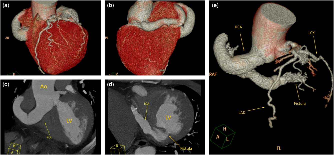
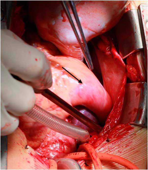
A38-year-old man was admitted to our hospital for chest distress and recurrent palpitations. No cardiac murmur and other abnormal respiratory sounds were found. His heart rate was 84 beats per minute, and his blood pressure was 126/79 mmHg. Electrocardiography showed sinus rhythm and no significant ST segment or T wave anomaly. His chest radiography was normal. Transthoracic echocardiography found an aneurysmal right coronary artery and an abnormal blood flow to the left ventricle posterior to the mitral valve leaflet. CT demonstrated an aneurysmal right coronary artery fistula into the left ventricle (Fig 1; Supplementary video 1). As transcatheter closure failed, median sternotomy was performed to suture the fistula orifice by partial coronary arterial incision (Fig 2). After the surgery, the patient recovered uneventfully.
( a – e ) Coronary CT showing the large and tortured right coronary artery (RCA) coursing the atrioventricular groove and finally into left ventricular wall near the posterior valve of the mitral valve with an aneurysm of 21-mm diameter. Ao=aorta; AR=anterior-right; FL=foot-left; LAD=left anterior descending coronary artery; LCX=left circumflex coronary artery; LV=left ventricle; PL=posterior-left; RAF=right-anterior-foot.

Intra-operative image showed the giant aneurysmal right coronary artery (black arrow) origin from the aorta.

Discussion
Coronary artery fistulas are rare anomalous terminations of the coronary arteries, accounting for ~0.2–0.4% of congenital cardiac anomalies. Coronary artery fistulas arise from the right coronary artery in ~50% of patients, from the left coronary artery in ~42% of patients, and from both the right coronary artery and the left coronary artery in ~5% of patients. Fistulous drainage occurs into the right ventricle in 41%, the right atrium in 26%, the pulmonary artery in 17%, the left ventricle in 3%, and into the superior caval vein in 1%.Reference Zenooz, Habibi and Mammen 1 Appropriate diagnosis of coronary artery fistulas is important, and early treatment by transcatheter or surgical closure provides the best results.
Acknowledgements
None.
Financial Support
This research received no specific grant from any funding agency or from commercial or not-for-profit sectors.
Conflicts of Interest
None.
Ethical Standards
Informed consent was obtained from the patient. The authors assert that all procedures contributing to this study comply with the ethical standards of China and with the Helsinki Declaration of 1975, as revised in 2008, and has been approved by the Ethics Committee of West China Hospital.
Supplementary Material
To view supplementary material for this article, please visit https://doi.org/10.1017/S1047951117000865