INTRODUCTION
Ahyong & Ng (Reference Ahyong and Ng2009) established the Indo-West Pacific family Aphanodactylidae (Pinnotheroidea) for Aphanodactylus Tesch, Reference Tesch1918, Gandoa Kammerer, Reference Kammerer2006, Gustavus Ahyong & Ng, Reference Ahyong and Ng2009 and Uruma Naruse, Fujita & Ng, Reference Naruse, Fujita and Ng2009. Recently, Naruse & Maenosono (Reference Naruse, Maenosono, Komatsu, Okuno and Fukuoka2012) established a new genus, Takedactylus, from Japan and assigned it to the Aphanodactylidae (see also Naruse et al., Reference Naruse, Fujita and Ng2009; Ng & Naruse, Reference Ng and Naruse2009; Ng et al., Reference Ng, Ahyong and Komai2010).
In this present paper, the poorly known genus, Selwynia Borradaile, Reference Borradaile and Gardiner1903, described from the Maldives, is reported as a senior subjective synonym of Aphanodactylus Tesch, Reference Tesch1918. Selwynia was originally classified as a goneplacid and later a pilumnid (see Borradaile, Reference Borradaile and Gardiner1903; Tesch, Reference Tesch1918; Ng, Reference Ng1987). The type species, Selwynia laevis Borradaile, Reference Borradaile and Gardiner1903, is re-diagnosed and shown to be distinct from other western Pacific Aphanodactylus species. The material of Aphanodactylus, Gustavus and Uruma examined has been listed by Ahyong & Ng (Reference Ahyong and Ng2009). The following abbreviations are used: CUMZ, Cambridge University Museum of Zoology; G1, male first gonopod; G2, male second gonopod; P, pereiopod. Measurements provided (in millimetres) are of the maximum carapace width and length, respectively. The terminology used follows Ahyong & Ng (Reference Ahyong and Ng2009).
RESULTS
SYSTEMATICS
Family APHANODACTYLIDAE Ahyong & Ng, Reference Ahyong and Ng2009
Aphanodactylidae Ahyong & Ng, Reference Ahyong and Ng2009: 34.
Gustavini Števčić, Reference Števčić2011: 129.
REMARKS
Ahyong & Ng (Reference Ahyong and Ng2009) assigned Gustavus Ahyong & Ng, Reference Ahyong and Ng2009, to their Aphanodactylidae. However, Števčić (Reference Števčić2011: 129) established Gustavini, within the Asthenognathinae Stimpson, Reference Stimpson1858 (nec Astenognathidae), with Gustavus as the type genus and provided a diagnosis for his new tribe. But Števčić (Reference Števčić2011) did not explain why this new tribe was warranted, why he allocated Aphanodactylidae subfamilial status within the Asthenognathidae and why he considered Asthenognathidae as a distinct family. Ng et al. (Reference Ng, Guinot and Davie2008: 226) had assigned Asthenognathinae to the Varunidae H. Milne Edwards, Reference Milne Edwards1853, and the rationale for this was discussed at length by Davie & N.K. Ng (Reference Davie and Ng2007) and Ng et al. (Reference Ng, Guinot and Davie2008: 252). Considering the number of taxa assigned to this family and the atypical features of many aphanodactylids, the action of Števčić (Reference Števčić2011) in recognizing a separate tribe for Gustavus Ahyong & Ng, Reference Ahyong and Ng2009, is not understood. Therefore, Aphanodactylidae is maintained here as a distinct family in Pinnotheroidea De Haan, Reference De Haan and von Siebold1833 (Ahyong & Ng, Reference Ahyong and Ng2009).
The synonymy of Selwynia Borradaile, Reference Borradaile and Gardiner1903, and Aphanodactylus Tesch, Reference Tesch1918, as proposed here, does not affect the status of Aphanodactylidae Ahyong & Ng, Reference Ahyong and Ng2009, as Article 40.1 of the International Code of Zoological Nomenclature (ICZN, 1999) requires that the original family name be retained.
Selwynia Borradaile, Reference Borradaile and Gardiner1903
Selwynia Borradaile, Reference Borradaile and Gardiner1903: 430.
Aphanodactylus Tesch, Reference Tesch1918: 283.
Type species: Selwynia laevis Borradaile, Reference Borradaile and Gardiner1903, by monotypy.
DIAGNOSIS
See diagnosis for Aphanodactylus in Ahyong & Ng (Reference Ahyong and Ng2009: 36).
REMARKS
Selwynia was established for S. laevis Borradaile, Reference Borradaile and Gardiner1903, on the basis of a single male specimen from Hulule, Male Atoll, in the Maldive and Laccadive Archipelagoes. Borradaile (Reference Borradaile and Gardiner1903: 430–431, figure 112) assigned this species to the Rhizopinae Stimpson, Reference Stimpson1858. Tesch (Reference Tesch1918: 53) retained Selwynia in his concept of Rhizopinae, and this classification was followed by Serène (Reference Serène1964, Reference Serène1968) and Ng (Reference Ng1987: 98). Moreover, in his redefinition of the subfamily, Ng (Reference Ng1987) commented that ‘Selwynia is very unusual amongst known rhizopines (indeed among known pilumnids) for an almost complete absence of hair, strongly built chelipeds and shortness of the ambulatory dactylus. Until the type can be re-examined however, it is tentatively placed in the Rhizopinae’ (see also Ng et al., Reference Ng, Guinot and Davie2008: 144).
Although Tesch (Reference Tesch1918) did not examine any specimens of Selwynia, he did provide a detailed key to the Rhizopinae that included this genus (Tesch, Reference Tesch1918: 53), and additionally established a new rhizopine, Paraselwynia, which he believed was close to Selwynia. In his discussion for Paraselwynia, Tesch (Reference Tesch1918: 74–75), made extensive reference to the description and figures of Selwynia in Borradaile (Reference Borradaile and Gardiner1903). However, when Tesch (Reference Tesch1918: 283) treated the Pinnotheridae in the same work, he established a new genus Aphanodactylus, with one species, A. sibogae, from Indonesia, without reference to Selwynia. The similarities between Aphanodactylus and Selwynia are marked, especially in the general smoothness of the carapace and the short ambulatory dactyli. Tesch (Reference Tesch1918) did not consider the relationship between Aphanodactylus and Selwynia probably because the former was regarded as a pinnotherid while the latter was a rhizopine assigned to the Goneplacidae. This same ‘disconnect’ is apparent in Ahyong & Ng (Reference Ahyong and Ng2009), Ng & Naruse (Reference Ng and Naruse2009) and Ng et al. (Reference Ng, Ahyong and Komai2010), who all reviewed Aphanodactylus with the inclusion of new species.
The type of Selwynia laevis Borradaile, Reference Borradaile and Gardiner1903, was recently re-examined and the affinity of Selwynia Borradaile, Reference Borradaile and Gardiner1903, with Aphanodactylus Tesch, Reference Tesch1918, became apparent. The four known species of Aphanodactylus, A. sibogae Tesch, Reference Tesch1918 (from Indonesia), A. edmondsoni Rathbun, 1932 (from Hawaii), A. loimiae Konishi & Noda, Reference Konishi and Noda1999 (from Japan) and A. panglao Ng & Naruse, Reference Ng and Naruse2009 (from the Philippines), are now assigned to Selwynia.
Selwynia laevis Borradaile, Reference Borradaile and Gardiner1903
(Figures 1–3)
MATERIAL EXAMINED
Holotype male (7.1 × 5.6 mm) (CUMZ I.63872), Hulule, Male Atoll, Maldives, coll. J. S. Gardiner, 1900.
DIAGNOSIS
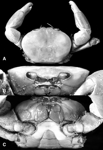
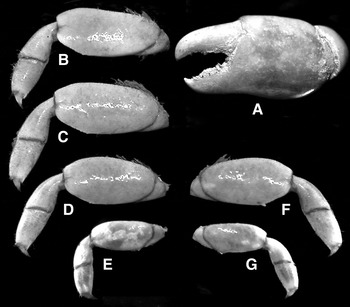
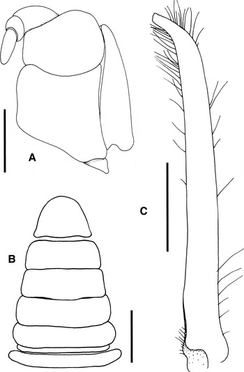
Holotype male: carapace broader than long, width to length ratio 1.27 (Figure 1A); front distinct, bilobed (Figure 1A); third maxilliped ischium longer than merus (Figure 3A); ambulatory legs (P2–P5) relatively short, extensor margin of merus unarmed; distal third of flexor margin of P2–P4 with 1 small denticle or spine and/or small granules, proximal third of P5 with 1 denticle and several granules (Figure 2B–G); male abdomen relatively narrow, first and second somites longitudinally short, telson triangular with rounded tip, sinuous margins (Figures 1C & 3B); G1 simple, slender, proximal part straight, distal fifth curved inwards towards median part of thoracic sternum (Figure 3C).
Selwynia laevis, holotype male (7.1 × 5.6 mm) (CUMZ I.63872): (A) overall dorsal view; (B) frontal view of cephalothorax; (C) buccal cavity, anterior thoracic sternum and abdomen.

Selwynia laevis, holotype male (7.1 × 5.6 mm) (CUMZ I.63872), pereopods: (A) left chela; (B–E) left first to fourth ambulatory legs, respectively; (F, G) right third and fourth ambulatory legs, respectively.

Selwynia laevis, holotype male (7.1 × 5.6 mm) (CUMZ I.63872): (A) left third maxilliped; (B) male abdomen; (C) right G1 (ventral view). Scale bars: A, C = 0.5 mm; B = 1.0 mm.

DESCRIPTION
Holotype male: carapace subovate, broader than long, width to length ratio 1.27; dorsal surface smooth, regions poorly demarcated, with 2 faint pairs of small swellings on hepatic part (Figure 1A). Front distinct, divided into 2 broad, rounded lobes in dorsal view, without lateral lobule, confluent with supraorbital margin; deflexed, forming vaguely triangular structure in frontal view (Figure 1A, B). Orbit ovate; supraorbital margin entire, smooth; infraobital margin entire, with poorly developed inner orbital tooth; no gap between cornea and external orbital angle, infraorbital margin, just reaching basal antennal article; eye mobile, short, peduncle stout, cornea large, pigmented (Figure 1A, B). External orbital angle short, small, directed anteriorly, cristate; anterolateral margin arcuate, entire, contiguous with posterolateral margin; posterolateral margin gently convex; posterior carapace margin almost straight (Figure 1A). Epistome longitudinally narrow; posterior margin with low median triangular lobe, lateral margins gently sinuous (Figure 1B). Antenna with short, stout basal article, not reaching distolateral angle of carapace, flagellum relatively short, enters orbit; antennules folding almost transversely (Figure 1B).
Third maxillipeds covering ~3/4 or more of buccal cavern when closed; ischium longer than merus, extended proximo-laterally, with small projection at distomesial angle; merus subovate, about 2/3 length of ischium, distal margin concave to fit palp (carpus, propodus and dactylus); exopod slender, tapering distally, reaching proximal 2/3 of merus; flagellum distinct, reaching mesial margin of merus (Figures 1C & 3A).
Chelipeds relatively strong, slightly unequal in size (Figure 1A). Merus triangular in cross-section, ventro-outer margin granulated, dorsal margin rugose, dorsal margin and outer surface setose; carpus smooth, inner angle rounded (Figure 1A). Chela with outer surfaces smooth, fingers shorter than palm; fingers with denticulated cutting edges, base with short setae (Figure 2A).
Ambulatory legs (P2–P5) relatively short; P3 longest, P5 shortest; most of surfaces glabrous, setose on proximal anterior margins and anterior margins of carpi and propodi (Figure 2B–G). Merus as long as or subequal to combined length of carpus, propodus and dactylus; extensor margin unarmed, flexor margin with 1 small denticle/spine and/or small granules, armed as follows: distal third of P2 with 1 denticle and several small granules, distal third of P3 with several granules, distal third of P4 with 1 short spine or denticle with several granules, proximal third of P5 with 1 denticle and several granules (Figure 2B–G). Propodi with flexor margins gently serrated, distal part with small spines; dactyli short, claw-like (Figure 2B–G).
Thoracic sternites 1 and 2 completely fused; sternites 2 and 3 separated by distinct straight suture; sternites 3 and 4 fused but demarcated by low transverse depression, sternites 4–8 laterally demarcated by distinct sutures (Figure 1C); sutures medially interrupted, no medial longitudinal suture; press button of abdominal locking mechanism on distal quarter of sternite 5; penis emerging from near anterior border of sternite 8.
Abdomen with all somites and telson distinct, mobile, relatively narrow, first and second somites longitudinally short; lateral margins of somite 3 to telson gently convex to almost straight; telson triangular with rounded tip and sinuous margins (Figures 1C & 3B). G1 simple, slender, proximal part straight, distal fifth curved inwards towards median part of thoracic sternum (Figure 3C). G2 missing.
REMARKS
The generic affinities of the poorly known Pinnixa brevipes H. Milne Edwards, Reference Milne Edwards1853, which is based upon an inadequately preserved, deformed and soft (possibly newly moulted) lectotype female from Mayotte in the Comoros (cf. Ng & Naruse, Reference Ng and Naruse2009: 288), should be reconsidered with respect to the synonymy of Selwynia and Aphanodactylus. Serène (Reference Serène1964: 278) noted that Pinnixa brevipes should perhaps be classified in Aphanodactylus (see also Ng et al., Reference Ng, Guinot and Davie2008: 247). Ng & Naruse (Reference Ng and Naruse2009: 289) discussed the taxonomic status of Pinnixa brevipes at length. They concluded that in view of the more rectangular carapace shape which has the dorsal surface distinctly punctate, a relatively more elongate orbit and the fact that Aphanodactylus is not known from the Indian Ocean; the species should be provisionally placed in Gandoa Kammerer, Reference Kammerer2006. This genus is relatively unknown and described on the basis of a single female from Zanzibar in East Africa. Gandoa Kammerer, Reference Kammerer2006, is a replacement name for Voeltzkowia Lenz, Reference Lenz1905, the latter preoccupied by Voeltzkowia Boettger, 1893 (Reptilia). Ng & Naruse (Reference Ng and Naruse2009: 288) commented on the close similarity of Gandoa and Aphanodactylus and suggested the two genera should be kept separate until fresh specimens of the former can be found (see also Ahyong & Ng, Reference Ahyong and Ng2009: 39). Until the type species of Gandoa, Voeltzkowia zanazibarensis Lenz, 1905, can be re-examined, it would be premature to change the status of Pinnixa brevipes. Consequently, we leave it in Gandoa for the time being.
With regard to diagnostic characters of S. laevis the armature on the extensor and flexor margins of the meri of P2–P5 is comparatively weak and the arrangement of their denticles and granules is also different from other congeners. Selwynia panglao is unique in having the extensor margins of the male P2 ambulatory merus armed with strong spines and granules, with those on P3 and P4 denticulated (Ng & Naruse, Reference Ng and Naruse2009: figure 3d). In other Selwynia species (including S. laevis), the extensor margin is barely denticulated to smooth (cf. Konishi & Noda, Reference Konishi and Noda1999: figure 1e, f; Ahyong & Ng, Reference Ahyong and Ng2009: figures 1E & 2E; Ng et al., Reference Ng, Ahyong and Komai2010: figure 2B). With regard to the flexor margin of the male P2–P4 merus of S. laevis, the denticles are low and lining only the distal third. In the other species, the flexor margin of the P2–P4 merus is armed with 1–6 distinct spines or sharp denticles. The flexor margin of the P5 merus has a small spine in S. laevis but this margin is usually unarmed or with only a small spine in the other species (cf. Konishi & Noda, Reference Konishi and Noda1999: figure 1e, f; Ahyong & Ng, Reference Ahyong and Ng2009: figures 1E & 2E; Ng et al., Reference Ng, Ahyong and Komai2010: figure 2B). The G1s of S. sibogae, S. edmondsoni and S. loimiae are similar in that the distal part gently curves inwards towards the thoracic sternum (Edmondson, Reference Edmondson1962: figure 2d; Ahyong & Ng, Reference Ahyong and Ng2009: figure 1H; Ng et al., Reference Ng, Ahyong and Komai2010: figure 2d; unpublished data). In S. panglao, the distal part is strongly inclined inwards instead (Ng & Naruse, Reference Ng and Naruse2009: figure 3 g). The G1 in S. laevis is curved (Figure 3C), and thus intermediate between that of S. panglao and the other species.
ACKNOWLEDGEMENTS
The authors are grateful to Matthew Lowe (CUMZ) for entrusting the Borradaile material to us for study. Martyn Low helped with checking the material earlier on, and his help is gratefully acknowledged. Some of the photographs were taken by Phil Hurst of the Photographic Unit, NHM.