
Introduction
Affinity technology is a fundamental method that allows selective and efficient chromatographic purification of bioproducts by exploiting the specific and reversible interaction between a ligand, immobilized on a solid support, and the target molecule. To this aim, any other non-covalent interactions such as hydrogen bonding and hydrophobic interactions that may arise between any molecule in solution and any part of the derivatized affinity support should be minimized.
Due to the high cost of affinity materials, the industrial application of this technology is considered feasible only for the purification of high added value products such as biotherapeutics. Among those, monoclonal antibodies (mAbs) are the class of therapeutics that arouse more interest due to their importance in the cure of illnesses such as cancer, autoimmune diseases, rheumatoid arthritis, and several other diseases.[ Reference Reichert, Rosensweig, Faden and Dewitz 1 ]
The development of a platform for antibodies purification is due to the exploitation of the interaction between Protein A and the constant fragment, the so-called Fc moiety, of the antibody.[ Reference Shukla, Hubbard, Tressel, Guhan and Low 2 ] This specific interaction allows the purification of a whole class of mAbs with the same process, regardless of their individual antigenic activity.[ Reference Huse, Böhme and Scholz 3 , Reference Blain 4 ] Indeed, the high cost of this process step explains the enormous research effort in this area to find economical alternatives to Protein A affinity chromatography.[ Reference Gagnon 5 – Reference Karst, Steinebach, Soos and Morbidelli 9 ] Within this framework, the development of affinity membranes with synthetic ligands, that mimic the interaction between Protein A and the Fc moiety of immunoglobulins is being investigated.[ Reference Boi, Dimartino, Hofer, Horak, Williams, Sarti and Lindner 10 , Reference Boi, Algeri and Sarti 11 ] Although the major attention is commonly devoted to the effects of different ligands, the purification performance of an affinity membrane depends on several factors, somehow related one to the other, i.e. the membrane material and structure, the affinity ligand, the spacer arm, and the immobilization protocol applied. The desired characteristics of the membrane support material are well known, even if difficult to attain; indeed, the membrane requires an adequate mechanical resistance, high void fraction, high internal surface area, ease of functionalization, and low non-specific binding.[ 12 ]
While the effect of the support on the separation performance is well understood, the influence of the spacer molecule and the ligand-spacer complex have not been completely worked out, although they are among the key factors for obtaining a successful affinity purification. Several studies have dealt with the influence of the spacer arm, on the design of optimized spacer molecules, and on the ligand spacer complex.[ Reference Zamolo, Busini, Moiani, Moscatelli and Cavallotti 13 – Reference Zamolo, Salvalaglio, Cavallotti, Galarza, Sadler, Williams, Hofer, Horak and Lindner 15 ] In previous works performed by our research group, it is shown that the spacer arm influences significantly relevant properties like binding capacity, selectivity, and recovery.[ Reference Boi, Dimartino, Hofer, Horak, Williams, Sarti and Lindner 10 , Reference Boi, Busini, Salvalaglio, Cavallotti and Sarti 16 , Reference Boi, Dimartino and Sarti 17 ] However, a systematic investigation was not performed to experimentally inspect the behavior of the spacer arm by itself, its effect on non-specific binding and, therefore, on protein recovery.
To this aim, the effect of the spacer 1,2-diaminoethane (2LP) immobilized onto cellulosic membranes was investigated, with respect to protein binding. The 2LP molecule has been used in the past as a spacer for the immobilization of small synthetic ligands, like mimetics A2P and B14, and it is a simple and convenient spacer to be used for ligand screening.[ Reference Boi, Dimartino, Hofer, Horak, Williams, Sarti and Lindner 10 , Reference Zamolo, Salvalaglio, Cavallotti, Galarza, Sadler, Williams, Hofer, Horak and Lindner 15 ]
Indeed, A2P has been immobilized onto regenerated cellulose membranes using different spacer molecules, but with all the spacers investigated it was not possible to obtain a complete recovery of the adsorbed protein.[ Reference Boi, Busini, Salvalaglio, Cavallotti and Sarti 16 ] This behavior was ascribed to the ligand A2P since protein adsorption on the native membrane was negligible in all cases and the contribution of the spacer itself was never investigated.
In the present work, the spacer 2LP was immobilized on regenerated cellulose membranes and tested for adsorption with pure protein solutions, both in batch and in dynamic chromatographic cycles. The same 2LP molecule was used as a spacer to immobilize the A2P ligand and the A2P-2LP affinity membranes were used in batch and in bind and elute chromatographic cycles with pure immunoglobulin G (IgG) solutions to measure the amount of IgG that can be reversibly adsorbed and eluted. The obtained results were critically compared and the influence of the spacer arm on non-specific protein adsorption was demonstrated.
Experimental
Materials and methods
Bovine serum albumin (BSA), lysozyme, glycine, 1,2-diaminoethane, ethanol, acetone and 2-ethanolamine, and all buffer chemicals were purchased from Sigma-Aldrich (Milan, Italy). Gammanorm, a polyclonal human IgG solution containing 59% IgG1, 36% IgG2, 4.9% IgG3, and 0.5% IgG4, was purchased from Octapharma (Stockholm, Sweden). Protein solutions were prepared daily by dissolving the appropriate quantity in 0.1 M phosphate buffered saline, PBS, pH 7.4. PBS was used as equilibration and washing buffer, while a solution of 0.1 M glycine pH 2.8 was used for the elution step. All buffer solutions were filtered prior to use through a 0.45 µm membrane filter using a vacuum apparatus, protein samples were additionally filtered through a 0.22 µm low protein adsorption syringe filter (Sartorius Stedim Biotech GmbH, Goettingen, Germany).
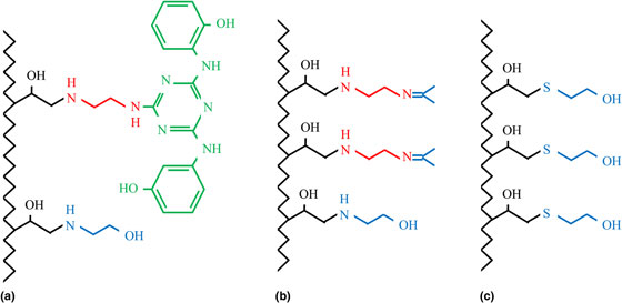
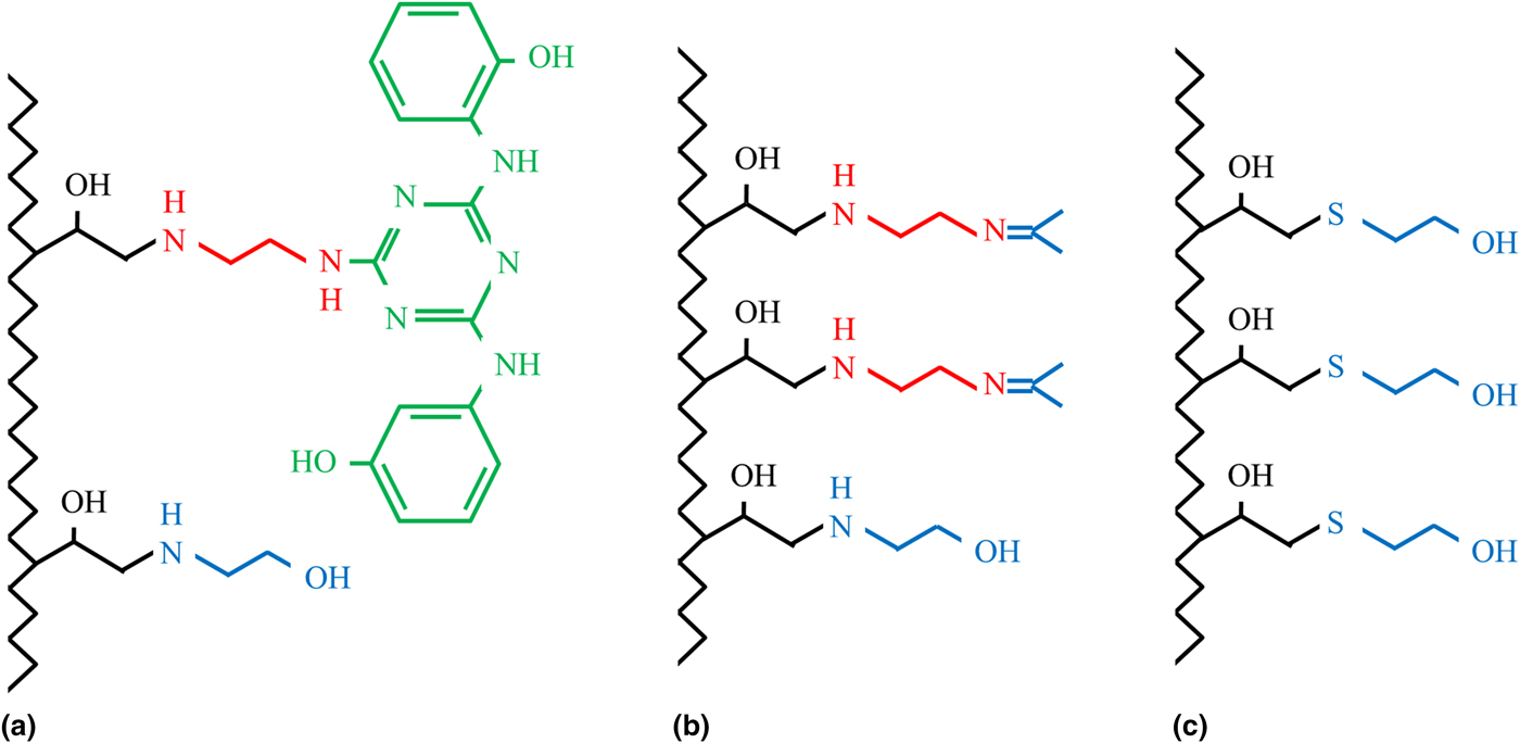
The ligand investigated in this study is the Protein A Mimetic Ligand™ A2P, designed and produced by Prometic Biosciences, Cambridge, UK. The structure of ligand A2P bound to the membrane through the spacer arm 2LP (1,2-diaminoethane) is reported in Fig. 1(a).
Chemical structure of membranes modified with (a) A2P (green)-2LP (red), (b) 2LP (red). In both cases the encapping of the residual epoxy groups is performed using 2-ethanolamine (blue) and of the residual amine with acetone (blue). (c) Chemical structure of the native membranes endcapped with 2-mercaptoethanol (blue).
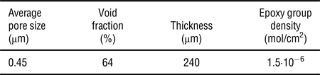
Sartobind Epoxy membranes, kindly provided by Sartorius Stedim Biotech GmbH (Goëttingen, Germany), were used as chromatographic supports for ligand immobilization. The membranes are composed of regenerated cellulose activated with epoxy groups; their fundamental characteristics are reported in Table I.
Epoxy-activated membrane properties.

The average pore size and epoxy group density are among the specifications provided by the manufacturer,[ 18 ] while the membrane void fraction and thickness were experimentally measured. The membrane thickness was measured using a micrometer (Absolute, Mitutoyo) and averaging the data over at least 10 points per membrane sheet; as a result, a thickness of 240 ± 5 µm was obtained. The void fraction was calculated by applying the method of moments: to this aim, independent chromatographic experiments were performed in non-binding conditions by feeding, at different flow rates, pulses of 10, 100 µL of a 5% v/v acetone solution.[ Reference Guiochon, Felinger, Shirazi and Katti 19 , Reference Herigstad, Dimartino, Boi and Sarti 20 ] The first moment measured at different flow rates was plotted against the theoretically calculated residence time and the porosity was calculated from a linear regression that was characterized by a R 2 value of 0.98.
Pure protein concentration was determined by UV absorption at 280 nm by using a UV-vis Spectrophotometer (UV-1601 Shimadzu Italia, Milan, Italy).
For the determination of IgG concentration, a Protein A cartridge was also used (Applied Biosystems, PA ImmunoDetection® Sensor Cartridge for Perfusion Immunoassay™ Technology) in HPLC analysis, while for protein mixtures (IgG and BSA, IgG and Lysozyme) an HPLC SEC column was used (BioSuite™ 250, 4 µm, UHR SEC, Waters, Milan, Italy). Both columns were mounted on a Waters Alliance 2695 separation module equipped with a module 2487 dual-wavelength absorbance detector (Waters, Milan, Italy).
Membrane modification
Affinity membranes were prepared by chemical immobilization of the A2P-2LP ligand complex on the membrane sheets that were cut into 1.5 and 2.6 cm diameter discs to be ready for use in batch and dynamic experiments. To investigate the effect of the spacer arm on non-specific protein binding the 2LP spacer was also immobilized without ligand on the epoxy activated membranes. Both types of membranes were prepared by adapting the protocol described by C. Boi et al., and the schematic drawing of the modified membranes is illustrated in Fig. 1.[ Reference Boi, Busini, Salvalaglio, Cavallotti and Sarti 16 ] Native Sartobind Epoxy membranes were also tested as a control after the endcapping of the epoxide active groups.
Preparation of A2P-2LP membranes: the epoxide activated supports were first suspended in ethanol, then soaked in a solution 10 mol-equiv. of the complex A2P-2LP in ethanol. The reaction mixture was shaken overnight (~15 h) in a thermostatic bath kept at 50 °C; the membranes were then washed several times using ethanol and water. The residual epoxy groups were endcapped with a solution of 0.1 M 2-ethanolamine in ethanol and kept at 50 °C overnight (~15 h). Finally, the membranes were thoroughly washed for 2 hours with ethanol and water, using an ultrasonic bath.
Preparation of 2LP membranes: the same protocol was used for the immobilization of the spacer arm 1,2-diaminoethane alone. In this case, an additional endcapping strategy needed to be adopted in order to deactivate the free amine groups on the spacer. This procedure was carried out after the 2-ethanolamine endcapping step, by suspending the membrane discs into pure acetone at room temperature for 24 h; subsequently, the membranes were washed in water.
Endcapping of the epoxy groups of native Sartobind Epoxy membranes: the membranes were first equilibrated in ethanol, and then kept in a solution of 5 mol/equiv. of 2-mercaptoethanol in ethanol at 50 °C for 15 h. Finally, the membranes were thoroughly washed with ethanol and water, using an ultrasonic bath.
Membrane characterization
The modified membranes, both the A2P-2LP type and the 2LP type, were preliminarily characterized in batch adsorption experiments by soaking the modified membrane discs in pure protein solutions of known concentration and kept under gentle agitation until equilibrium was reached. After the adsorption step, the membranes were first dabbed with paper to remove the excess of protein solution and then washed with PBS pH 7.4, keeping them under gentle agitation for 1 h. The elution step was carried out by immersing the membranes in the glycine buffer for 1 h, under gentle agitation. The protein concentration was determined at the beginning and at the end of the adsorption step, as well as at the end of the washing and elution steps, by measuring the absorbance of the solutions at 280 nm.
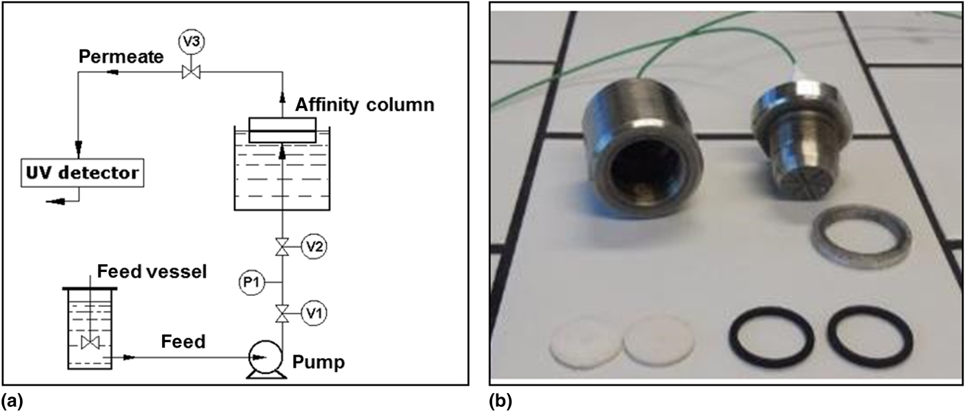
Dynamic binding experiments were performed on a Fast Protein Liquid Chromatography (FPLC) AKTA Purifier 100 (GE Healthcare, Milan, Italy). Experimental runs were conducted by placing a layered stack of 5–10 membrane disks of 2.6 cm diameter into a stainless steel housing suitably designed by our research group to ensure the correct flow distribution and the appropriate degree of compression of the membranes.[ Reference Morselli 21 ] The module is composed of a female and a male part; the membranes, two polymeric frits and two o-rings are placed in the female part; the male part presents a thread zone that ensures the closure of the module. The polymeric frits are placed at the inlet and at the outlet to guarantee an optimal distribution and collection of the flowing solution over the cross-sectional area of the membranes, while the o-rings are used to ensure the hydraulic seal of the cell. A schematic drawing of the chromatographic apparatus and a picture of the membrane module are reported in Fig. 2.
(a) Scheme of the chromatographic apparatus; (b) image of the membrane module.

The solute concentration in the effluent was measured on line by UV absorption at 280 nm for all solutions.
Results and discussion
Batch experiments
Batch experiments with pure protein solutions using the native Sartobind Epoxy membrane indicated that the membranes, treated with an appropriate blocking agent for the epoxide groups, such as 2-mercaptoethanol, do not contribute to a detectable non-specific adsorption. To exclude adsorption by ionic interactions, the tests were performed by using two proteins with very different isoelectric points, lysozyme (pI 11), and BSA (pI 4.7): both protein solutions, prepared in PBS pH 7.4, did not show a measurable adsorption on the native membranes. That is the amount of lysozyme and BSA adsorbed in separate experiments was zero. Moreover, when the native membranes were endcapped using 2-ethanolamine, 0.4 mg/mL of BSA was only adsorbed in the first chromatographic cycle performed, while lysozyme was not adsorbed at all.
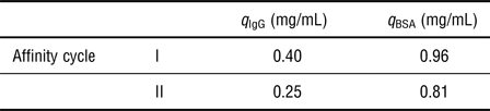
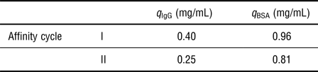
2LP modified membranes were tested for adsorption with pure solutions of BSA and IgG at a feed concentration of 0.5 mg/mL, performing two subsequent affinity cycles; in both cases there was a measurable adsorption, that is more relevant for BSA, as shown in Table II, indicating that the contribution of the 2LP spacer on protein binding is non-negligible. Indeed, non-specific adsorption may occur due to hydrophobic, ionic or van der Waals forces, but since the 2LP molecule has two amino moieties, that could favor ionic and van der Waals interactions, and two carbon atoms, that could favor hydrophobic interactions, all type of interactions cannot be excluded a priori. A detailed theoretical study, with the help of molecular modeling would be necessary to better understand the non-specific adsorption mechanisms, but it is beyond the scope of this work.
Batch adsorption on 2LP modified membranes.
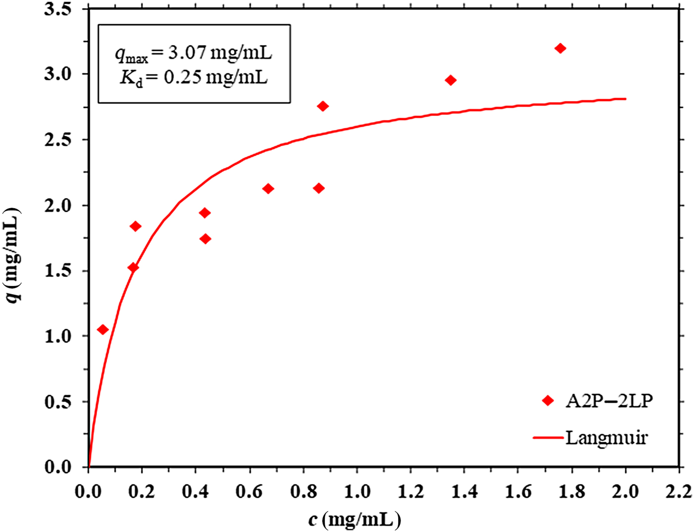
A2P-2LP affinity membranes were also tested for protein adsorption and an equilibrium isotherm for IgG was obtained. As it can be observed from Fig. 3, the experimental data can be successfully interpolated with the Langmuir model giving a static binding capacity, q max, of 3.07 mg/mL and a dissociation constant, K d, of 0.25 mg/mL. Note that these results represent one data set only, however they are consistent with the results of several experiments performed in previous works by our research group using A2P membranes, so they can be considered representative of the membrane behavior.[ Reference Boi, Dimartino and Sarti 17 ]
Equilibrium adsorption isotherm of IgG onto A2P-2LP affinity membranes.

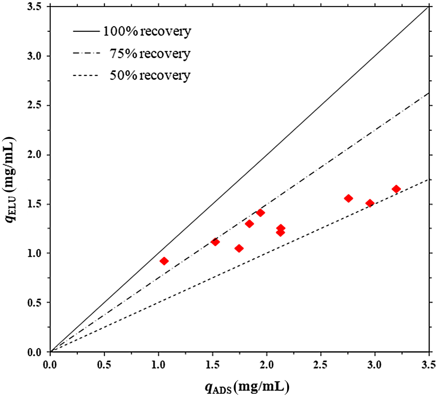
After adsorption, the affinity membranes were washed with PBS buffer and eluted with glycine buffer. In the concentration range inspected, the amount of IgG recovered with elution was always well below 100% and, usually, between 50 and 75% as shown in Fig. 4 in which the amount of IgG eluted is reported as a function of the amount of IgG adsorbed.
Recovery of IgG with elution versus the IgG adsorbed on A2P- 2LP affinity membranes.

The initial hypothesis that the non-specific adsorption on the 2LP spacer is responsible for the incomplete IgG recovery from the A2P-2LP affinity membranes is corroborated by an analysis of the adsorption and elution data. If it is considered that the average recovery with elution from the A2P-2LP affinity membranes is around 60% (Fig. 4) when the amount of IgG adsorbed, q, is around 2 mg/mL, the amount of IgG that can be eluted is around 1.6 mg/mL. This indicates that the amount of IgG that is non-specifically adsorbed on the membranes and cannot be recovered is about 0.4 mg/mL, which corresponds to the amount of IgG that non-specifically adsorbs on the 2LP spacer as reported in Table II.
Dynamic experiments
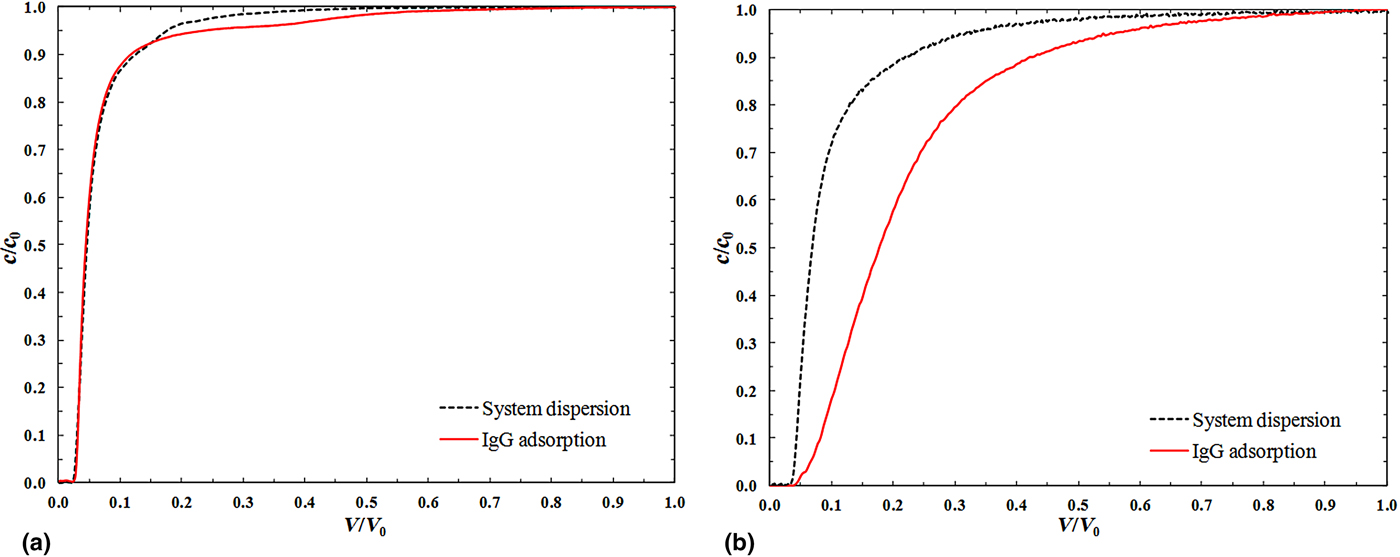
To better understand the contribution of non-specific binding, more detailed studies in a chromatographic set up have been performed. As customary for this type of characterization, the system dispersion was determined in independent experiments in non-binding conditions, in order to calculate the contribution of the system dead volumes and mixing effects on the dynamic binding capacity.[ Reference Boi, Dimartino and Sarti 17 , Reference Sarfert and Etzel 22 ] System dispersion curves were obtained for both 2LP type membranes and A2P-2LP affinity membranes by feeding a solution of IgG in PBS, using the membranes that were completely saturated during previous adsorption tests, in order to ensure experimental non-binding conditions. For every membrane type, system dispersion curves were compared with breakthrough curves in experiments performed at a constant flow rate of 1 mL/min. The curves were integrated and the amount of IgG adsorbed was calculated as a difference between the IgG adsorption curve and the system dispersion curve.
Results obtained in a typical experiment performed with 2LP type membranes are shown in Fig. 5(a), while results obtained with A2P-2LP affinity membranes are reported in Fig. 5(b). As it can be observed from Fig. 5(a), the area between the two curves is not negligible and represents the contribution of the 2LP spacer to IgG non-specific adsorption. For the example, reported in Fig. 5(a), the value of IgG non-specific adsorption has been calculated as 0.77 mg/mL, and it was of the same order of magnitude in all experiments performed. The 2LP membranes were subsequently washed with PBS and eluted with 0.1 M glycine pH 2.8, but the IgG adsorbed was not removed with the elution buffer.
Comparison between system dispersion curve and IgG adsorption on: (a) 2LP type membranes, (b) A2P-2LP membranes.

Integration of the curves reported in Fig. 5(b) gave the amount of IgG adsorbed by A2P-2LP affinity membranes, which was calculated as 3.37 mg/mL for the example under investigation. Therefore the amount of IgG that is non-specifically adsorbed is around 23% of the total IgG adsorbed on A2P-2LP affinity membranes.
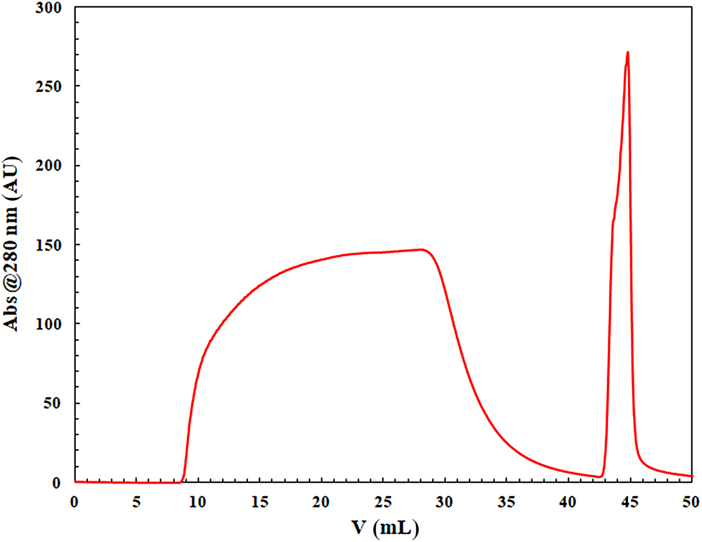
To investigate the effect of non-specific binding on IgG recovery, complete chromatographic cycles were performed with the A2P-2LP affinity membranes; a typical cycle is reported in Fig. 6.
Complete chromatographic cycle for IgG purification on a stack of 10 A2P-2LP affinity membranes. Feed solution 0.5 mg/mL IgG in PBS, flow rate 1 mL/min; q ADS = 1.85 mg/mL, q WASH = 0.08 mg/mL; q ELU = 1.06 mg/mL.

In the example under investigation the amount of IgG adsorbed per unit membrane volume, 1.85 mg/mL, was not completely recovered with elution; indeed the amount eluted, 1.06 mg/mL, was only 57% of the amount adsorbed. From a mass balance it is possible to calculate the amount of IgG that is not released with elution that corresponds to the quantity which is non-specifically adsorbed to the affinity membrane:
 $$q_{{\rm NON - SPECIFIC}} = q_{{\rm ADS}} - q_{{\rm WASH}} - q_{{\rm ELU}} $$
Using the data of the example shown in Fig. 5, q NON-SPECIFIC = 0.71 mg/mL, that is practically the same as the amount non-specifically adsorbed onto the membranes prepared with the 2LP spacer alone, that had a value of 0.77 mg/mL. Indeed, the glycine elution buffer applied to 2LP type membranes after adsorption was not effective to recover any detectable IgG amount. Since the native membranes do not contribute to non-specific adsorption, it is concluded that the amount of IgG that is non-specifically adsorbed by the A2P affinity membrane it is due to a strong non-specific binding to the 2LP spacer arm.
Therefore, a new immobilization strategy, using a different spacer molecule, needs to be developed for A2P to completely unveil its potential in IgG purification.
Conclusions
Results obtained in preliminary batch experiments using the 2LP-Sartobind Epoxy membranes have shown that the quantity of protein adsorbed in the presence of the spacer arm 2LP alone is not negligible, while it was for the original unmodified membrane. These results were confirmed in dynamic experiments in a chromatographic set up indicating that the contribution of the spacer 2LP to non-specific protein adsorption is relevant and must be accounted for. Therefore, when the spacer 2LP is used as a linker for ligand immobilization it influences the performance of the affinity membrane. Thus the indication is obtained that the limited recovery of the IgG adsorbed on A2P-2LP affinity membranes is not only due to the strong binding between protein and ligand but also to an irreversible binding on the 2LP spacer. Indeed, the A2P ligand could be better exploited using a different immobilization protocol and a different spacer molecule.
Acknowledgments
The authors acknowledge the Italian Ministry of University and Research.