Introduction
Mesoporous silica-based materials are extensively produced by expensive and not so easily scalable processes.[ Reference Alothman 1 ] Nevertheless, mesoporosity is specifically useful for precise biomedical applications[ Reference Luelf, Devaux, Prasetyanto, De Cola, Torres and Bottari 2 ] and chemical purposes.[ Reference Pagliaro 3 ] Mesoporous silica exhibits properties of high surface area, tunability of the pore sizes in the micro/nanometer range, and adaptable surface chemistry which, together with biocompatibility, make this material suitable for drug-delivery and sensing applications.[ Reference Slowing, Trewyn, Giri and Lin 4 ] Engineering bioglasses or composites with hydroxyapatite (silicon-doped hydroxyapatite) has become the current focus in biomaterial science.[ Reference Jugdaohsingh 5 ] In particular, silica supports for cell-adhesion deliver silicic acid in biologic systems, which stimulates condrocitic and osteocitic growths, also promoting the osteo-mineralization.[ Reference Arumugam, Ireland, Brooks, Rushton and Bonfield 6 ] For this reason, combination of silica-derived compounds with bioglasses improves the bone cells proliferation/adhesion onto surfaces around the implanted material.[ Reference López-Álvarez, Solla, González, Serra, León, Marques and Reis 7 ] Cell growth is also investigated in relation with different silica morphologies/geometries. For regenerative medicine the surface topography is strictly associated with the behavior of the cells in contact with the biomaterial: micrometer and nanometer scale topographies modify different aspects of cell behavior such as cell adhesion, morphology, proliferation, and differentiation, also related to local factors, which are present in the microenvironments.[ Reference Lohmann, Tandy, Sylvia, Hell-Vocke, Cochran, Dean, Boyan and Schwartz 8 – Reference Lüthen, Lange, Becker, Rychly, Beck and Nebe 13 ] Among all cell types, osteoblasts-like cells seem to be able to recognize differences in roughness and topographies on biomaterial surfaces.[ Reference Schweikl, Müller, Englert, Hiller, Kujat, Nerlich and Schmalz 14 ] It appears that micro-rough surfaces inhibit cell proliferation promoting cell differentiation,[ Reference Martin, Schwartz, Hummert, Schraub, Simpson, Lankford, Dean, Cochran and Boyan 15 , Reference Kieswetter, Schwartz, Humrnert, Cochran, Simpson, Dean and Boyan 16 ] while nanopatterned surfaces enhance bone cells adhesion.[ Reference Price, Ellison, Haberstroh and Webster 17 ] Only a mesoscale topography could improve both proliferation and adhesion (with partial effect on differentiation).[ Reference Zinger, Anselme, Denzer, Habersetzer, Wieland, Jeanfils, Hardouin and Landolt 12 , Reference Zinger, Zhao, Schwartz, Simpson, Wieland, Landolt and Boyan 18 ]
The nanostructured biosilica from diatoms macroalgae represents a cheap and natural biochemically nanotextured material, generated in vivo by a complex biomineralization process. Ten of thousands of diatoms with different mineral structures are observed in Nature. The formation of new amorphous silica occurs in specialized intracellular compartments, named the SDV (silica deposition vesicles), which act as cellular reaction vessels, and the process is absolutely under genetic control assuring a complete reproducibility of the structure formation of the amorphous silica.
Diatomaceous earth (DE) is produced by the accumulation and compaction of the siliceous parts of the diatoms, upon death of the organisms. It represents a very low-cost source of silica in which all the silica elements (valves, girdles, crowns, and hard silica particles), exhibit a high level of mesoscale, ranging from big micro-structures belonging to giant fossil diatom valves (centric and pennates diatoms), to small nanostructures exhibited by other smaller species (always centric), till rod-like structures (pennates or hemi-girdle structures, with whole or fractured clasps structures) and a poorer nanopatterning.
To the best of our knowledge, one example of separation (by size and morphology) of different components from diatomite has been reported: the starting material is a bidispersed coscinodiscus diatomite easy to separate using the method via ethanol sedimentation,[ Reference Zhang, Wang, Pan and Cai 19 ] but not useful for the hugely polydisperse DE.
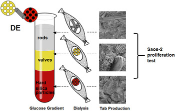
Here we propose a method of separation based on common glucose gradient (already used for protein separation) and dialysis purification (useful for silica nanoparticles). The aim of this work was to obtain different biosilica morphologies by a glucose gradient by means of Stokes sedimentation,[ Reference Carlisle, Brito, Gladysz, Ricci and Koopman 20 ] performed after acid/oxidative treatment of DE. The different morphologies obtained are made of rods, empty valves, and hard silica particles. We fully characterized these materials and we used them to produce fluorescent two-dimensional biosilica-on-glass (which we call here “tabs”) via chemical/thermal bonding of biosilica on glassy supports. These supports exhibited good resistance in water, ethanol, and soft scraping. We moved to a preliminary biologic application, testing osteoblasts-like cells biologic manner for this new biosilica scaffold. The scheme of the entire process is reported in Fig. 1.
Schematic representation of separation methodology: glucose gradient/dialysis approach on activated DE and SEM images of the corresponding fluorescent “tabs” obtained by the three fractions, finally tested on Saos-2 cells.

Experimental details
Purification of commercial DE
Based on a literature protocol,[ Reference Vona, Leone, Ragni, Palumbo, Evidente, Vurro, Farinola and Cicco 21 ] an activation process of DE was exploited in order to eliminate the clay components, constituted by oxides, and exhibit the −OH groups (activation) on the biosilica surface.
This method has been already used for biosilica extracted from living coltures.[ Reference Cicco, Vona, De Giglio, Cometa, Mattioli-Belmonte, Palumbo, Ragni and Farinola 22 , Reference Jiang, Luo, Liu, Deng, Jing, Bai and Li 23 ] A sample of 150 mg of DE (Sigma-Aldrich) was inserted in a glass Pyrex falcon containing 400 µL of distilled water and mixed for 5 min at room temperature under magnetic stirring at 400 rpm, in order to hydrate and to activate the biosilica; then the temperature was increased to 80 °C and 1 mL of HCl (37%) was added and left under stirring for 10 min. Later 1 mL of H2SO4 (98%) was added, leaving under stirring for 10 s. Subsequently 3 mL of H2O2 were put dropwise on the mixture leaving under magnetic stirring for 4 h. To isolate the activated diatoms from acids, washing steps were made using a bench top centrifuge at a fixed rotor (1000 rpm for 10 min); these steps were performed in distilled water, then with a mixture of acetone and distilled water (ratio 1:1) and finally ethanol, and then the pellet was dried in a vacuum pump.
Diatoms separation by glucose gradient and dialysis
The Stokes sedimentation in glucose gradient was used to separate the different morphologies of DE. The protocol is similar than the one used for protein separation.[ Reference Hanson, Schulenberg, Patton and Capaldi 24 , Reference Hanson, Carrozzo, Piemonte, Tessa, Robinson and Capaldi 25 ] A total of four solutions at different concentrations of D-(+)-glucose (Sigma Aldrich) in water were produced [43%, 30%, 20%, and 4% (w/v)] and 6 mL of each were placed one on the other drop wising into a gradient column, forming a discontinuous gradient. An amount of 120 mg of activated DE (suspended in 400 µL of distilled water) were added on the top of the gradient and, after 10 min, the various layers were collected in separate tubes and washed several time using centrifuge (10 min, 2000 rpm for 4%, 1500 rpm for the 20%, 1000 rpm for 30%, 43% called 43%f fraction, and a spontaneous pellet under the 43%, called 43%p) obtaining final sample pellets of the five fractions. The separation of glucose gradient was carried out for 10 times in order to have a sufficient amount of fractions. After washing steps, the contents of the five fractions were added to a nitrocellulose membrane, closed and placed in water under magnetic stirring for 12 h in order to remove the residual glucose. The protocol used is similar than the one reported for silica nanoparticles purification by dialysis.[ Reference Pálmai, Nagy, Mihály, Varga, Tárkányi, Mizsei, Szigyártó, Kiss, Kremmer and Bóta 26 ] All the fractions were collected and washed using the same centrifugation step used after the gradient, and then suspended in acetone and dried. The fractions made of 4% and 20% were grouped together in the portion of the silica rods, the 30% and 43% fractions were combined in the portion of the silica valves, and then the pellet of 43% fraction was made of hard silica particles.
Characterization methods of the biosilica fractions
These three combined samples (silica rods, valves, and particles) were analyzed using contrast phase microscopy evaluating the number of whole valves (from centric species)/field evaluation, and Fourier transform infrared spectroscopy (FTIR) for chemical analyses of functional groups (see Supplemental Data). To test if all processes of acid–oxidative treatment, separation via gradient and purification via dialysis did not affect the nanostructure, we evaluated architectural parameters such as empty valves, in a way similar to that reported in literature.[ Reference Lang, Del Monte, Rodriguez, Dockery, Finn and Pandit 27 ] Morphological analysis of silica valves were performed via optical microscopy (in reflection), together with the statistical evaluation of average diameter/pore and number of pores/area (see Supplemental Data).
Biosilica “tabs” preparation
The next step was to produce solid biosilica-based supports (also referred here as “tabs”) and to make a preliminary biocompatibility assay with adherent Saos-2 cells. For the production of tabs, three round glass slides per fractions (also treated via acid–oxidative treatment used for the DE, for 1 h) were used as supports. On each slide placed on the plate at 50 °C, a mixture containing 20 mg of each biosilica morphology (rods, valves, and hard silica particles), 100 µL of ethanol, 0.4 µL of APTES (amminopropyl-triethoxy-silane, Sigma Aldrich), 10 µL of sodium metasilicate, 6 µL of a carboxy-fluorescein solution in ethanol NaOH (stock solution: 3 mg fluorescein, 1 µL of NaOH 1 M, 1000 µL of ethanol) was added. The presence of the dye allowed the production of fluorescent tabs. Once the ethanol was evaporated, a protocol of synterization (already used for synterization of nanoparticles[ Reference Ohtsuka, Seki and Takeoka 28 ] and biosilica[ Reference Gale, Jeffryes, Gutu, Jiao, Chang and Rorrer 29 ]) was carried out: 10 min at 60 °C, 10 min at 100 °C, 20 min at 150 °C, 30 min at 200 °C, 2.5 h at 300 °C. After cooling at room temperature, the tabs were washed using distilled water and then ethanol to remove any excess of not sticked biosilica. The “tabs” were dried at 60 °C and observed by scanning electron microscopy (SEM) analyses and confocal microscopy. Water adsorption rate (WAR) measurements were determined to investigate tabs wettability depending on their different morphologies (see Supplemental Data).
MTT viability test
To test the Saos-2 cell viability onto the obtained biosilica tabs an MTT (3-(4,5-dimethylthiazol-2-Yl)-2,5-diphenyltetrazolium bromide) assay was performed. Saos-2 cells grown in the culture medium McCoy (10% fetal bovine serum) in an incubator 37 °C/95% CO2 were used (cell concentration per well = 1.5 × 10⁴ cells/mL). Tabs were tested in triplicate together with three blank slides subjected to the same oxidative acid treatment of DE. Cells in the multi-wells were left for 24 h in incubation and then MTT assay was carried out: the medium was removed and 200 µL of MTT solution (5 mg/mL in DMEM without phenol red) and 1.8 mL DMEM (Dulbecco's Modified Eagle's Medium) were added to all the wells with cells, and incubated at 37 °C for 4 h. Then the supernatant was removed and the formazan crystals dissolved in 2 mL of solution (4%, 1 N HCl in isopropanol). The difference between the absorbance at 570 and 690 nm was measured by spectrophotometry analyses.
Fluorescence staining test
488 Phalloidin fluorescent staining (SIGMA) was used to stain the actin fibers of Saos-2 grown in the same conditions of the cells for the MTT test and fixed using PFA 4%. Cells were then coated with Triton X-100 solution for 30 min, then washed with PBS and coated with 400 µL of phalloidin solution for 30 min. After phosphate buffered saline (PBS) washings the samples were observed under an optical fluorescent microscope, and DAPI (4',6-diamidino-2-phenylindole) solution was used to contrast nuclei. Qualitative analyses have been reported only for 96 h growth.
Results and discussion
DE constituents splitting by a glucose gradient and dialysis
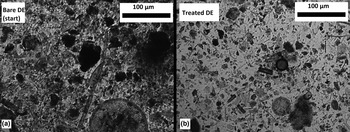
DE is a highly polydisperse material with several impurities consisting of a mixture of various metal oxides (Al, Fe, etc.). Before using such a biosilica, a cleaning procedure was required. As shown by contrast phase microscopy analysis (Fig. 2), after the acid–oxidative treatment the nanostructured biosilica composition was not affected, and most of hard silica particles (black particles) were eliminated in comparison with the bare DE.
Optical microscopy of: (a) bare DE (before cleaning procedure); (b) treated DE (after acid/oxidative treatment).

The glucose gradient was realized by four aqueous solutions at different concentrations of D-(+)- (43%, 30%, 20%, and 4% (w/v)], which were stratified in a glass column, as described in the “Experimental” section.
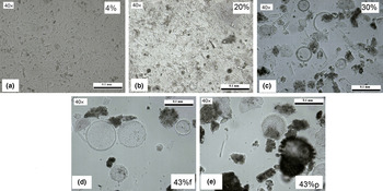
Roughly, 150 mg of biosilica were put on the top of the column and, after 5 min of elution time, four fractions were recovered in addition to a precipitate produced at the bottom of the 43% fraction. The five fractions were subjected to a dialysis process by a nitrocellulose membrane and after several washing steps, five samples were collected and analyzed by contrast phase microscopy. As shown in Fig. 3, the SEM analysis evidenced that in the glucose portions at lower concentrations, a plenty of silica rods was present, while the fractions with higher glucose concentrations were mainly composed of diatoms valves or fragments with higher surface area. Hard silica particles were founded in the precipitate at the bottom of the 43% glucose concentration.
Contrast phase microscopy images of the samples recovered after the glucose gradient elution. (a) 4%, (b) 20%, (c) 30%, (d) 43%f, and (e) 43%p.

After the dialysis process, the 4% and 20% glucose concentrations fractions were collected in a single sample (“silica rods”) for their morphological similarity; for the same reason, the 30% and 43% glucose concentrations fractions were grouped in the “silica valves” sample and the precipitate of the 43% glucose was the “hard silica particles” sample. The calculated yields after DE separation and dialysis (related to 150 mg of starting material) were: 2% valves, 19% for rods, and 8% for hard silica particles.
FTIR analyses (Supp. Inf., Fig. S1) confirmed the siliceous structure of the samples and the presence of inorganic impurities in the hard silica particles.
Tabs production and surface characterizations
The three different DE morphologies were used to produce the fluorescent biosilica-on-glass supports (tabs) via chemical/thermal bonding of the three samples on glass slides, as described in the “Experimental” section. The tabs exhibited good resistance in water, ethanol, and soft scraping.
A more detailed examination of the different morphological composition of the tabs was achieved by SEM (Fig. 4).
SEM images of the fluorescent tabs: (a) silica rods; (b) silica valves; (c) hard silica particles. Insets are referred to (*, **) marked elements in main pictures.

For the silica rods, the SEM image showed mainly fragments rods-like from pennates (in the inset the characteristic clasps) and girdle debris [Fig. 4(a)]; whole valves and fragments with the characteristic mesoporous structure appeared in the valves [Fig. 4(b)]; the hard silica particles showed an amorphous composition of biosilica fragments, mainly valves, intimately blended likely with metal oxides that clearly reduce the surface porosity [see the inset of Fig. 4(c)]. A further confirmation of the different porosity of the three tabs was provided by WAR measurements, which gave the specific information about the surface wettability of these supports and their hydrophilicity.
We assumed that lower wettability values registered for the silica rods and valves tabs were attributed to the presence of a great number of not obstructed nanopores after the cleaning (acid/oxidative) processes. As our clean diatomite can be compared with a bioactive materials (such as bioglass, ceramics, mesoporous silica, and hidroxyapatite), where an interconnected macroporous network with a nanometric pores system is present (and nano-amounts of air could be intimately entrapped) we can desume that full of air-filled cavities are present in our diatom-based mesoporous material.
For these reasons, our “tabs” exhibited different values of WAR (see Fig. S3, Supplemental Data). Lower WAR values were registered for the silica rods and valves tabs due to the presence of a great number of not obstructed nanopores, while the presence of biosilica debris or valves with obstructed nanopores in the hard silica particles tab exhibited higher wettability likely because of the absence of air inside nanostructured layers (nanostructures obstructed by silica–metal oxides co-composites). The advantageous tab fluorescence was observed by confocal microscope (Fig. S4, Supplemental Data): a higher fluorescence was detected in tabs containing valves and silica clusters with respect to the tabs containing rods or elongated fragments.
MTT assay
In our previous work,[ Reference Cicco, Vona, De Giglio, Cometa, Mattioli-Belmonte, Palumbo, Ragni and Farinola 22 ] we already demonstrated that osteoblast-like cells better grow on a mesoporous surface instead of a smooth one. In this study, starting from a single polydisperse material composed of various levels of porosity (valve, crown, and girdle), we were able to separate the different nanotextured biosilica portions, and investigate whether the osteoblast-like cells behavior changed.
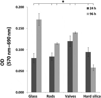
The MTT viability test highlighted that osteoblasts-like cells (Saos-2) showed a regular growth on the silica tabs, without showing significant toxicities in single culture conditions (24 and 96 h, Fig. 5).
Histogram of MTT values (expressed as difference of adsorbance 570–690 nm) for Saos-2 cells grown for 24 and 96 h onto “tabs” (rods, valves, and hard silica particles). Flat glass was used as positive control. All data are statistically evaluated and differences were considered statistically significant for P < 0.05 (*marked for correlation).

The silica valves tab exhibited the best biocompatibility (>60% with respect to glass control) followed by the silica rods tab, especially in terms of increase of growth over time (from 24 to 96 h). The cells’ growth on the mesoporous valves is slightly better than one occurring on the nanostructures of the rods. The hard silica particle's tab showed a decrease in growth over time likely due to the obstruction of the pores, data confirmed by the strong cell degradation shown after fluorescence staining.
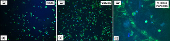
Information about Saos-2 cells morphology and behavior on rods, valves, and hard silica particles’ tabs were obtained by the fluorescence staining.
As showed in Fig. 6 (experiment for 96 h), cells grown on the valves tabs are more dense (in average population) than those ones grown on the rods and on hard silica particles tabs. In the rods and hard silica particles tabs, cells are not so clearly visible in the distinctive and different staining among nuclei and actin stress fibers. Hard particles sample exhibits strong green cells debris maybe due to cells degradation occurred on these silica particles.
Staining of Saos-2 grown on rod (a), valves (b), and hard silica particles (c) tabs with DAPI (blue staining for nuclei) and Atto-Phalloidin (green staining for actin stress fibers) after 96 h. The symbol “*” indicates the position of nuclei.

Conclusions
All the materials, which are currently used or proposed as scaffold in regenerative medicine, are usually bioglass or silica mesoporous materials, entirely fabricated in laboratories. The increasing demand of multifunctional biomaterials with specific physical, chemical, and biologic properties is often in conflict with the complexity and high costs of the tissue engineered products leading to a strong gap between the research and clinics. In our work, we proposed a low-cost scaffold based on shells of diatom microalgae, a natural biosilica-based material with a mesoporous highly ordered structure. Moreover, the possibility of chemical modification of the diatom surface and “in vivo” doping the diatom cells with organic molecules (that is, dye, growth factors, etc. covalently bound on the surface or entrapped in biosilica) considerably extends the functionality of the scaffold, leading in our opinion to a “new generation” material for biomedical applications.
A low-cost source of biosilica from diatoms is provided by DE, natural material that may generate a research incipit for building bio-inspired scaffold.
In this study, a Stokes sedimentation method based on glucose gradient and dialysis on activated polydisperse DE has been proposed, to obtain more omogeneous diatomic fractions enriched by components of the same morphology and texturization. Subsequently the three obtained morphologies (rods, empty valves, and hard silica particles) were fully characterized and used for producing fluorescent “tabs”, on which the growth of Saos-2 cells was tested. The silica valves promoted the osteoblast-like cells growth better than the silica rods, underlining the role of the dissimilar nanotexturization of a scaffold surface.
Supplementary Material
The supplementary material for this article can be found at https://doi.org/10.1557/mrc.2017.27