INTRODUCTION
The urohyal is an unpaired median bone located ventral to the basibranchial region within the hyoid and branchial arches, and is important in the mechanism of mouth opening and closing in fishes (Arratia & Schultze, Reference Arratia and Schultze1990; Chollet-Villalpando et al., Reference Chollet-Villalpandoa, De La Cruz-Agüeroa and García-Rodrígueza2014). The description of this structure in different fish groups dates back to Cuvier (Reference Cuvier1835). The morphology of the urohyal is used for taxonomic and phylogenetic purposes for several fish species (Bianchi, Reference Bianchi1984; Murray & Attia, Reference Murray and Attia2004; Otero, Reference Otero2004; Mabee et al., Reference Mabee, Grey, Arratia, Bogutskaya, Boron, Coburn, Conway, Shunping, Naseka, Rios, Simons, Szlachciak and Wang2011; De La Cruz-Agüero & Chollet-Villalpando, Reference De La Cruz-Agüero, Chollet-Villalpando, del Moral, Martínez, Franco, Ramírez and Tello2012; Marceniuk et al., Reference Marceniuk, Menezes and Brito2012; Chollet-Villalpando et al., Reference Chollet-Villalpandoa, De La Cruz-Agüeroa and García-Rodrígueza2014). It is also used in feeding studies to quantify the food ingested by piscivorous fish and other aquatic animals (Hansel et al., Reference Hansel, Duke, Lofy and Gray1988; Scharf et al., Reference Scharf, Buckel, Juanes and Conover1997, Reference Scharf, Yetter, Summers and Juanes1998; Johal et al., Reference Johal, Esmaeili and Tandon2001; Gosztonyi et al., Reference Gosztonyi, Kuba and Mansur2007; Gonzalez-Zevallos et al., Reference Gonzalez-Zevallos, Kuba and Gosztonyi2010; Tombari et al., Reference Tombari, Gosztonyi, Echeverria and Volpedo2010; Perez-Comesaña et al., Reference Perez Comesaña, Clavin, Arias and Riestra2013). This can often be species-specific and used in various studies of diet in fishes (Kusaka, Reference Kusaka1974; Esmaeili & Teimori, Reference Esmaeili and Teimori2006; De La Cruz-Agüero & Chollet-Villalpando, Reference De La Cruz-Agüero, Chollet-Villalpando, del Moral, Martínez, Franco, Ramírez and Tello2012).
There are several qualitative descriptive studies of the urohyal in fishes, for example Aprieto (Reference Aprieto1974) on some species of Carangidae, Sato et al. (Reference Sato, Hasegawa and Yonezawa1988) on Clupeidae, and De La Cruz-Agüero & Chollet-Villalpando (Reference De La Cruz-Agüero, Chollet-Villalpando, del Moral, Martínez, Franco, Ramírez and Tello2012) on different species of Gerreidae. However, the quantitative information for the urohyal in most past studies is very limited, despite it often being diagnostic (Chollet-Villalpando et al., Reference Chollet-Villalpandoa, De La Cruz-Agüeroa and García-Rodrígueza2014) and variable (Kusaka, Reference Kusaka1974; Esmaeili & Teimori, Reference Esmaeili and Teimori2006).
The morphology of the urohyal of Iranian fishes is not well studied. Esmaeili & Teimori (Reference Esmaeili and Teimori2006) have described the urohyal of some freshwater fishes and they included in their study the urohyal of Tenualosa ilisha. Jahromi et al. (Reference Jahromi, Esmaeili, Teimori, Nokhatolfoghahai and Ostovani2010) studied the morphology of two scarid species, Scarus ghobban and S. persicus.
Accordingly, this study was initiated to provide an additional osteological description of the fish species inhabiting the Persian Gulf and Oman Sea. The data and observations from this study will facilitate future taxonomic analyses regarding the fish groups studied.
MATERIALS AND METHODS
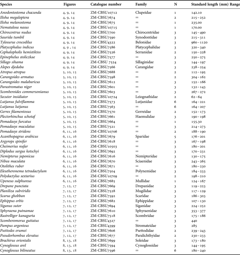
Urohyals were obtained from fishes collected from landing sites at Bandar Abbas and Bushehr cities, Iran. The fish specimens were kept on ice during the trip back to the laboratory where their total and standard lengths in mm were recorded. The flesh was removed by placing the fish specimen in boiling water for a few minutes and the urohyal extracted from the ventral side of the head, washed and stored dry in small envelopes. Families of species studied were arranged according to Eschmeyer (Reference Eschmeyer2014) and are presented in Table 1 together with the number of specimens for each species and size range. In total, 148 fish specimens of 49 species belonging to 43 genera and 28 families were studied. Eschmeyer (Reference Eschmeyer2014) and Fricke (Reference Fricke2014) were used for the updated taxonomic status of the species, spelling of species names, and taxonomic references. Digital images for the dorsal, ventral and left sides of the urohyals extracted from specimens were taken (as described by Kusaka, Reference Kusaka1974), using a Canon 7D camera. The graphical software Corel Draw X7 was used in the production of all figures. The description of the features of the urohyal was studied using the terminology of Chollet-Villalpando et al. (Reference Chollet-Villalpandoa, De La Cruz-Agüeroa and García-Rodrígueza2014) modified from Kusaka (Reference Kusaka1974). In this description the following abbreviations are used: Ba, basibranchial attachment; Co, condyle; De, dorsal extension; Dp, dorsal plate; Ha, hypohyal attachment; Lp, lateral plate; Pde, postero-dorsal edge; Pe, posterior edge; Rb, radial band; Ve, ventral extension; Vp, ventral plate (Figure 1). These features are defined as short (length < height), or broad if height > 1/2 length; long (length > height) or narrow if height < 1/2 length).
Left side view of a urohyal of Gerres filamentosus. Terminology of urohyal as follows: Ha, hypohyal attachment; Ba, basibranchial attachment; Ve, ventral extension; De, dorsal extension; De, dorsal plate; Pde, postero-dorsal edge; Rb, radial band; Lp, lateral plate; Pe, posterior edge; Ve, ventral edge; Vp, ventral plate; Co, condyle (modified from Kusaka, Reference Kusaka1974).

Number of samples (N), standard length range (SL, mm) and mean (m SL) of fishes collected from the Iranian marine waters and used in the urohyal morphology analysis.

All proportions of the urohyal were measured following Kusaka (Reference Kusaka1974), these are: DL, urohyal dorsal length; FL, urohyal frontal length; MH, urohyal maximum height; UH, urohyal height; UL, urohyal length; UW, urohyal width; VL, ventral length (Figure 2). Linear measurements were standardized as a function of the length of the urohyal and the angle values to eliminate the effect of fish size. Accordingly, the following features of the urohyal were calculated:
 $$\hbox{Size of urohyal} = \displaystyle{{\hbox{Length of the urohyal}} \over {\hbox{Head length}}} \times 100$$
 $$\hbox{Height of the lengthwise extension} = \displaystyle{{\hbox{Urohyal height}} \over {\hbox{Urohyal length}}} \times 100$$
 $$\hbox{Lateral development or spread} = \displaystyle{{\hbox{Urohyal width}} \over {\hbox{Urohyal length}}} \times 100$$
 $$\hbox{Aspect ratio} = \displaystyle{{\hbox{Urohyal length}} \over {\hbox{Urohyal width}}}$$
The values of the angles, ventral angle (VA, angle 3; ACB), condylar angle (CA, angle 1; BAC) and postero-dorsal angle (PDA, angle 2; ABC) were measured using an online digital screen protractor (ICONICO) (Figure 3). The urohyals are deposited in the collection of the Zoological Museum of Shiraz University (ZMCBSU), Biology Department, Shiraz, Iran (Table 1).
Dimension measurements of the left side view of a urohyal of Gerres filamentosus. DL, dorsal length; FL, frontal length; MH, maximum height; UH, urohyal height; UL, urohyal length; VL, ventral length (Kusaka, Reference Kusaka1974).

Angles of the left side view of a urohyal of Gerres filamentosus. Ventral angle (VA, angle 3; ACB); condylar angle (CA, angle 1; BAC); postero-dorsal angle (PDA, angle 2; ABC).

RESULTS
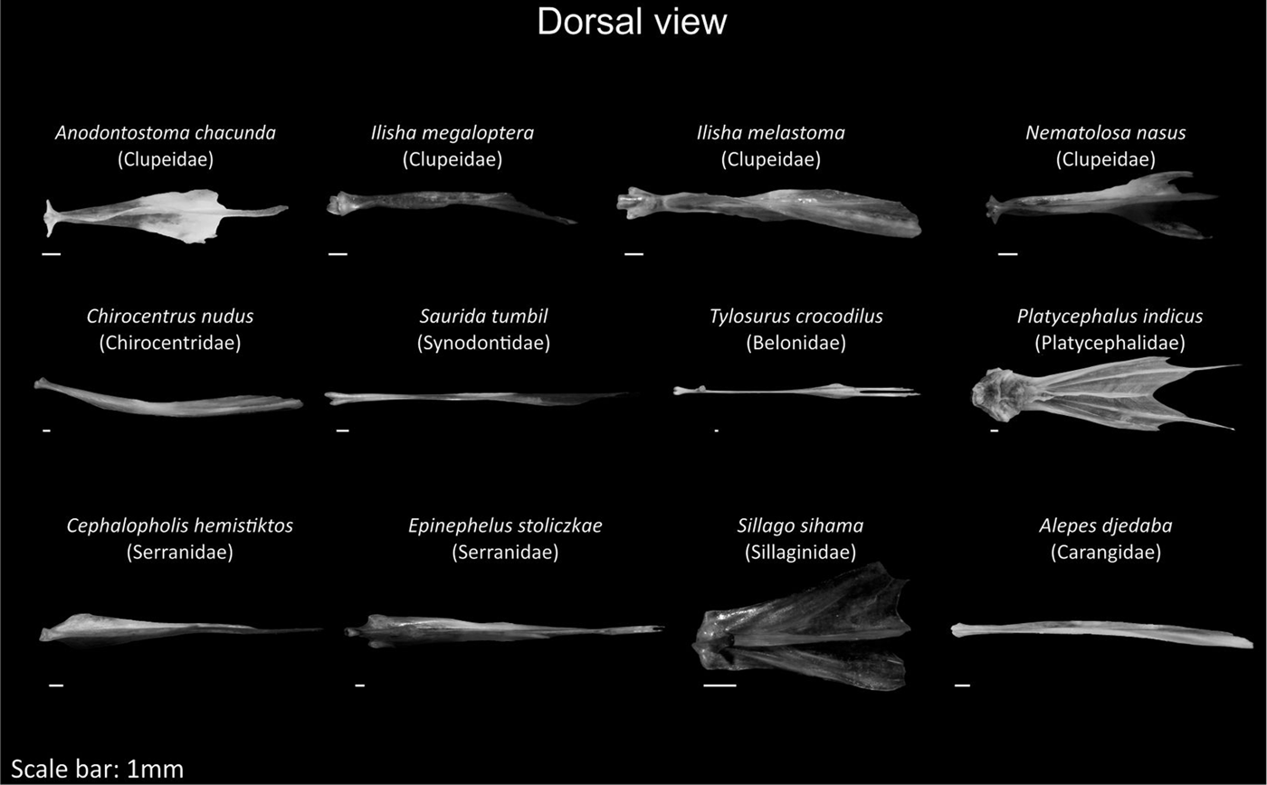
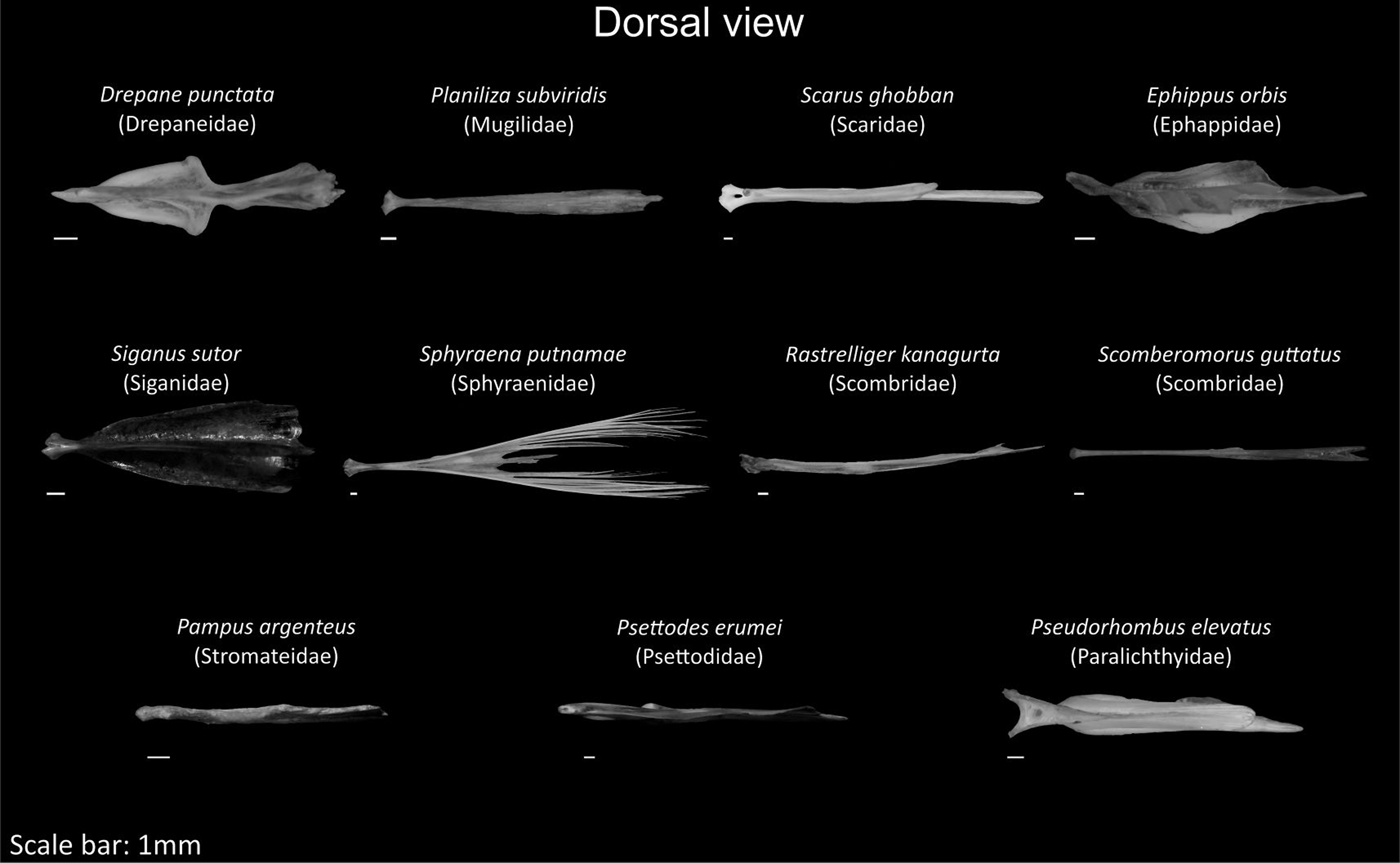
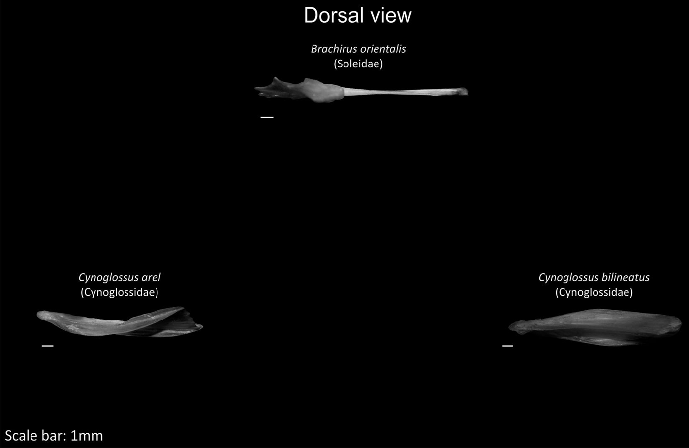
The urohyals are illustrated in Figures 4–18.
Dorsal view of the urohyal of Anodontostoma chaucunda (Clupeidae) to Alepes djedaba (Carangidae). Scale bar = 1 mm.

Dorsal view of the urohyal of Atropus atropus (Carangidae) to Pomadasys maculatus (Haemulidae). Scale bar = 1 mm.

Dorsal view of the urohyal of Pomadasys stridens (Haemulidae) to Upeneus sulphureus (Mullidae). Scale bar = 1 mm.

Dorsal view of the urohyal of Drepane punctata (Drepaneidae) to Pseudorhombus elevatus (Paralichthyidae). Scale bar = 1 mm.

Dorsal view of the urohyal of Brachirus orientalis (Soleidae) to Cynoglossus bilineatus (Cynoglossidae). Scale bar = 1 mm.

Ventral view of the urohyal of Anodontostoma chaucunda (Clupeidae) to Alepes djedaba (Carangidae). Scale bar = 1 mm.

Ventral view of the urohyal of Atropus atropus (Carangidae) to Pomadasys maculatus (Haemulidae). Scale bar = 1 mm.

Ventral view of the urohyal of Pomadasys stridens (Haemulidae) to Upeneus sulphureus (Mullidae). Scale bar = 1 mm.

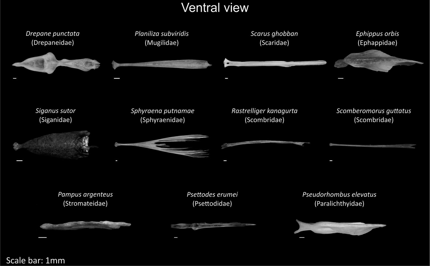
Ventral view of the urohyal of Drepane punctata (Drepaneidae) to Pseudorhombus elevatus (Paralichthyidae). Scale bar = 1 mm.

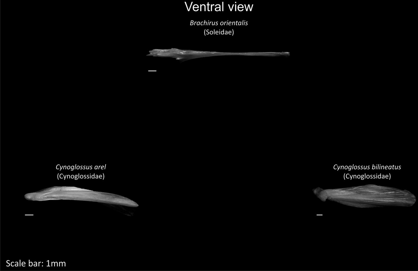
Ventral view of the urohyal of Brachirus orientalis (Soleidae) to Cynoglossus bilineatus (Cynoglossidae). Scale bar = 1 mm.

Lateral view of the urohyal of Anodontostoma chaucunda (Clupeidae) to Alepes djedaba (Carangidae). Scale bar = 1 mm.

Lateral view of the urohyal of Atropus atropus (Carangidae) to Pomadasys maculatus (Haemulidae). Scale bar = 1 mm.

Lateral view of the urohyal of Pomadasys stridens (Haemulidae) to Upeneus sulphureus (Mullidae). Scale bar = 1 mm.

Lateral view of the urohyal of Drepane punctata (Drepaneidae) to Pseudorhombus elevatus (Paralichthyidae). Scale bar = 1 mm.

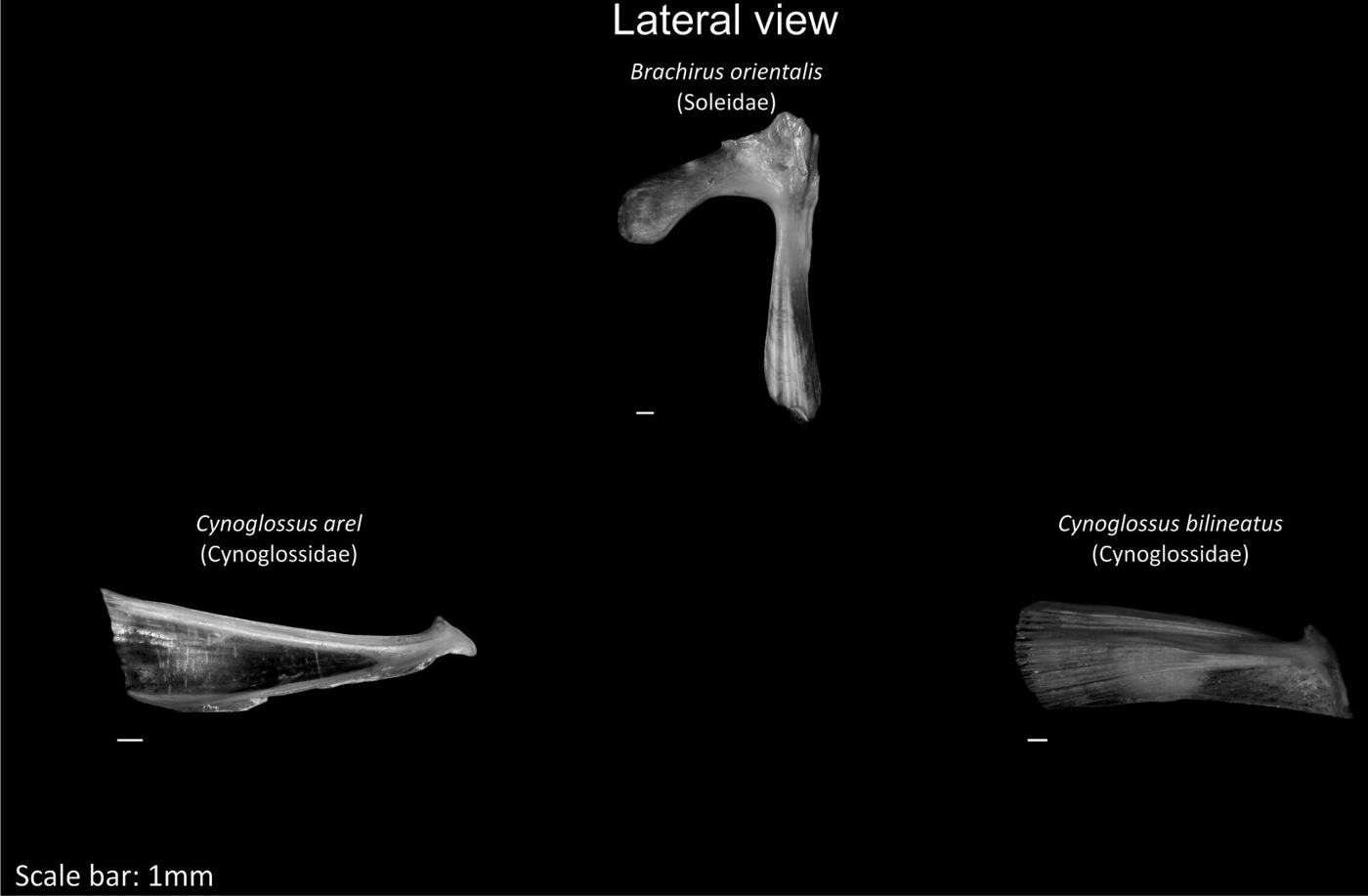
Lateral view of the urohyal of Brachirus orientalis (Soleidae) to Cynoglossus bilineatus (Cynoglossidae). Scale bar = 1 mm.

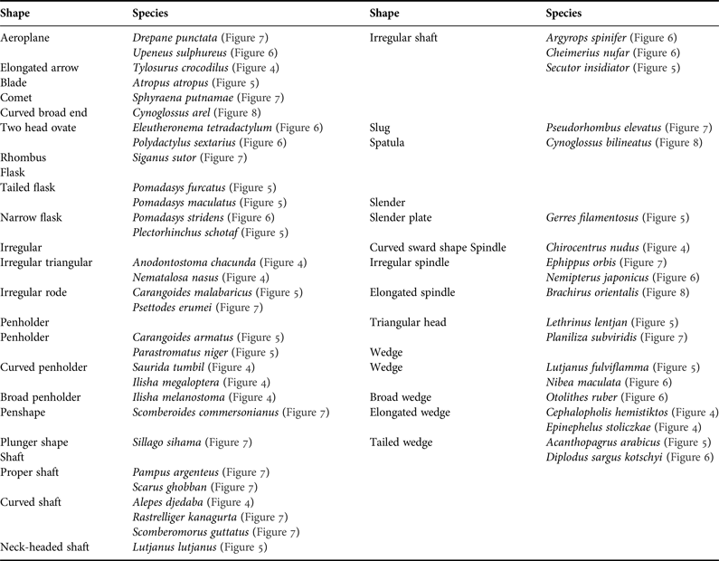
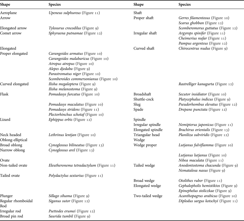
The dorsal, ventral and lateral sides of the urohyal are variable in shape in the examined species of fishes. The dorsal side shows 18 main shapes (Table 2, Figures 4–8). Out of this number there are seven shapes subdivided into several sub-shapes with the number of species represented by the shapes ranging from two to 10. The remaining 11 shapes are represented by one or two species. The shaft shape with its sub-shapes (10 species) is the most common among the fish species studied. The next most common shapes are wedge and penholder with their sub-shapes (six and seven, respectively). The slender shape category with its two sub-shapes holds two species. The flask shape and irregular shapes with their sub-shapes contain four species each.
Shape of urohyal bone as shown in the dorsal view.

The dorsal side of the urohyal of the studied species showed 18 main shapes. Such a high number of shapes originated from well diversified families involved in this morphological comparison. Some of the shapes succeeded in encompassing the species belonging to the same genera or families. For example, the four species of the family Haemulidae all have a flask shape. The four sparid species belong to four genera. Acanthopagrus arabicus and Diplodus sargus kotschyi have a wedge-tailed shape, while Argyrops spinifer and Cheimerius nufar have an irregular shaft shape. The two polynemid species Eleutheronema tetradactylum and Polydactylus sextarius have a two-headed ovate shape. The five clupeid species belong to four genera, two species of the genus Ilisha have a penholder shape, Anodontostoma chacunda and Nematalosa nasus have an irregular triangular shape, and Chirocentrus nudus Swainson, 1839 has a curved sword shape. On the other hand, several species belonging to different families have a similar shape of urohyal in dorsal view (Table 2).
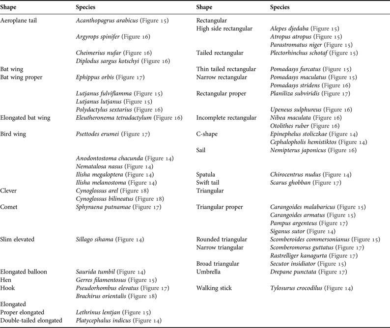
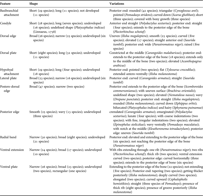
There are 16 shapes of the urohyal in ventral view (Table 3, Figures 9–13). In the lateral view, the urohyal showed 22 shapes (Table 4, Figures 14–18). Viewing the urohyal of the species examined in the present study, it was possible to recognize the following 11 features (Figure 1). These features showed considerable variations in shape between the studied species (Table 5, Figures 4–8).
Shape of urohyal bone as shown in the ventral view.

Shape of urohyal bone as shown in the lateral view.

Features of the urohyal bone with their shapes and variations.

Proportions
The majority of the species has a large urohyal (48 species), a medium to large size is found in Ilisha megaloptera, Ilisha melanostoma, and medium is seen in Tylosurus crocodilus.
The lengthwise extension has three criteria, these being slender, medium and high. The majority of the studied species falls in the medium criterion (30 species) followed by slender (13 species) and the high criterion (eight species).
For the lateral development spread of the studied species there are only two criteria, laterally unexpanded (23 species) and ordinary spread (12 species). The following species are shown to have criteria falling between the laterally unexpanded and ordinary spread criteria: Scomberoides commersonianus, Alepes djedaba, Lutjanus lutjanus, Acanthopagrus arabicus, Argyrops spinifer and Cheimerius nufar. For the species such as Anodontostoma chacunda, Plectorhinchus schotaf, Lutjanus fulviflamma, Polydactylus sextarius, Otolithes ruber, Nibea maculata, Sillago sihama, Diplodus sargus kotschyi and Brachirus orientalis, the urohyal showed a lateral spread intermediate between ordinary and wide.
The minimum and maximum values of the urohyal dorsal height are 0.18 and 0.34% in SL as shown in Drepane punctata and Scomberoides commersonianus respectively. The minimum value of the frontal length is 0.02% of SL and revealed by Nibea maculata, while the maximum value is 0.16% SL and shown by Upeneus sulphureus. The urohyal maximum height ranges from 0.02 and 0.19% SL for Platycephalus indicus and Siganus sutor respectively. The lower value obtained for the urohyal height is 0.01% SL shown by Sphyraena putnamae and the higher value was 0.34% SL revealed by Eleutheronema tetradactylum. The urohyal length showed lower value of 0.29% SL as in Ephippus orbis and higher value of 0.35% SL as in Scomberoides commersonianus. The minimum and maximum values shown by the ventral length of the urohyal are 0.02 and 0.28% SL as in Tylosurus crocodilus and Scarus ghobban respectively.
Angles
The three angles of the urohyal, condylar, ventral and postero-dorsal showed ranges of 4–79°, 18–179° and 3–155° respectively. The lowest value of the condylar angle (BAC) 4° is observed in Tylosurus crocodilus and the highest, 79° in Ephippus orbis. For the ventral angle (ACB), the lowest value 18° is found in Cynoglossus arel and the highest value in Tylosurus crocodilus. The postero-dorsal angle has values ranging from 3° as in Tylosurus crocodilus and 155° as shown in the urohyal of Cynoglossus arel.
DISCUSSION
This osteological study of the urohyal illustrates the wide range of characters present in the members of the studied families. This study must be considered preliminary because, out of 36 orders of Actinopterygii described (Eschmeyer, Reference Eschmeyer2014), only 22.2% of the known orders have been examined and out of a total of 144 families present in the order Perciformes (Eschmeyer, Reference Eschmeyer2014), only 14.6% of the known families have been examined. Nevertheless, 49 species belonging to 43 genera have been studied, so a reasonable survey was possible. It is possible to distinguish exclusive characters that are confined to particular taxa.
The present study enriches knowledge about the urohyal morphology, underlining its diagnostic value for teleostean taxonomy. The present research is the first comprehensive, comparative and quantitative assessment of the urohyal for some fish species from the marine waters of Iran, following the setting of its diagnostic characteristics by Arratia & Schultze (Reference Arratia and Schultze1990).
As the fish size effect has been eliminated, the variations in the morphology of urohyal between the studied species in the present work could reflect their natural variation. In several instances, fish species can be identified using the morphological differences in the shape of the urohyal (e.g. Kusaka, Reference Kusaka1974; Esmaeili & Teimori, Reference Esmaeili and Teimori2006). Accordingly, Aprieto (Reference Aprieto1974) has shown that the family Carangidae has revealed differences in the morphology of the skull and urohyal between individuals from wild and cultivated populations. Furthermore, the morphology of the urohyal was used in the description of a subspecies of the genus Merluccius (Lloris et al., Reference Lloris, Matallanas and Oliver2003), and recently Chollet-Villalpando et al. (Reference Chollet-Villalpandoa, De La Cruz-Agüeroa and García-Rodrígueza2014) have separated species of the family Gerreidae using the morphological characteristics of the urohyal.
In most previous studies on the urohyal, the description was considered from the qualitative point of view and no quantitative or comparative attempts were made (Andreata & Barbieri, Reference Andreata and Barbieri1981; Andreata, Reference Andreata1989; Gonzalez-Acosta et al., Reference Gonzalez-Acosta, De La Cruz-Agüero and Castro-Aguirre2005, Reference Gonzalez-Acosta, De La Cruz-Agüero and Castro-Aguirre2007; De La Cruz-Agüero & Chollet-Villalpando, Reference De La Cruz-Agüero, Chollet-Villalpando, del Moral, Martínez, Franco, Ramírez and Tello2012), but recently Chollet-Villalpando et al. (Reference Chollet-Villalpandoa, De La Cruz-Agüeroa and García-Rodrígueza2014) have shown the useful quantitative characters in separating some species of the family Gerreidae. In the present study both the quantitative and qualitative characteristics of the urohyal prove to be good taxonomic criteria to identify the species, and to solve their taxonomic problems.
The ventral side of the urohyal shows one sub-shape less than what it is observed in the dorsal side. The content of the shapes with subdivisions slightly varies between the dorsal and ventral sides. In the dorsal side, the number of species ranges from three to 10, while in the ventral side the number ranges three to 10. These trivial differences between the two sides might indicate that different species are conservative in the shape of their urohyal.
Looking at the distribution of the studied species, and according to the shape of their urohyal viewed ventrally, Sphyraena putnamae and Tylosurus crocodilus have the same general ventral shape of the urohyal. These two species belong to two different families, but the similarity in the shape of their urohyal might be due to the food type and feeding habits or similarities in muscle anatomy and function. Both species have fusiform bodies, posteriorly placed dorsal and anal fins, elongate jaws, large, conical teeth and piscivorous feeding habit (Lovejoy et al., Reference Lovejoy, Iranpour and Collette2004; Porter & Motta, Reference Porter and Motta2004). Such adaptation might make the ventral shape of these two species similar. On the other hand, the shape of the urohyal viewed ventrally succeeded in grouping some members of the same family in one shape grouping. For example, the four species of the family Haemulidae all have a flask shape, the six species of the family Carangidae have an elongated shape, and the two species of the family Lutjanidae have a wedge shape (Table 3, Figures 9–13). Members of some genera are grouped in sub-shapes within a similar main shape type, such as in the case of the two species of the genus Ilisha. For the four species of the family Sparidae, Argyrops spinifer and Cheimerius nufar were grouped in the irregular shaft sub-shape criteria, while the other two species, Acanthopagrus arabicus and Diplodus sargus kotschyi have a two-tailed wedge shape, and the two serranid species Cephalopholis hemistiktos and Epinephelus stoliczkae have an elongated wedge shape. The two species of the genera Cynoglossus and Scarus have different sub-shape types within the main shape division of oblong-elliptical and shaft respectively. Similarly, the two polynemid species Eleutheronema tetradactylum and Polydactylus sextarius, have non-tailed and tailed-ovate shape respectively. It is unlikely that the different shape groups obtained in the present study have a taxonomic basis as they comprise species belonging to different families; however, feeding and mouth opening mechanisms may have related origins (Chollet-Villalpando et al., Reference Chollet-Villalpandoa, De La Cruz-Agüeroa and García-Rodrígueza2014).
The lateral profile of the urohyal succeeded in placing the fish species in different shapes (Table 4, Figures 14–18). Some of these shapes contain solely species belonging to the same family as in the case of the four sparid species or to the same genus as seen in the two members of the genus Cynoglossus. These shapes can be considered a good taxonomic criterion to identify sparid and cynoglossid fish species. To accept this character as a taxonomic criterion for the families of these species, the morphology of more sparid and cynoglossid species is required. The lutjanid species fall in a sub-shape shared by other species, and the two species of sciaenids are present in a sub-shape not shared by other species (Table 4).
The lateral side of the urohyal shows more main shapes with several sub-shapes than the dorsal and ventral sides (Tables 2–4, Figures 14–18). This indicates that the lateral profile of the urohyal might be the best tool to identify fish species.
All the sides of the urohyal are designed to receive attachments for muscles serving different functions, but mainly for those assisting in the mouth opening mechanism (Kusaka, Reference Kusaka1974; Arratia & Schultze, Reference Arratia and Schultze1990). Therefore, the urohyal exhibits several morphological features that vary with the anatomy of the skull, mouth and the mechanism of feeding. Such morphological variations in the shape of the sides of the urohyal are noticed in some members of the family Gerreidae (Chollet-Villalpando et al., Reference Chollet-Villalpandoa, De La Cruz-Agüeroa and García-Rodrígueza2014), and they are observed in the urohyal of the fish species considered in the present study and might be related to the anatomy of the skull, mouth and the mechanism of feeding.
Kusaka (Reference Kusaka1974) suggested that the length of the urohyal relative to head length is between 20–50% for the fish species used in his study and there is a slight increase of 10% for some species. The results obtained in this study do not support the suggestion of Kusaka (Reference Kusaka1974) and show a range of 30.7–99.6%. As the study of Kusaka (Reference Kusaka1974) was preliminary, there is a possibility that some of the species examined in the present study that showed relative urohyal length values higher than 50–60% are not included in the study of Kusaka (Reference Kusaka1974). Large urohyal size is usually found in active fish species (Kusaka, Reference Kusaka1969a, Reference Kusakab, Reference Kusaka1974; Kusaka & Thuc, Reference Kusaka and Thuc1972). This criterion is found in the majority of the studied fish species which were active species. The urohyal of Ilisha megaloptera and Ilisha melanostoma is medium to large, and they are active species, but with their planktonic mode of feeding their urohyal is shown to fall between the medium and large criteria.
The present study shows that the deep bodied fish species, for example, in members of the families Sparidae, Drepanidae, Ephippidae, Haemulidae, Stromateidae and Leiognathidae have a short urohyal, while those with a slender head like members of the families Belonidae, Scombridae and Sphyraenidae have a long urohyal. This result agrees with the suggestion of Kusaka (Reference Kusaka1974). Moreover, Kusaka (Reference Kusaka1974) has related the development of the ventral spread of the urohyal to the activity of the fish and ranges from an undeveloped ventral side in the active fish species to well developed in the slow moving species. The urohyal of several species fall in between these two criteria. The present results support this suggestion in having the urohyal of the members of the families Belonidae, Scombridae and Synodontidae with reduced ventral side.
The value of the angles were successfully being used to determine the shapes of fish body structures like the otolith and to identify species accordingly (Chen et al., Reference Chen, Friesen and Iwasaki2011; Annabi et al., Reference Annabi, Said and Reichenbacher2013; Reichenbacher & Reichard, Reference Reichenbacher and Reichard2014; Teimori et al., Reference Teimori, Esmaeili, Erpenbeck and Reichenbacher2014) and bones (Brainerd & Patek, Reference Brainerd and Patek1998; Herrell et al., Reference Herrell, Adriaens, Verraes and Aerts2002). So far, no attempt has been made to use angles between the sides of fish urohyal to establish the characteristic of the urohyal and to separate species accordingly. In the present study, the value of three angles found between the three major sides of the urohyal were measured with an aim to evaluate this character as a taxonomic criterion to separate the examined fish species.
The values of angles of the different sides of the urohyal obtained for the fish species examined in the present study coincide with the shape of the urohyal of these species. They can define how the ventral and posterior edges are spread and how they differ in the height of the urohyal. As in other studies that used the values of angles in fish osteology (Brainerd & Patek, Reference Brainerd and Patek1998; Herrell et al., Reference Herrell, Adriaens, Verraes and Aerts2002) and as in the morphometry of the fish otolith (Reichenbacher et al., Reference Reichenbacher, Sienknecht, Kuchenhoff and Fenske2007; Chen et al., Reference Chen, Friesen and Iwasaki2011; Annabi et al., Reference Annabi, Said and Reichenbacher2013; Reichenbacher & Reichard, Reference Reichenbacher and Reichard2014; Teimori et al., Reference Teimori, Esmaeili, Erpenbeck and Reichenbacher2014), the angles proved a good support for the shape of the urohyal of the species in the present work.
The shape of the urohyal of the members of the families Clupeidae, Chirocentridae, Belonidae, Serranidae, Carangidae, Leiognathidae, Lutjanidae, Haemulidae, Nemipteridae, Polynemidae, Drepanidae, Siganidae, Sphyraenidae and Stromateidae in the present work looks similar to that shown by Kusaka (Reference Kusaka1974), with slight differences which are considered related to the species characteristics. These differences are: the shape of the urohyal of Alepes djedaba is slightly different from the shape of the other members of this genus; the postero-dorsal edge is elongated and high; the urohyal of Secutor insidiator is curved and straight; and the dorsal surface of the urohyal of Pampus argenteus is smooth and no broad spine is present.
Rao (Reference Rao1977) described the osteology of Saurida tumbil and illustrated the urohyal which looks similar to that obtained in the present study. The exception is slightly notched postero-dorsal edge in the present study vs broadly notched in S. tumbil, and the ventral plate is narrow in the specimen of the present study vs wide in the specimen of S. tumbil. Kusaka (Reference Kusaka1974) illustrated the urohyal of two species of the genus Saurida i.e. S. tumbil and S. undosquamis. Although these two species belong to the same genus, the shape of their urohyal looks completely different, and the shape given to S. tumbil is different from that obtained in the present study and from that obtained by Rao (Reference Rao1977). Since the illustrations provided by Kusaka (Reference Kusaka1974) are only sketches and the study is preliminary in nature, the differences could be due to such reasons.
The urohyal of Platycephalus indicus is thin and has not much surface for muscle attachments. This result is also observed by Gosline (Reference Gosline1996) who suggested that only few muscles are attached to the urohyal and the majority of muscles are attached to the hypohyal bone.
Kaga (Reference Kaga2013) has reviewed the family Sillaginidae and described the urohyal of four species of the genus Sillago. The urohyal of S. sihama shown by Kaga (Reference Kaga2013) is characterized in having low dorsal extension, short basibranchial attachment and straight hypohyal attachment. On the contrary, the urohyal of our specimen showed high dorsal extension, long basibranchial attachment and downward directed hypohyal attachment.
In general, the shape of the urohyal of Sillago sihama obtained in the present study looks similar to that of the other members of the genus Sillago reported by Kusaka (Reference Kusaka1974). However, there are some differences that characterize each species. In the present work, the urohyal of S. sihama differs from that of S. japonica in the length of the elongated basibranchial attachments (long in S. sihama vs short in S. japonica), dorsal and ventral edges (straight in S. sihama vs curved in S. japonica), and the posterior edge (broad in S. sihama vs broadly pointed in S. japonica).
Chollet-Villalpando et al. (Reference Chollet-Villalpandoa, De La Cruz-Agüeroa and García-Rodrígueza2014) described the urohyal of six species of the family Gerreidae belonging to four genera. The shape of the urohyal of Gerres filamentosus obtained in the present study falls within the shape ranges of the species described by Chollet-Villalpando et al. (Reference Chollet-Villalpandoa, De La Cruz-Agüeroa and García-Rodrígueza2014), but it conserves some characters that separate it from the remaining species of the family Gerreidae. Such similarities of the urohyal obtained for G. filamentosus are also seen when compared with the shapes shown by Kusaka (Reference Kusaka1969a, Reference Kusakab, Reference Kusaka1974) for other gerreid fish species.
Kusaka (Reference Kusaka1969a, Reference Kusakab, Reference Kusaka1974) studied and illustrated the urohyal of 16 species of the family Sparidae belonging to nine genera, Bianchi (Reference Bianchi1984) examined four species belonging to one genus, and the present study gives descriptions for the urohyal of four species belonging to four genera. It is possible to note that the shape of the urohyal of all these sparid species looks similar, and there are only slight differences between them. Such variations belong to the species characteristics which separate these species. Also the close resemblance in the urohyal in these sparid species reflects the success of the shape of the urohyal to recognize fish species.
The general shape of Nibea maculata obtained in the present study matches that of N. mitsukurii described by Kusaka (Reference Kusaka1974) except for some differences like the anterior side of the ventral plate narrow and indented and the basibranchial attachment directed upward. As both are members of the family Sciaenidae, the urohyal of N. maculata and Otolithes ruber look alike and also look similar to those sciaenid species described by Sasaki (Reference Sasaki1989).
The shape of the urohyal of the two Upeneus species described by Kusaka (Reference Kusaka1974) and that described by Kim (Reference Kim2002) match very well that of Upeneus sulphureus given in the present study. The species of the genus Upeneus shared with species of other mullid genera the shape of urohyal as shown by Kusaka (Reference Kusaka1969a, Reference Kusakab, Reference Kusaka1974) and Kim (Reference Kim2002).
The shape of the urohyal of Mugil cephalus L., 1775 given by Kusaka (Reference Kusaka1974), the shape of those six species of the four mugilid genera described by Antović & Simonović (Reference Antović and Simonović2006) and the Mugil species described by Keivany (Reference Keivany2014) look very close to that of Planiliza subviridis obtained in the present study. Such similarity indicates that the shape of the urohyal in the members of the family Mugilidae is conservative.
Jahromi et al. (Reference Jahromi, Esmaeili, Teimori, Nokhatolfoghahai and Ostovani2010) studied two species of the genus Scarus i.e. S. persicus and S. ghobban. In the present study, we used the same specimen of urohyal that Jahromi et al. (Reference Jahromi, Esmaeili, Teimori, Nokhatolfoghahai and Ostovani2010) have used. The idea behind using the same specimen is to add more information about the urohyal of this species such as morphometry, measuring angles and giving detailed description of the urohyal that are not provided by Jahromi et al. (Reference Jahromi, Esmaeili, Teimori, Nokhatolfoghahai and Ostovani2010). It was not possible to include the urohyal of S. persicus as the bone that was supposed to be a urohyal after close examination was revealed to be an epicleithrum.
Bellwood (Reference Bellwood1994) described the urohyal of 10 scarid species with one species belonging to the genus Scarus. The anterior part of the urohyal of these 10 species showed wide variation, while the posterior end appeared to be more conservative in its shape, and all the 10 species showed a similar general appearance of the posterior end, including S. ghobban described in the present work.
Kusaka (Reference Kusaka1969a, Reference Kusakab, Reference Kusaka1974) gave descriptions and illustrations of the urohyal of the members of the genera Rastrelliger and Scomberomorus. Kohno (Reference Kohno1984) studied the morphology of one scombrid species of the genus Gasterochisma. The shape of the urohyal of these three genera is closely related to that of the two scombrid species obtained in the present work.
From the five flatfish species dealt with in the present work, the urohyal of Brachirus orientalis and Pseudorhombus elevatus is curved (reversed L-shaped in Brachirus orientalis and reversed c-shaped in P. elevatus). Such curved shapes are also reported for the 13 flatfish species described by Kusaka (Reference Kusaka1974), for Tephrinectes sinensis and for other flatfish species like Citharichthys spilopterus and Hippoglossina macrops. The shape of the urohyal of the two cynoglossid species given in the present work looks completely different from that of other flatfish species. Such differences signify the possibility of using the shape of the urohyal in separating very close related species of flatfish.
The results obtained in the present study showed clearly the usefulness of the morphology of the urohyal to separate the fish species. Such findings can serve fish taxonomists as they add a new distinguishing criteria, fish biologists as they help to identify fish in the food of another fish and in aquatic birds and mammals and archaeologists in giving an idea about the feeding habits of humans in ancient gathering centres and their social life. The results of this study will shed light on the possibility of using other osteological parts of the fish body in similar comparison. Also, the present study has a comprehensive comparison of urohyal anatomy for a wide range of fish groups in one place, a coverage that researchers in several fields can be interested in.
ACKNOWLEDGEMENTS
Our sincere thanks are due to D. G. Smith, Smithsonian Institution for reading the manuscript and for his valuable advice and suggestions.