Introduction
Ultrastructural studies on the egg shell of acanthocephalans have shown the presence of four embryonic envelopes (Marchand, Reference Marchand1984b; Taraschewski, Reference Taraschewski2000; Nikishin, Reference Nikishin2001). Despite specific peculiarities of each shell, only the third envelope varies in the classes of acanthocephalans, being simple in the Eoacanthocephala and complex in the Archiacanthocephala. Furthermore, some species of the Eoacanthocephala possess a supplementary fifth envelope in the outer part of the shell, which is absent in the other classes, (Taraschewski et al., Reference Taraschewski, Peters and Latka1992; Taraschewski, Reference Taraschewski2000; Wongkham & Whitfield, Reference Wongkham and Whitfield2004). According to these authors, the supplementary envelope is the outermost one, but due to its morphological similarity with the outer envelope of the eggs of other acanthocephalan shells, this seems doubtful.
In the present study the fine morphology of the embryonic envelopes of Neoechinorhynchus crassus is described and their terminology in the Eoacanthocephala discussed.
Materials and methods
Females of Neoechinorhynchus crassus were collected from naturally infected Siberian whitefishes Coregonus lavaretus pidschian from the Pucheveem river (north-western Tschukotka). Living worms were fixed in 0.5 m glutaraldehyde in 0.1 m phosphate buffered saline (PBS) for two days, where 10–15 min after fixation they were cut into segments, 2–5 mm in length. The latter were washed twice, for 30 min each time, in PBS before being immersed for 5 h in 1% solution of osmium tetroxide in PBS, dehydrated in a graded series of ethanol, and embedded in a mixture of Epon and Araldite. Specimens were stained in uranylacetate in 70% alcohol for 1 day, and the sections of specimens were stained in lead citrate for 10–60 s. Specimens were viewed under a Tesla Bs- 500 electron microscope.
The nomenclature of Marchand (Reference Marchand1984a, Reference Marchandb) was used to designate embryonic envelopes.
Results
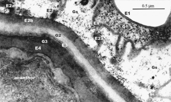
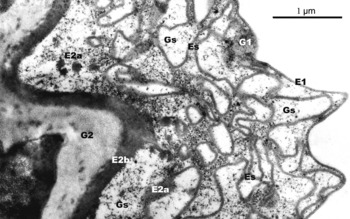
The mature egg shell of Neoechinorhynchus crassus consists of five envelopes (figs 1, 2). The outermost (El), which is 0.03–0.05 μm thick with a wavy outline, is formed of two electron-dense layers with a lighter space between them resembling a cytoplasmic membrane. The second envelope (E2) consists of a plate with distal extensions. The plate (E2b), 0.16–0.18 μm thick, is formed of parallel layers of electron-dense material with intermittent thin, less dense interlayers. The distal extensions of the main plate (E2a) are formed of fibrils, which are equal or a little smaller in diameter. Only the short basal parts of these fibrils possess a radial orientation, whereas other parts are oriented more or less parallel to the main plate.
The embryonic envelopes of Neoechinorhynchus crassus. The outer embryonic envelope (E1) is winding, with a thick three-layered membrane (arrows) and a rugose supplementary envelope (Es) located beneath; the second envelope (E2) consists of the plate E2b and distal extensions E2a, which are connected to the supplementary envelope (asterisk); the space G3 is filled with electron-dense material, and the cavity lined with granular material (double arrow). G1, space between the envelopes E1 and Es; Gs, space between the envelopes Es and E2b; G2, space between the envelopes E2b and E3 (it is beyond the visibility of the figure); E4, the fourth envelope.

The embryonic envelope of Neoechinorhynchus crassus. The outer embryonic envelope (E1) is three-layered, although the supplementary envelope (Es) is not defined; the elements of the second envelope (E2b and E2a) are layered. The third envelope (E3) is a three-layered membrane and the space G3 is filled with a dense material, but its structure is not defined. G1, space between the envelopes E1 and Es; Gs, space between the envelopes Es and E2b; G2, space between the envelopes E2b and E3, E4, the fourth envelope.

The space between the outer and second envelopes in different directions is occupied by an intensely sinuous envelope (Es), which is not found in other acanthocephalans (figs 123). This envelope is plate-like, 0.04–0.06 μm thick, and formed of a moderately electron-dense homogenous material. Spaces between the main plate and the elements El and E2b, Gl and Gs, contain irregularly distributed small-granular inclusions, more abundant in the Gl space (fig. 2). The sinuous envelope is more or less radially oriented, with deeply embedded areas in the shell connected to the E2a elements (fig. 1).
Tangential section of the embryonic envelopes of Neoechinorhynchus crassus near the pole of the egg. The supplementary envelope (Es) is characterized by numerous folds; the elements of E2a are smaller in diameter, than those in the lateral areas of the egg. E1, the first envelope; G1, space between the envelopes E1 and Es; Gs, space between the envelopes Es and E2b; E2b, the second envelope; G2, space between the envelopes E2b and E3 (it is beyond the visibility of the figure).

The G2 space, 0.09 to 0.13 μm in width, is filled with evenly distributed small-granular material of a moderate electron density.
The third envelope E3, is 0.02–0.03 μm thick, possesses a wavy membrane and is formed of two electron-dense layers, separated by lighter intervals (fig. 2) and is similar to the E3a envelope, described by different authors (see reviews of Taraschewski, Reference Taraschewski2000; Nikishin, Reference Nikishin2001) as representative of other classes of acanthocephalans. The G3 space is exclusively filled with dense homogenous material, with oval cavities, 0.22–033 × 0.10–0.19 μm, occasionally containing granular material (fig. 1).
The innermost envelope (E4), 0.14–0.22 μm thick, is formed of material with a similar density to that of E2b (figs 1, 2). The E4 envelope partially joins the surface of the acanthor leaving a fragmentary G4 space, which is filled with as dense material as in the G3 space.
Discussion
The structure of the embryonic envelopes of N. crassus, in general, corresponds to that of other representatives of the Eoacanthocephala (Marchand, Reference Marchand1984a, Reference Marchandb; Taraschewski et al., Reference Taraschewski, Peters and Latka1992; Wongkham & Whitfield, Reference Wongkham and Whitfield2004). However, an analysis of the morphology, histogenesis and the function of each envelope allows further specification as to the identification and nomenclature of the outer elements of the shell.
The seeming resemblance of the outer envelope (El), 0.03–0.05 μm, and the wavy three-layered membrane are characteristic of the majority of acanthocephalans (Nikishin, Reference Nikishin2001). The outer envelope possesses the same morphology as that of the Eoacanthocephala and of the five envelopes described, the outer one is considered to be E0 (Taraschewski et al., Reference Taraschewski, Peters and Latka1992; Wongkham & Whitfield, Reference Wongkham and Whitfield2004).
The similarity of the outer embryonic envelope of acanthocephalans is not restricted by its morphology. Wongkham & Whitfield (Reference Wongkham and Whitfield2004) demonstrated that, on contact with water, the envelopes of Pallisentis rexus degrade quickly and release the next one (El), which in the present study is referred to as the supplementary envelope (Es). This straightens and expands considerably, thereby preserving the egg's ability to float and increasing the probability of invasion into the intermediate host. Some representatives of the Palaeacanthocephala developing in aquatic intermediate hosts show a similar process, except that they lack a supplementary envelope and the E2a elements are released (Oetinger & Nickol, Reference Oetinger and Nickol1974; Uznanski & Nickol, Reference Uznanski and Nickol1976).
Finally, one more peculiarity of the outer envelope suggests that it is a remnant or derivative of a fertilization envelope, possibly modified to a certain degree (Stranack, Reference Stranack1972; Whitfield, Reference Whitfield1973; Marchand, Reference Marchand1984a; Peters et al., Reference Peters, Taraschewski and Latka1991; Nikishin, Reference Nikishin1995; Wongkham & Whitfield, Reference Wongkham and Whitfield2004). The present study considers that the outer envelope of the egg of acanthocephalans should be designated as E1.
Only some representatives of the Eoacanthocephala possess a supplementary envelope, which for the first time was found in Tenuisentis niloticus by Marchand (Reference Marchand1984b) as the element Elb. The components of Elb were recognized in four other species of Eoacanthocephala by Marchand, although these components could be the supplementary envelope or its fragments. Taraschewski et al. (Reference Taraschewski, Peters and Latka1992) described the supplementary envelope in Neoechinorhynchus rutili and underlined its significance for invasion of the aquatic intermediate host. For example, when eggs of Pallisentis rexus are immersed in water, the supplementary envelope straightens and the volume of the egg expands. This results in the eggs floating to the surface and in close proximity to the copepod intermediate host (Wongkham & Whitfield, Reference Wongkham and Whitfield2004). In all acanthocephalans studied, including N. crassus, the supplementary envelope is of a similar size and appears as an intensely winding, even rugose plate, formed by amorphous material of a moderate electron density, resembling a three-layered membrane. The ‘supplementary envelope’ is denoted as Es in figs 123 , compared with El (Taraschewski et al., Reference Taraschewski, Peters and Latka1992; Taraschewski, Reference Taraschewski2000; Wongkham &, Whitfield, Reference Wongkham and Whitfield2004), because this envelope is not an indispensable egg shell component in acanthocephalans.
Another peculiarity in the case of N. crassus is the presence in the second envelope (E2) or extensions (E2a) from the distal surface of the E2b plate. Among other eoacanthocephalans only Pallisentis golvani (Marchand, Reference Marchand1984b), Paratenuisentis ambiguus and N. rutili (Taraschewski et al., Reference Taraschewski, Peters and Latka1992) have been found to have E2a elements. In addition, E2a fibrils have also been found in other acanthocephalans, investigated by Marchand (Reference Marchand1984b), Taraschewski et al. (Reference Taraschewski, Peters and Latka1992) and Nikishin (Reference Nikishin2001). In all cases the elements of the second envelope are formed by a layer of material, resembling keratin (Marchand, Reference Marchand1984a, Reference Marchandb; Taraschewski et al., Reference Taraschewski, Peters and Latka1992; Wongkham & Whitfield, Reference Wongkham and Whitfield2004). The role of the E2b plate is likely to protect the egg from mechanical damage and water penetration (Whitfield, Reference Whitfield1973; Wongkham & Whitfield, Reference Wongkham and Whitfield2004). The function of the E2a elements could be analogous to that of some Palaeacanthocephala, where these elements are released following the destruction of the outer envelope, thus contributing to intermediate host invasion (Oetinger & Nickol, Reference Oetinger and Nickol1974; Uznanski & Nickol, Reference Uznanski and Nickol1976). However, as previously mentioned, this function is undertaken by the supplementary envelope, if present in the Eoacanthocephala.
The third envelope (E3) resembles a wavy three-layered membrane and as in the case of El and E2b elements is present in all acanthocephalans. However, the E3 envelope in representatives of two other classes is described as the E3a element by Taraschewski et al. (Reference Taraschewski, Peters and Latka1992) and Nikishin (Reference Nikishin2001).
One other peculiarity of the shell of N. crassus is the G3 space, which is filled by material with a high electron density, but its inner structure, origin and function remain unknown.
The fourth envelope (E4) in many acanthocephalans (Marchand, Reference Marchand1984b) is prominent with the least variability, but in N. crassus the structure of E4 was not well defined. In other acanthocephalans E4 is represented by several elements and has a layered appearance (Marchand, Reference Marchand1984a, Reference Marchandb; Wongkham & Whitfield, Reference Wongkham and Whitfield2004), although neither chitin nor keratin have been found (Peters et al., Reference Peters, Taraschewski and Latka1991; Taraschewski et al., Reference Taraschewski, Peters and Latka1992).
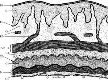
In conclusion, the morphological peculiarities of the egg shell of the Eoacanthocephala are summarized in fig. 4. The outer envelope (El), ranging in thickness 10–35 nm, is a three-layered membrane and represents a derivative of the fertilization envelope. The supplementary envelope (Es) is an exclusively winding plate and is present only in some acanthocephalans. The second envelope (E2) consists of two components, namely plate E2b and distal extensions E2a, which supports Es, although the latter are not always in class representatives. The components of E2 are fibrous and similar to keratin. The third envelope (E3), not more than 10 nm thick, is a wavy three-layered membrane and is analogous to the E3a element in the egg shell of classes Palaeacanthocephala and Archiacanthocephala. The fourth envelope (E4) is a layer of dense material (E4c), usually limited outwardly by a thin granular dense layer (E4a) and a less dense inner layer (E4b).
Schematic diagram of the structure of the egg shell of the Eoacanthocephala, where five embryonic envelopes are present. E1, the first envelope; G1, space between the envelopes E1 and Es; Es, supplementary envelope; Gs, space between the envelopes Es and E2b; E2a, extensions of the envelope E2b; E2b, the second envelope; G2, space between the envelopes E2b and E3; E3, the third envelope; G3, space between the envelopes E3 and E4; E4, the fourth envelope; G4, space between the envelope E4 and the surface of the acanthor.

Evidently, the composition and the structure of the shell of acanthocephalans are adapted to the survival of embryos in the environment and to the searching and capture of the eggs by the intermediate host. Thus, further studies are needed on acanthocephalans infecting intermediate hosts, which inhabit different ecological conditions.
Acknowledgements
The author thanks Ms A. Nikishina and Ms E. Shetinina for their help in translating the manuscript into English.