Introduction
Anastrepha Schiner is the most diverse genus of fruit flies (Diptera: Tephritidae) in the American tropics and subtropics with almost 250 described species (Norrbom et al. Reference Norrbom, Carroll, Thompson, White and Freidberg1999a, Reference Norrbom, Zucchi and Hernández-Ortiz1999b; Norrbom Reference Norrbom2004a; Norrbom and Korytkowski Reference Norrbom and Korytkowski2009). It is also the most economically important genus of fruit flies in this region, including a number of major fruit pests (Norrbom Reference Norrbom2004b). Despite its importance, many species remain undescribed. In this paper we describe a new species to make its name available for a forthcoming interactive identification system and key for the genus.
It is a great pleasure to dedicate this paper to the editors of the Manual of Nearctic Diptera. The senior author started his graduate program in 1980, a fortuitous time to begin the study of dipterology. The publication of the first volume of the manual the following year was a tremendous help to us and all dipterists. It provided a knowledge base so that all dipterists, and especially students, could recognize problematic taxa needing study. The morphology chapters were particularly important as the standardized terminology made communication about fly taxonomy so much easier. The editors of the manual should also be lauded for their efforts to build the Canadian National Collection and their enthusiasm for collecting flies, which fortunately has been quite contagious. One species of Tephritidae, Gymnocarena macalpinei Norrbom, was previously named for one of the editors (Norrbom Reference Norrbom2006), and a second, a species of Anastrepha close to Anastrepha loewi Stone, is proposed in this paper. The latter species is also redescribed.
Materials and methods
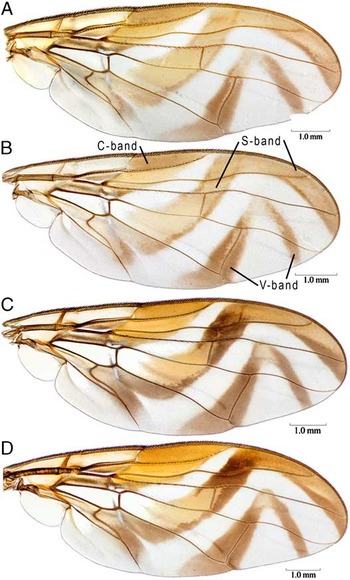
We follow the morphological terminology of White et al. (Reference White, Norrbom, Headrick and Carroll1999). The names for the wing bands follow Stone (Reference Stone1942) and are shown in Figure 4. The following measurements and ratios of Norrbom and Korytkowski (Reference Norrbom and Korytkowski2009) are also used. Wing length was measured from the base of the costa to the wing apex in cell r4 + 5; wing width was measured at the broadest part, distal to the apex of vein R1 to the margin of cell cu1. The position of the apex of vein R1 is the ratio of the distance from the base of the costa to the apex of vein R1 divided by wing length. The width of cell r4 + 5 at the level of dm-cu was measured on a line directly anterior to the junction of vein M and dm-cu. Its apical width was measured on a line from the apex of vein R4 + 5 and the junction of vein M and the wing margin. The width of the distal part of the S-band was measured perpendicular to the band at the apex of vein R2 + 3, and the width of cell r2 + 3 was measured along the same line; both measurements include the costa. Oviscape length was measured medially on the ventral side from the concavity in the base to the apex, including the medial lobe. The length of the aculeus tip was measured ventrally from the inner (proximal) margin of the sclerotized part to the extreme apex.
Label data for all examined specimens will be made available in the New World fruit fly specimen database on the Systematic Entomology Laboratory web site (see www.sel.barc.usda.gov:591/diptera/Tephritidae/TephIntro.html). A USNM barcode label was added to many specimens that previously lacked a barcode label. These labels do not indicate ownership; they are simply unique specimen identifier numbers. In the Type data and Specimens examined sections, the barcode number is listed following the depository acronym for each specimen or series.
Acronyms for institutions where specimens are deposited follow Norrbom et al. (Reference Norrbom, Carroll, Thompson, White and Freidberg1999a): CAS – California Academy of Science, San Francisco; CNC – Canadian National Collection, Ottawa; INBio – Instituto de Biodiversidad, Santo Domingo de Heredia, Costa Rica; MEUP – Museo de Entomología, Universidad de Panamá; MNHNP – Muséum National d'Histoire Naturelle, Paris; and USNM – National Museum of Natural History, Smithsonian Institution, Washington.
Anastrepha loewi Stone
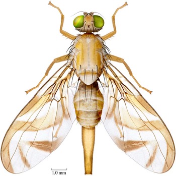
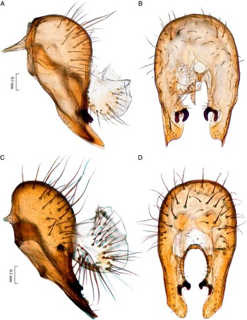
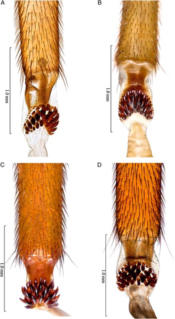
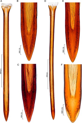
(Figs. 1, 3A–3B, 4A–4B, 5A–5B, 6A–6B, 7A–7C)
Anastrepha loewi: Female habitus, dorsal (Panama: Cerro Jefe, USNMENT00214581).

Anastrepha loewi Stone Reference Stone1942: 49 [description; Panamá, Venezuela]; Fernández Yépez Reference Fernández Yépez1953: 26; Foote Reference Foote1967: 12 [in catalog]; Steyskal Reference Steyskal1977: 14 [in key]; Caraballo Reference Caraballo1981: 87; Norrbom et al. Reference Norrbom, Carroll, Thompson, White and Freidberg1999a: 80 [in catalog, Colombia]; Norrbom Reference Norrbom2004a: 06604470 [in database].
Anastrepha integra: Hendel Reference Hendel1914: 15. Misidentification; see Stone Reference Stone1942: 49.
Diagnosis
Males of this species can be easily distinguished from other species of Anastrepha by the large brown spot on the gena in combination with the head and thorax lacking other brown markings. The V-shaped, medial indentation in the posterodorsal margin of the epandrium, the shape of the surstyli, and the length of the phallus (4.40 mm) are also useful diagnostic characters. Females similarly differ from most other species of Anastrepha (except Anastrepha woodi) in having a small genal spot, but it is sometimes faint and may be overlooked. The following combination of characters will also distinguish females of this species: subscutellum and mediotergite entirely yellow to orange; C-band and S-band of wing separate or only very narrowly connected along costal margin; oviscape 3.24–3.69 mm long, 1.03–1.04 times as long as mesonotum; aculeus tip 0.30–0.32 mm long, 0.16–0.18 mm wide, nonserrate.
Description
Mostly yellow to orange, with white to pale yellow markings (sometimes poorly differentiated). Setae dark red brown to black.
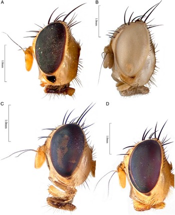
Head: Yellow to orange except brown ocellar tubercle, and gena with brown spot bordering eye, small and pale brown (sometimes very faint) in female (Fig. 3B), large and dark brown in male (Fig. 3A). Two to three frontal setae; two orbital setae, posterior seta well developed. Ocellar seta weak, at most 1.5 times as long as ocellar tubercle. Facial carina, in profile, straight to slightly concave dorsally and medially. Antenna extended 0.60–0.75 distance to ventral facial margin.
Thorax (Fig. 1): Mostly yellow to orange, without brown markings, with following areas (often poorly differentiated) white to pale yellow: postpronotal lobe and lateral margin of scutum bordering it; paired sublateral scutal vitta from transverse suture to posterior margin, including base of intra-alar seta; scutellum; dorsal margins of anepisternum and katepisternum; katepimeron; and most of anatergite and katatergite. Medial scutal vitta indistinct or absent. Subscutellum and mediotergite entirely orange. Mesonotum 2.98–3.54 mm long. Postpronotal lobe and notopleuron nonmicrotrichose; scutum nonmicrotrichose except postsutural lateral margin, lateral to supra-alar seta; scutellum entirely microtrichose; scutal setulae yellowish to orange. Chaetotaxy typical for genus. Katepisternal seta weak, short to moderately long, one-half to three-fourths as long as anepisternal seta, but weaker, pale to moderate red brown.
Legs: Entirely yellow to orange.
Wing (Figs. 4A–4B): Length 7.30–8.74 mm, width 2.78–3.50 mm, ratio 2.46–2.63. Apex of vein R1 at 0.61–0.64 wing length. Cell c 0.86–0.98 times as long as pterostigma; pterostigma 5.29–6.17 times as long as wide. Vein R2 + 3 strongly sinuous. Crossvein r-m at 0.68–0.72 distance from bm-cu to dm-cu on vein M. Vein M weakly curved apically; cell r4 + 5 1.08–1.16 times as wide at apex as at level of dm-cu. Cell bcu with distal lobe moderately long, length of bcu 1.66–1.72 times as long as anterior margin. Wing pattern mostly orange and brown. C-band mostly orange, in cells bc and c paler orange to subhyaline, in pterostigma orange brown, and in cell br with streak of brown; in cells r1 and r2 + 3 entirely orange, without brown on apical margin. C-band and S-band separated by hyaline band extended from cells bm and dm to costa distal to apex of vein R1, hyaline band not narrowed along veins R2 + 3 and R4 + 5, but narrowed anteriorly in cell r1, or with bands very narrowly connected only at costal margin. S-band with posterior margin of basal half narrowly brown, broadening in cell cu1, and on anterior margin in cells r1, r2 + 3, and sometimes partially in cell br; orange area in cells dm, br, and r4 + 5 extended anteriorly into cell r1; posterior margin with weak or no incision in cell cu1; distal section of band moderately broad, at apex of vein R2 + 3 0.65–0.74 times width of cell r2 + 3, even in width, well separated from apex of vein M, orange except margins and most or all of area in cell m moderate brown; hyaline area proximal to apex of band extending to vein R2 + 3. V-band complete, moderate brown except much of part in cell r2 + 3, proximal arm separated from S-band along vein R4 + 5 and in cell r2 + 3, with basal extension along wing margin extending half to three-fourths distance to vein A1 + Cu2; distal arm slender, connected to proximal arm and extending to vein R4 + 5. Cell r4 + 5 with hyaline area between V-band and vein M.
Abdomen: Mostly orange, without brown markings.
Male terminalia: Epandrium with acute medial notch in posterior dorsal margin; in lateral view 0.7 times as long as high. Lateral surstylus moderately long, extended beyond prensisetae by 2.0–2.5 times length of prensiseta; in lateral view (Fig. 5A) slender and slightly posteriorly curved; in posterior view (Fig. 5B), lateral margin straight, keel-like, projecting posteriorly, ending in slender lobe; mesal margin convex, then strongly concave. Proctiger with ventral and lateral sclerotized areas connected. Phallus 4.40 mm long, 1.48 times as long as mesonotum; glans 0.60 mm long, 0.14 times as long as phallus.
Female terminalia: Oviscape 3.24–3.69 mm long, 1.03–1.04 times as long as mesonotum, straight in lateral view, entirely orange; spiracle at basal 0.31–0.33. Eversible membrane (Fig. 6A–6B) with 15–20 long, relatively stout, hook-like scales in two to three irregular rows in subtriangular pattern. Aculeus (Fig. 7A) slightly ventrally curved in lateral view, 3.25–3.40 mm long, 1.00–1.01 times oviscape length; in ventral view base strongly expanded, 0.25–0.30 mm wide; shaft 0.13–0.18 mm wide at midlength; tip (Figs. 7B–7C) 0.30–0.32 mm long, 0.09–0.10 times aculeus length, 0.16–0.18 mm wide, 1.69–2.00 times as long as wide, 0.090–0.095 mm wide in lateral view, 0.51–0.59 times ventral width, in ventral view gradually tapered to blunt apex, lateral margin convex, nonserrate. Spermathecae not examined.
Distribution
Anastrepha loewi is known from Colombia, Costa Rica, Panamá, and Venezuela.
Biology
The host plants and other aspects of the biology of this species are unknown.
Type data
Holotype ♀ (USNM), PANAMA: Panamá: La Campana [8°43′N, 79°54′W], Muñoz Grove, fruit fly trap, 20.xii.1938, J. Zetek 4309, Paratype: PANAMA: Panamá: La Campana, Muñoz Grove, fruit fly trap, 29.vi.1938, J. Zetek 4177, 1♂ (USNM USNMENT00051703). [both examined]
Other Specimens Examined
COLOMBIA: “CORDILLERE vers’ occid.”, Río Yurumangui, 1933, E. Aubert de la Rue, 1♂ (MNHNP USNMENT00051702). COSTA RICA: Heredia: 3 km S of Puerto Viejo, Estacion Biológica La Selva, 10°26′N, 84°1′W, 50–150 m, bosque primerio M/00/015, 12.ii.1993, INBio-OET, 1♀ (INBio INBioCRI001273365). PANAMA: Panamá: Parque Nacional Chagres, Cerro Jefe, Don Leo [9°12′55″N, 79°22′40″W], McPhail trap 522, 26.i.2001, C.A. Korytkowski & C. Campo, 1♀ (USNM USNMENT00214581); Altos de Pacora, trampa 50, 2005, C.A. Korytkowski, 1♀ (MEUP).
Anastrepha woodi sp. nov.
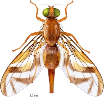
(Figs. 2, 3C–3D, 4C–4D, 5C–5D, 6C–6D, 7D–7F)
(A) Anastrepha woodi: Female habitus, dorsal (Costa Rica: Estación Barbilla, INBioCRI0003370579).

Head, lateral: (A–B) Anastrepha loewi male (Colombia: Rio Yuromangui, USNMENT00051702) and female (Panama: Cerro Jefe, USNMENT00214581). (C–D) Anastrepha woodi males (Costa Rica: Estación Barbilla, INBioCRI0003320310, INBioCRI0003370580).

Wing: (A–B) Anastrepha loewi (holotype; Panama: Cerro Jefe, USNMENT00214581). (C–D) Anastrepha woodi (Costa Rica: Estación Barbilla, INBioCRI0003370579, INBioCRI0003370580).

Male terminalia, lateral and posterior: (A–B) Anastrepha loewi (Panama: Muñoz Grove, USNMENT00051703). (C–D) Anastrepha woodi (Costa Rica: Estación Barbilla, INBioCRI0003370580).

Eversible membrane: (A–B) Anastrepha loewi (holotype; Panama: Cerro Jefe, USNMENT00214581); (C–D) Anastrepha woodi (Costa Rica: Estación Barbilla, INBioCRI0003370579; Colombia: Anchicayá, USNMENT00214391).

Anastrepha loewi: (A) aculeus (Panama: Cerro Jefe, USNMENT00214581); (B–C) aculeus tip (Panama: Cerro Jefe, USNMENT00214581; Costa Rica: La Selva, INBioCRI001273365). Anastrepha woodi: (D) aculeus (Colombia: Anchicayá, USNMENT00214391); (E–F) aculeus tip (Colombia: Anchicayá, USNMENT00214391; holotype, INBioCRI0004186678).

Diagnosis
This species runs to A. loewi Stone in the key of Steyskal (Reference Steyskal1977), but it differs in having the scutum mostly to entirely microtrichose (mostly nonmicrotrichose in A. loewi), the basal half of the S-band with the orange area not extending anteriorly beyond vein R4 + 5, the male with the genal brown spot small, similar in size to that of the female (the spot is large in the male of A. loewi), the aculeus tip with minute serrations (nonserrate in A. loewi); and the lateral surstylus with the apex more elongate medially (without slender apicolateral lobe as in A. loewi).
Description
Mostly yellow to orange, with white to pale yellow markings (sometimes poorly differentiated). Setae dark brown to black.
Head (Figs. 3C–3D): Yellow to orange except brown ocellar tubercle, and gena with small, pale brown spot (sometimes very faint) bordering eye in both sexes. Two frontal setae; two orbital setae, posterior seta well developed. Ocellar seta weak, at most 1.5 times as long as ocellar tubercle. Facial carina, in profile, straight to slightly concave dorsally and medially. Antenna extended 0.60–0.75 distance to ventral facial margin.
Thorax (Fig. 2): Mostly yellow to orange, without brown markings, with following areas (often poorly differentiated) white to pale yellow: postpronotal lobe and lateral margin of scutum bordering it; paired sublateral scutal vitta from transverse suture to posterior margin, including base of intra-alar seta; scutellum; dorsal margins of anepisternum and katepisternum; katepimeron; and most of anatergite and katatergite. Medial scutal vitta indistinct or absent. Subscutellum and mediotergite entirely orange. Mesonotum 3.48–4.22 mm long. Postpronotal lobe and notopleuron nonmicrotrichose; scutellum entirely microtrichose; scutum entirely microtrichose or sometimes (Costa Rica specimens) with elongate postsutural nonmicrostrichose or sparsely microtrichose area between dorsocentral and intra-alar lines, occasionally (one Costa Rican male) with similar presutural area; scutal setulae yellowish to pale brown. Chaetotaxy typical for genus. Katepisternal seta weak, short to moderately long, one-third to two-thirds as long as anepisternal seta, but much weaker, pale to moderate brown.
Legs: Entirely yellow to orange.
Wing (Figs. 4C–4D): Length 8.35–10.10 mm, width 3.11–3.98 mm, ratio 2.53–2.68. Apex of vein R1 at 0.59–0.66 wing length. Cell c 0.85–1.05 times as long as pterostigma; pterostigma 5.37–6.17 times as long as wide. Vein R2 + 3 moderately to strongly sinuous. Crossvein r-m at 0.69 distance from bm-cu to dm-cu on vein M. Vein M moderately curved apically; cell r4 + 5 0.85–0.96 times as wide at apex as at level of dm-cu. Cell bcu with distal lobe moderately long, length of bcu 1.54–1.89 times as long as anterior margin. Wing pattern mostly orange and brown. C-band mostly dark orange, including pterostigma; in cells bc and c paler orange to subhyaline; in cell br with streak of brown or mostly brownish except extreme base; in cells r1 and r2 + 3 often with apical margin narrowly brown. C-band and S-band broadly separated by hyaline band extended from cells bm and dm to costa distal to apex of vein R1, hyaline band not narrowed along veins R2 + 3 and R4 + 5, narrowest anteriorly in cell r1. S-band brown in cell cu1 except basally, posterior margin narrowly to broadly brown, orange area in cells dm, br, and narrowly in r4 + 5 not extended anteriorly beyond vein R4 + 5; dark brown between R4 + 5 and costa or at least middle of cell r1; posterior margin with weak to strong incision in cell cu1 (possibly due to slightly to strongly teneral condition of type specimens); distal section of band moderately broad, at apex of vein R2 + 3 0.65–0.74 times width of cell r2 + 3, even in width or slightly broadening in cell r2 + 3, separated from apex of vein M, orange except posterior half in cell r2 + 3 and all of area in cell m moderate brown; hyaline area proximal to apex of band extending to vein R2 + 3 or narrowly into cell r1. V-band complete, moderate brown except apex, proximal arm separated from S-band along vein R4 + 5 and in cell r2 + 3, with basal extension along wing margin extending half to two-thirds distance to vein A1 + Cu2; distal arm slender, connected to proximal arm and extending to vein R4 + 5. Cell r4 + 5 with hyaline area between V-band and vein M.
Abdomen: Mostly orange, without brown markings.
Male terminalia: Epandrium with acute medial notch in posterior dorsal margin; in lateral view 0.7 times as long as high. Lateral surstylus moderately long, extended beyond prensisetae by 2.0–2.2 times length of prensiseta; in lateral view (Fig. 5C) slender and slightly posteriorly curved, with small subapical lobe; in posterior view (Fig. 5D), lateral margin straight, keel-like, projecting posteriorly, then distinctly mesally curved, margin slightly concave, then continuing to blunt apex; mesal margin convex. Proctiger with ventral and lateral sclerotized areas connected. Phallus 4.20–4.66 mm long, 1.10–1.20 times as long as mesonotum; glans 0.70–0.75 mm long, 0.16–0.17 times as long as phallus.
Female terminalia: Oviscape 3.01–3.92 mm long, 0.87–1.07 times as long as mesonotum, straight in lateral view, entirely orange; spiracle at basal 0.30–0.37. Eversible membrane (Figs. 6C–6D) with 20–25 long, moderately stout, hook-like scales in three irregular rows in subtriangular pattern. Aculeus (Fig. 7D) ventrally curved in lateral view, 2.62–3.88 mm long, 0.83–0.99 times oviscape length; in ventral view base strongly expanded, 0.23–0.27 mm wide; shaft 0.14–0.16 mm wide at midlength; tip (Figs. 7E–7F) 0.26–0.32 mm long, 0.08–0.10 times aculeus length, 0.14–0.18 mm wide, 1.49–2.29 times as long as wide, 0.06–0.07 mm wide in lateral view, 0.34–0.50 times ventral width, in ventral view gradually tapered to blunt apex, lateral margin convex, finely serrate on apical 0.24–0.43. Spermathecae elongate ovoid.
Distribution
Anastrepha woodi is known only from Colombia, Costa Rica, and Panama.
Biology
The host plants and other aspects of the biology of this species are unknown.
Type data
Holotype ♀ (INBio INBioCRI0004186678), COSTA RICA: Limón: Veragua Rainforest, Restaurant, LN 212220 625230, 400–440 m, Colecta Libre, 8.x.2008, R. Villalobos. Paratypes: COLOMBIA: Valle del Cauca: 70 km E of Buenaventura, Anchicayá, 400 m, 17–20.ii.1970, D.M. Wood, 1♀ (CNC USNMENT00214391). COSTA RICA: Cartago: Parque Nacional Barbilla, Estación Barbilla, LN 218279 596387, 500–600 m, 20–30.x.2000, E. Rojas, 1♂ (USNM INBioCRI0003320310); same, 500 m, 10–24.viii.2001, E. Rojas, 1♂ (INBio INBioCRI0003370580) 1♀ (USNM INBioCRI0003370579); same, 500 m, 7–23.vii.2002, E. Rojas, 1♂ (INBio INBioCRI0003513931). Limón: Veragua Rainforest, Restaurant, LN 212220 625230, 400–440 m, Colecta Libre, ix.2008, R. Villalobos, 1♂, 2♀ (INBio INBioCRI0004163777, INBioCRI0004163778, INBioCRI0004163804). PANAMA: Chiriquí: Cordillera Central, 50 km N of Chiriquí, 17–30.vi.1995, J. & K. Ribardo, 1♀ (CAS USNMENT00104586).
Etymology
This species is named in honor of D. Monty Wood, collector of one of the type specimens and a leader in the development of dipterology in Costa Rica, where most of the type series originated.
Acknowledgments
We sincerely thank the curators and institutions who kindly loaned or provided specimens for study, particularly Jeff Cumming (CNC) and Manuel Zumbado (INBio). Jerrett McCormick, George Venable, and Lucrecia Rodriguez helped to produce some of the illustrations, and Terrence Walters (USDA, APHIS, CPHST) secured funding for some of the illustration work. We also thank Roberto Zucchi, Michael Gates, Norman Woodley, and Brad Sinclair for their comments on previous drafts of the manuscript.