Introduction
The plant genus Panax L. (Araliaceae), on the whole, holds one of the most important places in oriental medicine as many of the 18 species comprising this taxon are medicinally valuable (Wen & Zimmer, Reference Wen and Zimmer1996; Wen & Nowicke, Reference Wen and Nowicke1999; Lee & Wen, Reference Lee and Wen2004). Many aspects of biology, biochemistry and population genetics in Panax species were widely elucidated in Proceedings of the Panax Congresses (Reference Bailey, Whitehead, Proctor and Kyle1994, Reference Weber, Zeuske and Imhof1998, Reference Baek2002, Reference Oh and Choi2006), in a number of books (Grushvitsky, Reference Grushvitsky1961; Zhuravlev & Kolyada, Reference Zhuravlev and Kolyada1996) and in papers (Bai et al., Reference Bai, Brandle and Reeleder1997; Koren et al., Reference Koren, Potenko and Zhuravlev2003; Cruse-Sanders & Hamrick, Reference Cruse-Sanders and Hamrick2004; Hong et al., Reference Hong, Lau, Leo, Liu, Yang, Koh and Hong2005; Zhuravlev et al., Reference Zhuravlev, Reunova, Kats, Muzarok, Oh and Choi2006). However the information regarding the phylogenetic position of Panax in Araliaceae is restricted and contradictory.
Following studies of Araliaceae based on sequence data, Panax was found to be genetically close to Aralia. In a phylogenetic tree, based on sequence data set of the nuclear entire ITS region, Panax and Aralia being combined in the ‘Aralia group’ were placed together in one of the two major clades obtained (Plunkett et al., Reference Plunkett, Wen and Lowry2004; Artyukova et al., Reference Artyukova, Gontcharov, Kozyrenko, Reunova and Zhuravlev2005). On the other hand, a study of genetic similarity, based upon comparison of random amplified polymorphic DNA (RAPD) patterns, suggests that the Panax species are rather far from the Aralia species and are either clustered along with the non-aralian species or distanced from both the aralian and non-aralian Araliaceae species (Zhuravlev et al., Reference Zhuravlev, Artyukova, Kozyrenko and Reunova2003).
It should be stressed that only a few authors that have used morphological characters in the description of infrafamilial groups have treated Panax as a member of the tribe Araliaceae (Takhtadjan, Reference Takhtadjan1987). In fact, most researchers such as Bentham (Reference Bentham, Bentham and Hooker1867), Harms (Reference Harms1898), Calestani (Reference Calestani1905), Hutchinson (Reference Hutchinson1967), Tseng & Hoo (Reference Tseng and Hoo1982) and Grushvitsky et al. (Reference Grushvitsky, Skvortsova, Ha and Arnautov1985) placed Panax into the tribe Panaceae, which does not include Aralia.
To sum up, based on the sequence results and some morphological data, it could be suggested that Panax is a close relative to Aralia. However, in accordance with RAPD and a dominating amount of classic morphological data, Panax seems to be distant from Aralia. Despite the fact that the second view is supported by more arguments, both hypotheses may have to be examined and more phylogenetically applicable characteristics should be found to contribute to the discussion of the intriguing question of Panax relationships in Araliaceae.
As some investigations have convincingly demonstrated that the morphological and ultrastructural features of pollen are of taxonomic value (Erdtman, Reference Erdtman1966; Lanza et al., Reference Lanza, Marsilio and Martinelli1996; Cooper et al., Reference Cooper, Osborn and Philbrick2000; Schols et al., Reference Schols, Furness, Wilkin, Smets, Cielen and Huysmans2003; Stafford & Knapp, Reference Stafford and Knapp2006), the study of these aspects is worthwhile to be undertaken in regard to Panax comparison to aralian and non-aralian taxa. In spite of the fact that the pollen morphology of several araliacean species has been already investigated (Petrovskaya-Baranova, Reference Petrovskaya-Baranova1959; Erdtman, Reference Erdtman1966; Tseng, Reference Tseng1971; Tseng & Shoup, Reference Tseng and Shoup1978; Tseng et al., Reference Tseng, Chuang and Hsieh1983; Shang & Callen, Reference Shang and Callen1988; Yong-quan & Jia-heng, Reference Yong-quan and Jia-heng1989; Henwood, Reference Henwood1991; Koren et al., Reference Koren, Krylach, Zaytseva, Zhuravlev, Weber, Zeuske and Imhof1998), there is only one report concerning the pollen ultrastructure of some Panax and Aralia published by Wen & Nowicke (Reference Wen and Nowicke1999). According to these authors observations, a close affinity of Panax and Aralia is suggested. However, these electron microscopic data were restricted by pollen morphology and exine ultrastructure, although specific characteristics of the vegetative cytoplasm have not been analysed. Besides, since pollen morph diversity was discovered by light microscopy in Panax ginseng (Koren et al., Reference Koren, Krylach, Zaytseva, Zhuravlev, Weber, Zeuske and Imhof1998) and some other araliacean species (Reunov, unpublished data), it would be useful to find out whether this diversity is congruent to the pollen heteromorphism (see for review Till-Bottraud et al., Reference Till-Bottraud, Joly, Lachaise and Snook2005) or rather connected with gradual pollen modification occurring during flower maturation. Anyway, the comparative investigation of Panax various pollen morphs with those in other Araliaceae seems to be useful from a phylogenetic perspective.
The present study is aimed at subsequent examination of pollen in premature and mature anthers to compare the features of pollen diversity development and to find the comparable characteristics of pollen ultrastructure in P. ginseng as a representative of Panax, Aralia elata as a representative of Aralia and Oplopanax elatus as a representative of a non-aralian genus. We hope that some new findings will be useful for discussion of the aralian or non-aralian Panax affinity.
Materials and Methods
Species and areas
The flowers of P. ginseng C.A. Meyer were collected from plants transferred from the taiga of Russian Primorye to the breeding farm. The flowers of A. elata (Miq.) Stamen were collected from trees growing in nature in different regions of Russian Primorye. The flowers of O. elatus (Nakai) Nakai were collected in the Mountain-Taiga Station of Primorsky Krai of Russia. All specimens were gathered during the summer of 2005.
The samples
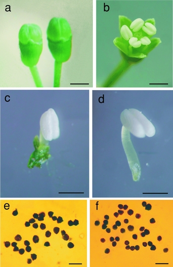
To study the steps of pollen development in each species, the closed buds (Fig. 1a) and open flowers (Fig. 1b), that presented in the same inflorescence at the same time, were collected during the pollination period. The closed bud stamens (Fig. 1c) and open flower stamens (Fig. 1d) were removed and fixed for up to 24 h in 2.5% glutaraldehyde in 0.1 M sodium cacodylate buffer, pH 7.4 and for 2 h in 2% osmium tetroxide in the same buffer. After washing in the buffer the samples were dehydrated in up to 70% ethanol and further underwent subsequent treatment for light microscopy and transmission electron microscopy.
The buds, flower and pollen of P. ginseng. (a) The buds; (b) an open flower; (c) the bud stamen; (d) an open flower stamen; (e) the pollen from bud anther; (f) the pollen from open flower anther. Bars: (a, b) 2 mm; (c, d) 1 mm; (e, f) 50 μm.

Pollen measurements by light microscopy
The experiments were repeated for three buds and three open flowers for two P. ginseng plants, two A. elata trees, and two O. elatus plants. One stamen from each bud and one stamen from each flower were randomly chosen for investigation. The anthers fixed as above were placed into a large 70% ethanol droplet on an object-plate coverslip, crushed by pincers, and the pollen stock was mechanically withdrawn. Drops of pollen ethanol mixture were allowed to dry at room temperature and studied by Polyvar light microscope. Measurements of polar and equatorial axes from 100 pollen grains from each anther were made using an ocular micrometer. The average of polar and equatorial axes from pollen grain was taken to be its average diameter. This diameter averaged from 100 pollen grains of each bud anther or flower anther was an average pollen grain diameter for the early and the late anthers, respectively. The results obtained for the early and the late anthers were analysed by Microsoft Excel program using Student's t-test.
Scanning electron microscopy
The anthers fixed up to 24 h in 2.5% glutaraldehyde in 0.1 M sodium cacodylate buffer, pH 7.4 were placed into a large fixative droplet on a poly-l-lysine-coated Thermanox coverslip and then crushed using pincers. The pollen stock was mechanically withdrawn. Drops of pollen fixative mixture were allowed to dry at room temperature. The Thermanox coverslips with the pollen attached were washed several times in a buffer, postfixed in 2% osmium tetroxide in the same buffer for 2 h, dehydrated in a graded ethanol series, transferred to acetone, and critical-point dried in carbon dioxide. Dried samples were mounted onto aluminum stubs and gold coated before examination with a Leo-340 scanning electron microscope.
Pollen pattern counting by scanning electron microscopy
The calculations were done for three buds and three open flowers for two P. ginseng plants, two A. elata trees, and two O. elatus plants. One stamen from each bud and one stamen from each flower were randomly chosen for investigation. The pollen stock of each stamen was put onto individual aluminum stubs and treated as above. At each stub, 100 pollen grains were investigated by examination with a Leo-340 scanning electron microscope, the pollen patterns were identified and the frequency of the latter was calculated. The results were analysed by the Microsoft Excel program using Student's t-test.
Transmission electron microscopy
The anthers were fixed for 24 h in 2.5% glutaraldehyde in 0.1 M sodium cacodylate buffer (pH 7.4) and for 2 h in 2% osmium tetroxide in the same buffer. Following dehydration in a graded series of ethanol and acetone, the material was embedded in Epon-Araldite. Serial sections were cut on an Ultracut-E (Reichert) ultramicrotome using a diamond knife, stained with uranyl acetate and lead citrate, and examined with a JEM 100 S transmission electron microscope.
Results
Panax ginseng
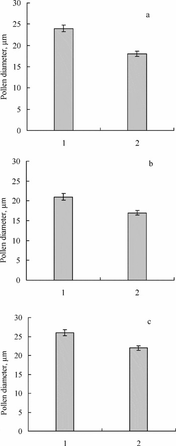
It was shown that both the anthers from closed buds (early anthers) and the anthers from open flowers (late anthers) contained pollen grains varying in size. The maximum average diameter of pollen grains in early and late anthers was about 27 μm, while the minimal diameter was 16 μm. The larger pollen grains were typical of early anthers whereas smaller pollen prevailed in the late anthers (Fig. 1e, f). Consequently the early anthers contained pollen of an average diameter of about 24 μm, while in the late anthers it was about 18 μm (Fig. 2a). Taking into account that size diminution seems to go with prereproductive pollen modification, the pollen grains were further examined in the course of their average diameter decrease by both scanning electron microscopy and transmission electron microscopy.
Average diameters of bud and open flower pollen in P. ginseng (a), A. elata (b) and O. elatus (c). 1, the bud pollen; 2, the pollen of the open flower.

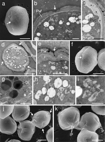
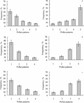
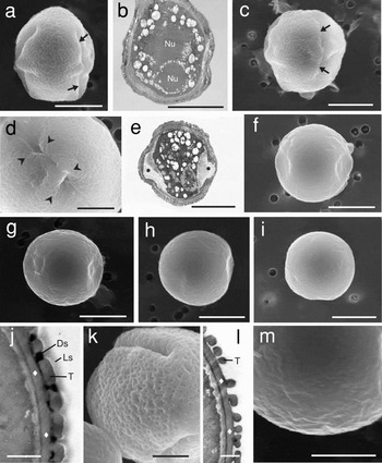
The biggest pollen grains had an average diameter between 27 and 25 μm and were oval shaped from the mesocolpium-centred equatorial view. These grains when viewed are covered by a sculptured wall with irregularly shaped perforations and have apertures (Fig. 3a). At this stage, the apertures, in most cases, belong to an exine that does not form an invagination and is separated from the pollen vegetative cytoplasm by a thin intine layer (Fig. 3b). However, in some grains a thickened intine was observed between the vegetative cytoplasm and exine (Fig. 3c). Some grains were remarkable due to the presence of enlarged intine areas that appeared typically between the apertures and vegetative cytoplasm which seemed to shrink under aperture areas (Fig. 3d). The exine was frequently invaginated into these areas making apertures deeper (Fig. 3e). As our calculations have shown, the oval-shaped pollen pattern prevailed in the early anthers and was equivalent to a frequency of ~41% of the whole pollen stock, but the average amount of such pollen was equal to ~6 % when calculated in the late anthers (Fig. 4a).
The pollen typical of P. ginseng buds. (a) Mostly a mesocolpium-centred equatorial view of the oval-shaped pollen pattern corresponding to size range between 27 and 25 μm in diameter (scanning electron microscopy, SEM); (b) radial section of the oval-shaped pollen grain with vegetative cytoplasm that is adjacent to the pollen wall; a thin intine layer is seen between the pollen wall and vegetative cytoplasm (transmission electron microscopy, TEM); (c) radial section of the oval-shaped grain with intine enlargement arising between the vegetative cytoplasm and pollen wall (TEM); (d) radial section of pollen grain with a vegetative cytoplasm shrinking under aperture areas (TEM); (e) radial section across aperture; note an invaginating pollen wall during aperture deepening (TEM); (f) mostly aperture-centred equatorial view of linear-rhombic pollen pattern corresponding to average diameter between 24–22 μm (SEM); (g) the electron-dense inclusion pattern; (h) the electron-lucent inclusion pattern; (i) the bush-like inclusion pattern; (j) the regularly rhombic grains (21–20 μm); one of them shows the polar view and three apertures (SEM); (k) the equatorial aperture edge projections typical of regularly rhombic grains (SEM); Pvc, pollen vegetative cytoplasm; Pw, pollen wall exine; In, inclusion; Di, the electron-dense inclusion pattern; Li, the electron-lucent inclusion pattern; Bi, bush-like inclusion pattern; P, the equatorial aperture edge projection; the arrows show the pollen apertures; the asterisks show the enlarged intine areas. Bars: (a, d, f, j, k) 10 μm; (b, c, g, h) 1 μm; (i) 0.5 μm; (e) 3 μm.

Frequency of pollen patterns in the bud (left) and open flower (right) anthers. (a) P. ginseng. 1, an oval-shaped pollen (corresponds to sizes between 27–25 μm); 2, a linear-rhombic pollen (24–22 μm); 3, a regularly rhombic pollen (21–20 μm); 4, a cupola-shaped pollen (19–18 μm); 5, a rounded pollen (17–16 μm). (b) A. elata. 1, an oval-shaped pollen (25–24 μm); 2, a regularly rhombic pollen (23–19 μm); 3, an irregularly rhombic pollen (18–17 μm); 4, a rounded pollen with projection (16–15 μm). (c) O. elatus. 1, an oval-shaped pollen (28–26 μm); 2, a regularly rhombic pollen (25–22 μm); 3, a rounded pollen with projection (21–20 μm); 4, an ovoid pollen with projection (19–18 μm).

Pollen with a slightly modified morphology has been found in morphs of 24 and 22 μm. The pollen changed its shape from oval shaped to linear rhombic when seen from the mesocolpium-centred view (Fig. 3f). Due to progressive pollen wall exine invaginations the linear-rhombic pollen was marked by apertures that seemed longer than those in oval-shaped pollen (Fig. 3a). About 28% of the pollen marked by this morphotype was found in the early anthers, although in the late ones its frequency was only ~11% of the whole pollen stock (Fig. 4a).
An abundance of inclusions is a characteristic feature of vegetative cytoplasm. They are the round electron-dark inclusions that are remarkable by a combination of both more and less dark matter (Fig. 3g), round electron-lucent inclusions (Fig. 3h) and bush-like grey coloured inclusions of various shapes (Fig. 3i). These three inclusion patterns have been constantly observed in vegetative cytoplasm of all pollen patterns from the largest to the smallest.
During the following reduction in average diameter (21–20 μm), which occurred as a result of vegetative cytoplasm shrinkage and aperture invagination progress, pollen still had three apertures (Fig. 3j), however, its shape transformed from linear rhombic to regularly rhombic (Fig. 3k). In addition, its general morphology appeared to be modified by paired symmetrical projections formed with aperture edges located in the equatorial area of the pollen body (Fig. 3k). The number of this morphs was similar in both the early and late anthers with about a 14% occurrence (Fig. 4a).
Afterwards, pollen transformed its contour shape from regularly rhombic to cupola shaped (Fig. 5a). This pattern is characteristic of pollen that corresponded to an average diameter between 19 to 18 μm. In some pollen of this category the vegetative cytoplasm does not undergo shrinkage and is more likely to adjoin to the pollen wall (Fig. 5b). However, it seems that some cupola-shaped pollen lose their new cupola-like appearance due to the apertures being feebly marked (Fig. 5c). The equatorial projections of such apertures were often found to be connected (Fig. 5d) and the electron-dense pollen vegetative cytoplasm at this stage obviously undergoes shrinkage again followed by the appearance of large lateral intine areas (Fig. 5e). Cupola-shaped pollen was found to be ~10% in the early anthers but was calculated as ~18% in the late anthers (Fig. 4a).
The pollen typical of open flowers of P. ginseng. (a) Mesocolpium-centred equatorial view of the cupola-shaped pollen pattern corresponding to average diameter between 19 and 18 μm (SEM); (b) the cupola-shaped grain with electron-dark vegetative cytoplasm adjoining the pollen wall (TEM); (c) mostly the mesocolpium-centred equatorial view of the cupola-shaped pollen that loses its cupola-like shape (SEM); (d) the accreting aperture edges (SEM); (e) the cupola-shaped pollen with enlarged intine areas and shrinking vegetative cytoplasm (TEM); (f–i) the rounded pollen pattern (17–16 μm), (f) rounded pollen with equatorial concavities (SEM); (g) rounded pollen with concavity disappearing in the left area (SEM); (h) rounded pollen grain having a remnant of concavity in right area (SEM); (i) the smallest rounded pollen grain lacked any concavities (SEM); (j, k) the radial section across the mesocolpium and an oblique polar view of a large grain by TEM and SEM respectively; (l) the radial section across the mesocolpium by TEM; note the vegetative cytoplasm (left electron-dense substance) that is not shrinking but adjoins to the pollen wall; (m) the surface of a small grain by SEM. Nu, nucleus; T, tectum; Ds, an electron-dense substance interspersed among the columellae; Ls, light substance above the tectum, the rhombs show the bottom layer of pollen exine; the arrows show pollen apertures; the arrowheads show the accreting aperture edges; the asterisks show the enlarged intine areas that arise concurrently with pollen vegetative cytoplasm shrinkage. Bars: (a–c, e–i) 10 μm; (d, m) 5 μm; (j) 1 μm; (k, l) 0.5 μm.

The further diminished pollen variant (17–16 μm) is rounded. An essential feature of such pollen is the presence of equatorial concavities arising from wall invagination into the intine spaces, which appeared simultaneously with the vegetative cytoplasm shrinkage that occured earlier (Fig. 5f). These concavities seemed gradually to disappear on the surface of rounded pollen, which undergoes subsequent diminution of the average diameter (Fig. 5g, h). The smallest ping-pong ball-like pollen grains with a diameter of about 16 μm were almost round and lacked any concavities (Fig. 5i). It was typically found for rounded pollen that its vegetative cytoplasm shrink completely and adjoined the pollen wall (Fig. 5l). As our calculations have shown the rounded pollen pattern was minimal (~7%) in the early anthers and maximum (~51%) in the late anther (Fig. 4a).
The comparative study of the pollen wall showed that the exine of relatively large pollen grains (27–20 μm) consisted of three components and included: (1) the columellate ectexine containing short columellae, thick tectum, and thick foot layer; (2) an electron-dense substance interspersed among the columellae; and (3) the outer friable layer which consisted of an electron-lucent substance (Fig. 5j). This wall construction provides the characteristic sculptured appearance that is typical of large pollen grains (Fig. 5k). However, in the exine of smaller pollen (19–16 μm), the outer electron-lucent layer, as well as the electron-dense substance, were no longer observed. The foot layer is a component of the ectexine at this stage and, the columellae and tectum are less prominent (Fig. 5l). Due to this modification, the wall of small pollen was found to be thinner, the surface appeared to have less ornamentation (Fig. 5m).
Aralia elata
Observations showed that both the bud and open flower anthers contained abundant pollen that varied in size. The average diameters of variations found were between 25 and 15 μm in both types of anthers. However, larger pollen grains were typical of early anthers, whereas a smaller pollen size was a characteristic feature of the late anthers. Consequently, the average diameter of bud pollen was about 21 μm, but in the late anthers it was ~17 μm (Fig. 2b). As size diminution is probably a feature of prereproductive pollen modification, the pollen patterns were further considered in line with average diameter decrease by both scanning electron microscopy and transmission electron microscopy.
The largest pollen grains (25–24 μm average diameter) were oval shaped when seen from the mesocolpium-centred equatorial view. The sculptured surface with perforations is marked by apertures that are not very deep (Fig. 6a). Despite this, the pollen pattern dominates in the early anthers (~54%) however there was less patterning (~7%) in the late anthers (Fig. 4b).
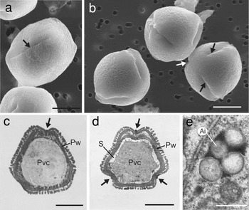
The pollen typical of A. elata buds. (a) An oblique mesocolpium-centred equatorial view of the oval-shaped pollen pattern corresponding to an average diameter of between 25 and 24 μm (SEM); (b) oblique polar of the smaller regularly rhombic grains between 23 and 19 μm in diameter (SEM); (c, d) radial sections of whole grains; note the progression of pollen vegetative cytoplasm shrinkage and pollen wall exine invagination (TEM); (e) the inclusion (TEM). Pw, pollen wall; Pvc, pollen vegetative cytoplasm; S, space for wall invaginations formed by intine enlargement; Ai, aralian inclusion; the arrows show the pollen apertures. Bars: (a–c) 10 μm; (d) 5 μm; (e) 0.5 μm.

The pollen grains of smaller average diameter (23–19 μm) were marked by a regularly rhombic shape and by three apertures that seemed to be deeper than in the previous pollen pattern (Fig. 6b). It was shown by comparative study of the two mentioned pollen patterns that in pollen in the 25–24 μm range the vegetative cytoplasm adjoined to the pollen wall (Fig. 6c). However, in pollen of between 23 and 19 μm the pollen vegetative cytoplasm underwent shrinkage because sufficient space in the intine area appeared for wall invaginations (Fig. 6d). It is obvious that the apertures became deeper because of the progressive invagination of the pollen wall exine (Fig. 6c, d). The number of pollen of this pattern between 23 and 19 μm in diameter was calculated as ~26% and ~11% respectively for the early and late anthers (Fig. 4b).
Many inclusions, appearing as several electron-lucent globules embedded into the electron-dark matter (Fig. 6e) were characteristic of vegetative cytoplasm in all A. elata pollen morphs from the largest to the smallest.
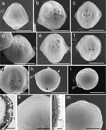
The next morphological pattern was typical for pollen with an average diameter ranged between 18 and 17 μm. These pollen grains were found to be marked by relatively deep apertures and an irregular rhombic shape when seen from the mesocolpium-centred equatorial view (Fig. 7a). As a rule, at this stage, the aperture edges are remote from each other, and there is quite a large gap between them (Fig. 7b). Some of the pollen grains seem to revert to an irregularly rhombic contour shape due to the apertures becoming shorter and the aperture edge projections approaching closer to each other (Fig. 7c) and undergoing contact (Fig. 7d). Contact of the whole edges for some pollen grains was not found, however a certain kind of initial bridge was seen between the remote edges (Fig. 7e). It seems obvious that the accretion of aperture edges is the final step in irregularly rhombic pollen modification prior to the next pollen pattern (Fig. 7f, g). Around ~12% of irregularly rhombic pollen were present in early anthers although ~32% were found in the late anthers (Fig. 4b).
The pollen typical of open flowers of A. elata. (a) Mesocolpium-centred equatorial view of the irregularly rhombic pollen pattern that have an average diameter between 18 and 17 μm (SEM); (b) aperture-centred equatorial view of the irregularly shaped rhombic pollen grain with a large gap between aperture edges (SEM); (c) the grain losing its irregularly rhombic contour due to adjacent aperture edges (SEM); (d) a similar grain with contacting aperture edges (SEM); (e) a similar grain with aperture edges connected by an initial bridge (SEM); (f) a similar grain with accreting aperture edges (SEM); (g) a similar grain with accreted aperture edges (SEM); (h, i) a rounded pollen with a projection (16–15 μm), (h) a grain with an aperture during accretion (SEM); (i) the smallest grain (SEM); (j, k) the radial section across mesocolpium and an oblique polar view of a large grain by TEM and SEM, respectively; (l) the radial section across the mesocolpium by TEM; note the vegetative cytoplasm (left electron-dense substance) that is not shrinking but adjoining the pollen wall; (m) the surface of smaller grain by SEM. G, the gap between aperture edges; Ib, an initial bridge; P, the projection formed by apertures retarding in their accretion; T, the tectum; Co, the columellae; the rhombs show the foot layer of pollen exine; the asterisks show the aperture edge projections. Bars: (a–f, h, i) 10 μm; (g, m) 5 μm; (j, k) 3 μm; (l) 1 μm.

The pollen grains that corresponded to next morphologic pattern had average diameters of 16–15 μm. The pollen variants of this size are rounded with projection formed by one of three apertures. Two apertures were usually found accreted while accretion of the third aperture was retarded from the other two (Fig. 7h). In the smallest rounded pollen (15 μm), the vestiges of ‘lagging’ aperture edge projections merely could be observed. It should be emphasized that this projection appeared comparatively large and obviously constitutes the bright morphological feature found in A. elata pollen (Fig. 7i). In the rounded pollen with a projection the vegetative cytoplasm did not undergo shrinkage and seems to be adjoined to the pollen wall (Fig. 7l). Despite only ~8% of rounded pollen with a projection being observed in the early anthers the number in the late anthers was calculated as ~48% (Fig. 4b).
Observation of the pollen between 25–17 μm showed that it contained only an columellate ectexine (Fig. 7j) and had a sculptured perforated surface (Fig. 7k). No additional substance filling the exine cavities between columellae and the outer friable layer was found in A. elata ectexine. In the pollen that had shrunk to 16–15 μm the exine becomes thinner and its columellae become shorter (Fig. 7l) and this pollen pattern appears to have less ornamentation (Fig. 7m).
Oplopanax elatus
An observation of the bud anthers showed that pollen varied in size. The average diameter of the largest grains was about 28 μm, while the smallest was measured as ~18 μm. The same pollen size range was found in the open flower anthers. However, the larger grains were typical of early anthers, whereas smaller pollen prevailed in the late anthers. As a result, the average size of pollen in the early anthers was about 26 μm and in the late anthers it was about 22 μm (Fig. 2c). Due to the decrease of average diameters as an obvious characteristic of prereproductive pollen modification the morphotypes were examined according to size diminution by both scanning electron microscopy and transmission electron microscopy.
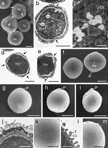
The large pollen grains of diameter 28–26 μm had an oval shape and were marked by apertures (Fig. 8a) that can be described as slight invaginations of pollen wall exine (Fig. 8b). The occurrence of this pollen morphotype was found to be ~44% in the early anthers although it was only ~9% in the late anthers (Fig. 4c).
The pollen typical of buds and open flowers of O. elatus. (a) The large oval-shaped pollen variation (corresponding to average diameter ranged between 28–26 μm), medium-sized regularly shaped rhombic pollen variation (25–22 μm) and small rounded pollen variation with projection (21–20 μm) found both in buds and open flowers (SEM); (b) the radial section of an oval-shaped grain; (c) the electron-dense and electron-lucent inclusions; (d, e) the radial sections of the grains showing the progression of pollen vegetative cytoplasm shrinkage and intine enlargement (electron-lucent layer) (TEM); (f) the regularly rhombic grains with accreting apertures (SEM); (g) the rounded grain with a projection formed by aperture edges retarding in their accretion (SEM); (h) the rounded grain with a more sharp projection (SEM); (i) the smallest pollen corresponding to 19–18 μm average diameter that is ovoid and has a projection (SEM); (j), (k) the radial section across mesocolpium and the surface of large pollen by TEM and SEM, respectively; (l) the radial section across mesocolpium by TEM; note the vegetative cytoplasm (electron-dense substance) that is not shrinking but adjoin to the pollen wall; (m) the surface of small grain by SEM. Lp, large pollen (oval shaped); Mp, medium pollen (regularly rhombic); Sp, smaller pollen (rounded with projection); Nu, nucleus; Di, electron-dark inclusion; Li, electron-lucent inclusion; Pvc, pollen vegetative cytoplasm; Aa, accreting aperture; P, the equatorial projection formed by aperture edges; Ds, an electron-dense substance interspersed among the columellae; Ls, light substance above the electron-dense substance interspersed among the columellae; T, tectum; Co, columellae; the asterisks show the enlarged intine areas arising parallel to pollen vegetative cytoplasm shrinkage; the rhombs show the foot layer of exine; the arrows show the pollen apertures. Bars: (a, b, d–i) 10 μm; (c, l) 0.5 μm; (j) 2 μm; (k, m) 5 μm.

The pollen vegetative cytoplasm was filled with an electron-dense substance rich in numerous inclusions. However, we were able to observe only two patterns of inclusions. These were the electron-dense inclusions with an oval–round shape and the electron-lucent inclusions marked by an irregular shape (Fig. 8c). These inclusion patterns have been observed throughout all morph variants including the smallest pollen grains.
It seems obvious that subsequent diminution of pollen occurs in parallel with pollen vegetative cytoplasm shrinkage followed by the appearance of enlarged intine areas situated under apertures that enable progressive invagination of the latter (Fig. 8d, e,). The medium pollen grains of a somewhat smaller average diameter (25–22 μm) had regularly rhombic contour shape and deeper apertures (Fig. 8a). Some samples were observed with the apertures that were no longer deep but accreting, the equatorial projections of the aperture edges are typical of this pollen stage (Fig. 8f). The regularly rhombic pattern was found to correspond to ~28% of grains in the early anthers and comprise ~13% in the late anthers (Fig. 4c).
The pollen of smaller size (21–20 μm) due to vegetative cytoplasm shrinkage had a different pattern from that previously found and possessed a more rounded shape and the visible absence of apertures (Fig. 8a). However, the remnant of an equatorial projection of one of the three apertures still could be seen (Fig. 8g). Following size diminution, the grains had a more pointed projection (Fig. 8h). The frequency of this type of pollen, characterized as rounded with projection, was ~17% in the early anthers and ~35% in the late ones (Fig. 4c).
The pattern in the smallest pollen (the average diameter is about 19–18 μm) was an ovoid morphology, covered by a wrinkled wall and was distinctive as having a single sharp projection (Fig. 8i). It was regularly observed that the vegetative cytoplasm of ovoid pollen does not undergo shrinkage and adjoined the pollen wall (Fig. 8l). There was a clear-cut distinction in the amount of ovoid pollen that was comparatively small (~11%) in the early anthers but strictly larger (~43%) in the late anthers (Fig. 4c).
The separate study of the pollen wall showed a significant difference in the construction of the exine in large and small pollen. Thus, the large grains (28–21 μm) were characterized by a thick tectum and an infratectum of thick columellae with the electron-dark substance over the foot layer and electron-lucent substance situated above (Fig. 8j). This structure of the exine provides a porous appearance typical of large pollen (Fig. 8k). The ectexine of smaller grains (20–18 μm) still contains the foot layer and has remnants of tectum and columellae (Fig. 8l). This type of wall had ornamentation (Fig. 8m).
Discussion
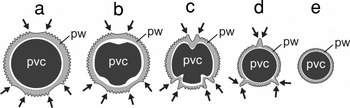
As our study has shown both the bud anthers as well as open flower anthers of P. ginseng, A. elata and O. elatus taken during the reproductive period were characterized by a diversity of pollen size. However, larger pollen was typical of the bud anthers, whereas smaller pollen sizes were more characteristic of the open flower anthers. Thus, for the early and late anther pollen stock comparison we could conclude that in every species the gradual diminution of pollen size that occurred in the anthers during their maturation was confined to its pollination time. Hence, the subsequent examination of pollen grains in the course of average diameter decrease was used here as a method permissive to highlight any features of pollen modification that presumably occurred during the plant's reproductive period. As a result of this method application it was found that size diminution is not random but rather connected with pollen metamorphosis. In accordance with our observations for P. ginseng, A. elata and O. elatus the largest pollen grains in every species were covered by a sculptured wall and marked by apertures. The smallest pollen patterns had no distinctive apertures and were covered by a smooth wall. Between these variants the pollen morphology varies considerably due to pollen undergoing intensive modification. Apparently, shrinkage of the pollen vegetative cytoplasm is one of the modifications occurring during pollen diminution and actually permits this process (Fig. 9a–e). This shrinkage underlies intine widening under exine aperture areas, aperture deepening (Fig. 9b, c) and accretion of the aperture edges (Fig. 9c, d). The disappearance of aperture edge formed projections is total in P. ginseng (Fig. 9e) but partial in the other two species. One more modification is the gradual diminution of the pollen wall thickness (Fig. 9a–e). The thick sculptured wall of large pollen changed regularly into the thin and less sculptured variant. Thus, the vegetative cytoplasm shrinkage, intine enlargement, exine aperture deepening and accretion as well as modification of the exine seem to be the mechanisms of ‘pollen size-reducing metamorphosis’ that occurs in P. ginseng, A. elata and O. elatus during these species reproductive period. As comparison of pollen stock from early and mature anthers has not been studied before this pollen modification, which presumably starts in the buds prior to their opening and progresses after bud opening has not been described in previous reports and is a novel addition to pollen formation in Araliaceae. The subcellular mechanisms of processes that carry out this metamorphosis still have to be elucidated.
A schematic drawing of pollen size reducing metamorphosis, which is universal in P. ginseng, A. elata and O. elatus. (a) The large pollen pattern; (b) the intermediate pollen pattern undergoing intine enlargement and vegetative cytoplasm shrinkage, (c) the intermediate pollen pattern undergoing the vegetative cytoplasm shrinkage as well as aperture deepening; (d) the intermediate pollen pattern undergoing the vegetative cytoplasm shrinkage and aperture accretion; (e) the smallest pollen pattern marked by a shrunk vegetative cytoplasm, thin pollen wall and accreted apertures. pvc, pollen vegetative cytoplasm, pw, pollen wall exine; the electron-lucent layer marks intine; the arrows show the apertures.

It should be stressed that some data that describe high intraspecific pollen size variation in Panax and Aralia could be found in previous publications. Koren et al. (Reference Koren, Krylach, Zaytseva, Zhuravlev, Weber, Zeuske and Imhof1998) examined 200 pollen grains from each of 20 P. ginseng plants and found pollen varying in morphology, lacking one, two or three apertures, having sizes 1.5-fold smaller or larger than that of the triangular-rounded morphotype with three apertures. Although Wen & Nowicke (Reference Wen and Nowicke1999) examined only 10 grains from each species, the length of pollen's polars and equatorial axes varied widely. Moreover, some of the figures in Wen & Nowicke's (Reference Wen and Nowicke1999) report support aperture deepening, e.g. Figs. 11, 26 and the accretion of the aperture edges, e.g., Figs. 1, 9, 20, 51 and 62. However, these authors did not specifically refer to these conditions, possibly because size-reducing metamorphosis had not yet been considered. It would be of interest in future work to examine whether this pattern of metamorphosis is typical for all Araliaceae taxa or if some other variants are possible. Nevertheless, at this stage of investigation the metamorphosis character seems to be universal, therefore could hardly be useful as a characteristic for Araliaceae phylogeny. The value of studying pollen metamorphosis appears clear as the morphology of intermediate and final pollen morphotypes is clarified.
In the present report, pollen diversity was analysed and compared between P. ginseng, A. elata and O. elatus. It was found that the initial large ‘oval-shaped’ morphotype is similar between species and could hardly be used for taxonomic purpose. However, further modification was marked by divergence of ways of transformation. Indeed, the first transformation step the close ‘regularly rhombic’ contour shape was been observed in A. elata and O. elatus, although P. ginseng was distinct in having a ‘linear-rhombic’ pollen contour. After the third modification step the pollen from each species was specific in its outline shape i.e. ‘regularly rhombic’ in P. ginseng, ‘irregularly rhombic’ in A. elata and ‘rounded with projection’ in O. elatus. The fourth modification step was the final one for A. elata and O. elatus pollen, which transformed into ‘rounded with projection’ and ‘ovoid with projection’ respectively, although the pollen of P. ginseng at that stage still took on its intermediate ‘cupola-like’ shape before its fifth and last modification step given rise to final ‘rounded’ pollen. It seems likely that every species is peculiar in having a specific intermediate morphs corresponding to definite dimensional ranges. Based on our morphometric study it could be suggested that intermediate morphs are generally stable in their morphology and undergo only an alteration in their proportions together with a gradual size decrease of large morphs and an increase in the smaller morphs that are involved with anther maturation (Fig. 4a–c). Probably, the amount of intermediate pollen patterns and their morphology might be involved in interspecific comparison. It should be stressed that based on these characteristics P. ginseng seems to be rather to be different to close to A. elata and O. elatus.
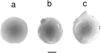
The final pollen to the present report had the smallest grains. It was particularly interesting to compare these pollen patterns because it was expected to be useful in resolving dispute on aralian or non-aralian Panax affinity (see above). As it is shown, the smallest pollen grains of P. ginseng and A. elata have a rounded shape but the pollen of the latter is notably different in having a somewhat smaller size and a cone-like projection (Fig. 10a, b). The size and shape of O. elatus pollen are so different from the previous mentioned that the originality of this seems beyond doubt (Fig. 10c). Based on these findings one can conclude that the smallest pollen grains of P. ginseng, A. elata, and O. elatus are species specific. It would be too speculative to suggest a close proximity of Panax and Aralia relying on smallest pollen comparison as the aralian morphotype is distinct in having such a distinctive feature as the cone-like projection.
The comparative view of the smallest pollen patterns in P. ginseng (a), A. elata (b), O. elatus (c). Bar: 5 μm.

Taking into account that pollen morphology has been applied to support the phylogenetic proximity of Panax and Aralia by Wen & Nowicke (Reference Wen and Nowicke1999), it should be stressed that pollen patterns used by these authors were definitely marked by a thick/sculptured wall and colporate apertures. Indeed, since Erdtman (Reference Erdtman1966) described the pollen of 30 species from 20 genera of Araliaceae as mostly three colporate, usually reticulate, with sexine thickened at the poles such patterns seem have been accepted as the dominant pollen types in Araliaceae. However, if the principle of size-reducing metamorphosis could be taken into account, it might be possible that those patterns were more likely at the early stages of metamorphosis. Probably, other pollen variants differing in morphology might also be found for species involved in Wen & Nowicke's (Reference Wen and Nowicke1999) study and careful investigation would be desirable to clarify the morphs appropriate for taxonomical comparison.
One more concern that seems to be interesting is the co-existence of different pollen morphs in the mature anthers of P. ginseng, A. elata and O. elatus. Pollen heteromorphism as a product of different morphotypes within an individual is broadly distributed in plants (see for review Till-Bottraud et al., Reference Till-Bottraud, Joly, Lachaise and Snook2005) although the reason for this still has to be discovered. However, P. ginseng, A. elata and O. elatus pollen heteromorphism most likely is the consequence of the transformation of large pollen grains to smaller ones during the anther maturation and it is not clear whether all these morphs are available for reproduction. In accordance with heteromorphism theory (see for review Till-Bottraud et al., Reference Till-Bottraud, Joly, Lachaise and Snook2005) it would be tempting to speculate that the presence of different morphs in mature anthers of P. ginseng, A. elata and O. elatus may be necessary for species reproductive success. But, this hypothesis must be examined by experimentally controlling the various morphs ability for fertilization.
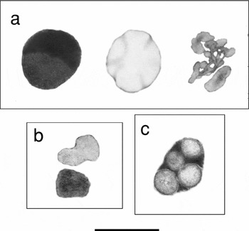
It was found that the pollen vegetative cytoplasm of the three species examined is peculiar in having the inclusions, which are invariable during pollen-size reducing metamorphosis but differ between species in ultrastructure and pattern numbers. P. ginseng has three patterns of inclusions (Fig. 11a), there are two patterns of inclusions in O. elatus (Fig. 11b) and only one inclusion type is characteristic of A. elata (Fig. 11c). All inclusion patterns do not overlap among species, testifying to the fact that studied species are equally distanced one from another.
The comparative view of pollen inclusions found in P. ginseng (a); O. elatus (b); and A. elata (c). Bar: 1 μm.

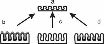
The same could be concluded after consideration of pollen walls. Despite the fact that walls of the smallest pollen grains appear to be similar in the three species (Fig. 12a) there is a clear-cut difference in the construction of the exine of largest pollens' patterns. Hence, in regard to the ultrastructure, the largest pollen wall exines of P. ginseng (Fig. 12b) differs from those in A. elata (Fig. 12c) and O. elatus (Fig. 12d) and any phylogenetic affinity is unlikely to be suggested.
A schematic drawing of the pollen wall exine which has similar construction in the small pollen of three species (a); but seems to be different in the large pollen of P. ginseng (b); A. elata (c); and O. elatus (d). The arrows show the direction of exine modification.

So, as a result of this study some new pollen characteristics such as the morphology of smallest pollen grains, amount and morphology of intermediate pollen patterns, inclusion types and the structure of largest pollen's exine were found to be applicable for effective taxonomic comparison. We suggest that an expanded study of all pollen morphotypes when applied to other species using these additional pointers would be useful to add to the ongoing phylogenetic discussion of aralian or non-aralian Panax affinity.
Acknowledgements
We are very grateful to Drs A.N. Prilutskiy and P.G. Ostrogradskiy for their kind help in material collection. Many thanks to Ms Yulia Reunova for her assistance in flower photography and to Mr Denis Fomin for his technical assistance in TEM units. This work was supported by the Russian Science Support Foundation for A.A. Reunov, by grants from the Far East Branch of the Russian Academy of Sciences (no. 06-I-P11-028 and no. 06-I-P11-030) and by ‘Leading Schools of Thought’ of the President of Russian Federation (no. NSH6923-2006.4) for Yu.N. Zhuravlev.