Studies of infants of diabetic mothers have demonstrated a two- to sixfold increased risk of congenital anomalies,Reference Becerra, Khoury, Cordero and Erickson 1 with specific patterns of malformations, such as holoprosencephaly, renal/urinary defects, vertebral/rib defects, and laterality defects that occur more often in infants of diabetic mothers than in infants born to non-diabetic mothers.Reference Kucera 2 – Reference Martinez-Frias, Bermejo, Rodriguez-Pinilla, Prieto and Frias 4 The Baltimore-Washington Infant Study has reported an increased risk of cardiac anomalies in infants of diabetic mothers, including outflow tract anomalies, laterality defects, and cardiomyopathy.Reference Loffredo, Wilson and Ferencz 5 Considering specific malformation syndromes, caudal dysplasia sequence has shown a distinct link with maternal diabetes.Reference Passarge and Lenz 6 In addition, several cases of infants of diabetic mothers presenting with the features of DiGeorge anomalyReference Gosseye, Golaire, Verellen, van Lierde and Claus 7 – Reference Kumar, Sapire, Lockhart, McCombs, Hawkins and Van Mierop 11 and of oculo-auriculo-vertebral spectrumReference Wang, Martinez-Frias and Graham 12 have been reported. Infants with DiGeorge anomaly born from diabetic mothers can also display clinical manifestations that are part of the caudal dysplasia sequence spectrum, including vertebral/renal anomalies or club feet.Reference Gosseye, Golaire, Verellen, van Lierde and Claus 7 On the other hand, finding DiGeorge anomaly features in a series of patients reviewed as caudal dysplasia sequence seems rare.Reference Bohring, Lewin and Reynolds 13
We have previously reported infants of diabetic mothers with clinical characteristics of DiGeorge anomaly associated with renal agenesis, but lacking skeletal defects of the caudal region, which is generally found in caudal dysplasia sequence.Reference Digilio, Marino, Formigari and Giannotti 10 Among the present personal series of 204 patients with DiGeorge anomaly/Velo-cardio-facial syndrome (OMIM 192430) and nine patients with caudal dysplasia sequence, evaluated between January 1992 and May 2011, we observed a neonate, born to a mother with poorly controlled insulin-dependent diabetes, in whom features of DiGeorge anomaly and caudal dysplasia sequence were coexisting.
Clinical report
The proband, a female, was the first child of a mother affected by insulin-dependent diabetes since the age of 14 years. Family history was otherwise unremarkable, and consanguinity was denied. The baby was born by Caesarean section at the 37th week of gestation. The pregnancy was complicated by poorly controlled blood sugars. Insulin treatment was used for glycaemic control, although the mother did not make regular use of it. Birth weight was 3980 grams (greater than 97th centile), length 46 centimetres (less than 3rd centile), and head circumference 35 centimetres (50th centile). Apgar scores were 6 and 7 at 1 and 5 minutes, respectively. Clinical examination at birth showed macrosomia, generalised oedema, smaller and uniformly hypoplastic lower limbs, and equinovarus feet deformity. No obvious facial dysmorphic features were apparent.
Two-dimensional color Doppler echocardiography showed truncus arteriosus type A1 of Van Praagh, with hypertrophic cardiomyopathy involving the left ventricle and the interventricular septum. Cerebral ultrasonography disclosed a large intracranial subependymal haemorrhage in the germinal matrix with increased vascular resistances. Bilateral hypoplastic kidneys with persistence of foetal lobulation were detected by renal ultrasound (longitudinal diameter: 36 millimetres; parenchymal average thickness: 8 millimetres). Skeletal X-ray examination showed multiple vertebral malformations in the thoracic and lumbosacral spine, with incomplete development of the sacrum, and decreased length of all lower limb bones.
Laboratory investigations showed severe hypoglycaemia (23 milligrams per decalitre) in the first days of life, insulinaemia below the normal range (4.3 microinternational units per millilitre), normal insulin growth factor-1 levels (125 nanograms per millilitre), reduced proteins, and mild hyperbilirubinaemia (total bilirubin: 1.87milligrams per decalitre; direct bilirubin: 0.17 milligrams per decalitre). Serum calcium (8.8 milligrams per decalitre) and parathyroid hormone (15 picograms per millilitre) were normal despite the absence of parathyroid glands (see autopsy). The T-cell number was below the normal range: CD3 35% (normal value 58–75%); CD4 20% (normal value 29–47%); CD8 14% (normal value 17–33%). Standard chromosome analysis on circulating lymphocytes showed a normal 46,XX karyotype. Fluorescence in situ hybridisation analysis using the LSI N25 probe specific for 22q11.2 (Vysis, Abbott Laboratories, Chicago, Illinois, United States of America) revealed an unremarkable pattern.
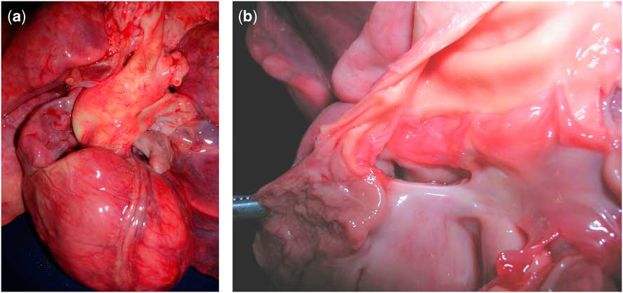
The patient died on the 45th day of life of cardiorespiratory insufficiency. The autopsy showed marked cerebral atrophy with cystic lesions in the frontal region, bilaterally. Heart examination showed truncus arteriosus type A1 (Fig 1a) with juxtatruncal ventricular septal defect (Fig 1b). Both ventricles displayed severe wall hypertrophy, confirming hypertrophic cardiomyopathy. Absence of the thymus and parathyroid glands was noted. Kidneys were mildly hypoplastic, lacking abnormal parenchymal differentiation.
(a) External appearance of truncus arteriosus type A1, anterior view. The great arteries seem almost completely divided and normally interrelated. This aspect is seen in truncus only when the aortopulmonary septum is quite well formed. (b) Truncus arteriosus from left ventricular aspect. The three leaflets truncal valve shows mild myxoid features. Note the juxtatruncal ventricular septal defect.
Discussion
Clinical features in the present patient are compatible with the co-occurrence of both DiGeorge anomaly and caudal dysplasia sequence (Table 1). DiGeorge anomaly is an aetiologically heterogeneous developmental field defect related to neural crest cells migration anomaly, clinically characterised by the association of conotruncal heart defect, hypoplasia or absence of the thymus and immune deficit, neonatal hypocalcaemia, and facial anomalies.Reference DiGeorge 14 The cardiac defects more commonly found in DiGeorge anomaly include tetralogy of Fallot, truncus arteriosus, and interrupted aortic arch.Reference Marino, Digilio and Toscano 15 Most patients with DiGeorge anomaly have a microdeletion of chromosome 22q11.2.Reference Greenberg 16 The frequency of chromosome 22q11.2 deletion in conotruncal malformation ranges between 7% and 48%.Reference Iserin, de Lonlay and Viot 17 , Reference Ziolkowska, Kawalec and Turska-Kmiec 18 In particular, truncus arteriosus, found in our patient, occurs in 34–46% of 22q11.2 deletion patients.Reference Goldmuntz, Clark and Mitchell 19 , Reference Anilkumar, Kappanayil, Thampi, Nampoothiri, Sundaram and Vasudevan 20 DiGeorge anomaly has also been reported in several recognisable malformation patterns or in association with the exposure to different teratogens, including maternal diabetes.Reference Gosseye, Golaire, Verellen, van Lierde and Claus 7 – Reference Kumar, Sapire, Lockhart, McCombs, Hawkins and Van Mierop 11 Renal and vertebral malformations have been found in infants of diabetic mothers with DiGeorge anomaly.Reference Gosseye, Golaire, Verellen, van Lierde and Claus 7 , Reference Wilson, Blethen and Vallone 9 , Reference Digilio, Marino, Formigari and Giannotti 10 Structural defects of the caudal region are cardinal features of caudal dysplasia sequence.Reference Welch and Aterman 21 These include incomplete development of the sacrum and of the lumbar vertebrae, skeletal limb anomalies secondary to neurologic impairment, and decreased leg movements. Caudal dysplasia sequence is a rare heterogeneous congenital defect resulting from an insult to the structures of caudal eminence. Suggested aetiologic and pathogenetic mechanisms include maternal diabetes,Reference Passarge and Lenz 6 genetic predisposition,Reference Finer, Bowen and Dunbar 22 environmental teratogenic factors,Reference Padmanbhan 23 and vascular hypoperfusion.Reference Stevenson, Jones and Phelan 24 In particular, it has been estimated that 16% of human cases of sacral agenesis are associated with maternal diabetes mellitus.Reference Passarge and Lenz 6 Associated anomalies affecting other systems are often present, including cardiovascular, neurological, gastrointestinal, and genitourinary system defects.
Clinical features of the present patient compared with the characteristics of DiGeorge anomaly and caudal dysplasia sequence.

aGreenberg, 1993
bBohring et al, 1999
The empiric risk of congenital heart defects to the offspring of insulin-dependent diabetic mothers is 18%.Reference Becerra, Khoury, Cordero and Erickson 1 The anatomic types of structural congenital heart defects observed in infants of diabetic mothers include hypertrophic cardiomyopathy,Reference Ferencz, Rubin, McCarter and Clark 8 mimicking asymmetric septal hypertrophy and conotruncal malformations, in particular truncus arteriosus and double outlet right ventricle.Reference Loffredo, Wilson and Ferencz 5 It has been speculated that diabetic women may transfer factors across the placenta to the embryo that alters the normal migration of neural crest ectomesenchymal tissue.Reference Ferencz, Rubin, McCarter and Clark 8 The diagnosis of truncus arteriosus type A1 in the present patient suggests a link between maternal diabetes, DiGeorge anomaly, and cardiac defects. The co-occurrence of congenital heart defects and hypertrophic cardiomyopathy in infants of diabetic mothers has been described previously.Reference Cheatham, McManus, Kugler and Hofshire 25 In conclusion, DiGeorge anomaly and caudal dysplasia sequence have been independently seen in association with maternal diabetes. Therefore, their co-occurrence in the present case argues for a relationship with maternal diabetes, which can cause a wide spectrum of defects, manifesting either as isolated DiGeorge anomaly or caudal dysplasia sequence, or DiGeorge anomaly associated with renal and vertebral malformations, or DiGeorge anomaly plus caudal dysplasia sequence. Truncus arteriosus with hypertrophic cardiomiopathy is likely a distinct type of congenital heart defect associated with DiGeorge anomaly or secondary to maternal diabetes.