Introduction
Skull base radiology plays a crucial role in the management of skull base pathology.Reference Gardner, Kassam, Rothfus, Snyderman and Carrau 1 Imaging modalities including computed tomography (CT), magnetic resonance imaging (MRI), angiography and positron emission tomography allow excellent three-dimensional (3D) visualisation of the complex anatomy and pathology. Tissue characteristics of the tumour, its relationship to the adjacent structures, and its site of origin can be characterised, enabling an accurate differential diagnosis. This is indispensable to the surgical team's operative plan. The application of advanced computer software to the imaging data can create realistic 3D images, enable virtual surgical planning and integrate the images for use in intra-operative navigation.Reference Zhao, Yu, Li, Xu and Li 2
In our institution, a team of dedicated neuroradiologists with a strong interest in skull base radiology work closely with a multidisciplinary team of clinicians managing these disorders. Contemporary imaging modalities that include 1.5 T and 3 T MRI are routinely used for investigations of the patient, and for intra-operative navigation in the appropriate surgical setting.
We have recently managed cases where the contemporary protocols for pre-operative MRI scanning were unsatisfactory in delineating the relationship of the pathology in question to the optic nerve and adjacent vascular structures at the orbital apex. Following consultation with the skull base radiology team, a targeted MRI study using high-resolution techniques was designed to acquire images in orthogonal planes centred parallel and perpendicular to the oblique course of the relevant optic nerve.
Targeted MRI studies were performed on the two cases discussed in the following section using a 3 T magnetic resonance (MR) platform (GE Healthcare, Fairfield, Connecticut, USA). Images were obtained with a matrix of 512 × 512 at 2.2 mm thickness in a coronal-oblique plane perpendicular to the long axis of the optic nerve. The MR platform was used to generate T2-weighted sequences with fat saturation (echo time = 83.3 ms, repetition time = 4205 ms) and post-gadolinium, T1-weighted, fat-saturated sequences (echo time = 7.2 ms, repetition time = 671 ms). These sequences were found to be the most useful for providing an anatomical roadmap for the endoscopic skull base surgeon in the approach to the optic nerve.
Case reports
Case one
A 39-year-old female patient presented to the neurosurgery department in 2008 with a 10-year history of ‘migraine-like’ headaches, right retro-orbital pain and poor night vision. In the three months prior to her presentation, the patient had noticed visual field changes, and ophthalmological examination findings were consistent with a right hemianopia. Using conventional CT and MR imaging, a planum sphenoidale meningioma was diagnosed.
The patient underwent right frontotemporal craniotomy for excision of the tumour. Intra-operatively, the tumour was located anterior to the optic chiasm, elevating the right optic nerve and reaching the right internal carotid artery. The tumour was debulked, leaving residual tumour at the entrance of the optic canal medial to the optic nerve. Histopathology confirmed the diagnosis of meningioma (World Health Organization (WHO) grade 1 – common, benign tumour). The patient's visual symptoms resolved post-operatively, with restoration of normal visual fields and visual acuity of 6/12 in the right eye.
Surveillance MRI conducted in 2012 revealed a strip of enhancing lesion measuring 7.5 × 4 mm along the superomedial aspect of the right anterior clinoid process, in keeping with residual meningioma. An MRI scan performed six months later revealed an increase in the lesion size to 9 × 4 mm; in addition, a second deposit was observed in the anterior wall of the pituitary fossa.
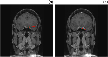
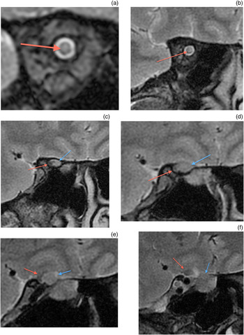
One month after the MRI scan, the patient (now 44 years of age) presented to the emergency department with headache and a sudden deterioration of vision in her right eye. A new visual field defect was identified (superonasal quadrantanopia), indicating compression of the right optic nerve. Whilst confirming the growth of residual tumour on serial scans, the MRI images failed to establish a clear relationship of the tumour mass to the optic nerve (Figure 1). A targeted MRI study using the imaging parameters mentioned above was undertaken, focusing on the right orbital apex (Figure 2). This demonstrated the tumour surrounding the pre-chiasmic optic nerve and extending to the orifice of the optic canal, causing obstruction of the subarachnoid space and resulting in oedema of the nerve.
Routine coronal, T1-weighted, gadolinium-enhanced magnetic resonance imaging scans of case one (a & b), demonstrating residual meningioma (red arrows) at the planum sphenoidale.

Coronal, T2-weighted, targeted magnetic resonance imaging scans of case one (a–f). Red arrows point to the optic nerve in its orbital, intracanalicular and intracranial course. Blue arrows denote the residual meningioma applied to the inferior, medial aspect of the optic nerve and the optic chiasm. Peri-tumoural oedema is evident in the distal (orbital) optic nerve, presumably due to compression of the proximal nerve by the meningioma.

Urgent transnasal endoscopic surgical decompression of the right optic nerve was undertaken. Intra-operatively, the meningioma was noted to have both intradural and extradural components, and was extending into the right sphenoid sinus with its dural tail extending along the right optic nerve into the optic canal. The optic nerve was exposed from the optic canal to the chiasm, and its dural sheath was split to decompress the nerve. Piecemeal removal of the tumour was accomplished. The pathological diagnosis confirmed WHO grade 1 meningioma.
The patient's symptoms improved significantly in the post-operative period, with restoration of normal visual fields in the right eye and a visual acuity of 6/7.5. A small residual lesion was detected on post-operative MRI between the left optic nerve and the internal carotid artery, and this remains under close surveillance.
Case two
A 32-year-old female patient was referred with a history of Graves' disease; after failed medical treatment for this condition, she underwent total thyroidectomy. She experienced a deterioration of her vision and headaches, resulting in a diagnosis of severe Graves' ophthalmopathy. The patient's symptoms had thus far been managed medically with the use of steroids and methotrexate. Visual deterioration prompted surgical intervention. Bilateral, external (two and a half wall) orbital decompression was performed by the oculoplastic surgeon.
Twelve months later, the patient presented to the out-patient clinic with deterioration in her visual acuity, bilateral orbital pain and headaches in the weeks leading up to the consultation. Physical examination showed noticeable lid lag and proptosis. The fundoscopy findings were consistent with optic neuropathy. Her visual acuity was 6/24 in the left eye and 6/60 in the right eye.
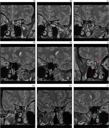
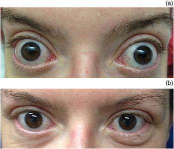
A similar targeted MRI study was undertaken to highlight the optic nerve compression, and the targeted sequences were utilised to pinpoint the area of most critical compromise (Figure 3). The patient underwent bilateral, endoscopic orbital decompression. Following complete removal of the medial wall and partial removal of the orbit floor, the periorbita was incised. Orbital fat prolapsed into the ethmoidectomy cavity and was debulked. The procedure resulted in significant improvement in the degree of exophthalmos (Figure 4). The patient received adjuvant radiotherapy (20 Gy in 10 fractions) to the orbits.
Coronal, T2-weighted (without fat suppression), targeted magnetic resonance imaging scans of case two. Parts (a) through (e) show the left optic nerve (green arrows), and images (f) through (i) show the oedematous right optic nerve (green arrows). Both nerves are followed along their course from the orbit to the planum sphenoidale. Red arrows in image (f) depict the orbital fat prolapsed into the maxillary antrum following previous orbital decompression.

Photographs of case two demonstrating the degree of exophthalmos before (a) and after (b) endoscopic orbital decompression.

Following the completion of treatment, the patient's visual acuity was 6/48 in the left eye and 6/38 in the right eye. Her headache and diplopia resolved completely, and her vision was subjectively better.
Discussion
Advances in radiological imaging have allowed faster acquisition, fine sectioning and greater resolution of MR and CT images. A more detailed examination of the area of interest is possible, and clinicians can better appreciate the intimate relationship between the pathological process and critical neurovascular structures.Reference Gardner, Kassam, Rothfus, Snyderman and Carrau 1 , Reference Grindle, Curry, Kang, Evans and Rosen 3
Contemporary computer software programmes allow volume rendering and image fusion, to create an accurate 3D depiction of the sequential two-dimensional images.Reference Oishi, Fukuda, Ishida, Saito, Hiraishi and Fujii 4 The sequential imaging data can be manipulated by the radiation oncologists for planning of the radiotherapy fields.
Pre-operative virtual planning of the procedure can be performed with the help of manual contouring. Oishi et al. have termed this technology ‘3D multi-fusion volumetric imaging’.Reference Oishi, Fukuda, Ishida, Saito, Hiraishi and Fujii 4 Depth perception of these 3D images can be enhanced with the use of perspective projection, surface shading and by the addition of colour schemes to various structures.
Tang et al. studied 30 cases of intracranial meningiomas and examined the effectiveness of the Dextroscope Virtual Reality System (Volume Interactions, Singapore) for pre-operative virtual planning.Reference Tang, Sun, Gong, Mao, Wu and Zhang 5 The neurosurgeon was able to interact with the graphic display on a virtual planning workstation and simulate the operative procedure. The authors reported consistency between the workstation graphics and the intra-operative findings. Hence, the neurosurgeon was provided with useful information to plan the microsurgical removal of the meningioma.Reference Tang, Sun, Gong, Mao, Wu and Zhang 5
The benefit of such technology in predicting the surgical window required for access to the tumour has been demonstrated by Oishi and colleagues.Reference Oishi, Fukuda, Ishida, Saito, Hiraishi and Fujii 4 , Reference Oishi, Fukuda, Ishida, Saito, Hiraishi and Fujii 6 The technology has been shown to reduce workload when compared to standard planning of a surgical procedure. In addition, it is useful as a teaching tool for students and for the training of junior surgeons. At present, contouring is performed slice by slice, and can take 30–60 minutes per case. The complexity of skull base anatomy requires the process to be driven by the clinician and, therefore, it precludes automation.Reference Haerle, Daly, Chan, Vescan, Kucharczyk and Irish 7
There are ongoing efforts to improve the quality of MR image acquisition. There is an underlying need to accurately determine tumour characteristics such as its interface with normal tissue and the intra-tumoural soft tissue constitution. These issues have been addressed with the development of more powerful magnets, currently up to 7.0 T strength, although in current clinical practice it is the 3 T magnets that are utilised. The safety of 7.0 T MRI was studied in 24 subjects by Paek et al. and no serious adverse effects were reported.Reference Paek, Chung, Paek, Hwang, Sohn and Choi 8 Reported symptoms included transient vertigo, headache, back pain and neck stiffness. Exquisite images of the supratentorial tumours and associated vasculature, with high anatomical and contrast resolution, have been obtained with 7.0 T MRI.Reference Paek, Chung, Paek, Hwang, Sohn and Choi 8
The role of MRI in demonstrating the accurate anatomy of neural structures and the perineural spread of head and neck malignancy to the skull base and central nervous system is well documented.Reference Linn, Peters, Moriggl, Naidich, Bruckmann and Yousry 9 Magnetic resonance imaging can achieve high sensitivity and specificity in the identification of perineural metastasis pre-operatively.Reference Hanna, Vural, Prokopakis, Carrau, Snyderman and Weissman 10 The role of targeted MRI in establishing the spread of cutaneous head and neck cancer along the large nerves was reported by our group in 2011.Reference Gandhi, Panizza and Kennedy 11 A sensitivity of 100 per cent and a specificity of 94.4 per cent was achieved with the targeted scanning protocol. The MRI correctly identified the extent of disease spread along the nerve in 83.3 per cent of cases.Reference Gandhi, Panizza and Kennedy 11
The imaging protocols described in this study were specifically designed to show the relationship of the optic nerve at the orbital apex to the tumour in the first case. In the second case, the MRI delineated the point of maximal compression of the optic nerve and allowed effective decompression.
Conclusion
The high-resolution, targeted MR protocol described here allows excellent visualisation of the tumour–normal tissue interface, despite the proximity of the lesion to the bone of the skull base and air within the paranasal sinus. The tremendous value of such information to surgeons planning a functional, minimal access, ablative procedure cannot be overstated.