INTRODUCTION
Tropical marine sublittoral diatom biotas are ‘parts of the biosphere where diatoms are abundant and known very poorly indeed’ (Mann & Vanormelingen, Reference Mann and Vanormelingen2013, p. 416; Mann & Droop, Reference Mann and Droop1996). However, in recent years, studies of the coral reefs in Guam have begun to uncover a rich and diverse diatom flora (Navarro & Lobban, Reference Navarro and Lobban2009; Lobban & Jordan, Reference Lobban, Jordan, Smol and Stoermer2010; Lobban et al., Reference Lobban, Schefter, Jordan, Arai, Sasaki, Theriot, Ashworth, Ruck and Pennesi2012), including to date four new genera—Astrosyne (Ashworth et al., Reference Ashworth, Ruck, Lobban, Romanovicz and Theriot2012); Gato Lobban & Navarro (Reference Lobban and Navarro2013); Mastogloiopsis Lobban & Navarro (Reference Lobban and Navarro2012); and Perideraion Lobban & Jordan (Lobban et al., Reference Lobban, Ashworth, Arai, Jordan and Theriot2011)—and 17 new species. Some 250 species have been identified so far from Guam, mostly from farmer-fish territories (Jordan et al., Reference Jordan, Lobban and Theriot2009–2014; Lobban et al., Reference Lobban and Navarro2012), but many taxa remain to be identified, and this habitat is but one of those on the coral reefs and associated mangroves (Lobban & Jordan, Reference Lobban, Jordan, Smol and Stoermer2010). Farmer fish are certain species of damselfish (Pomacentridae) that maintain and defend seaweed turf ‘farms’ (Ceccarelli et al., Reference Ceccarelli, Jones and McCook2001; Ceccarelli, Reference Ceccarelli2007), and it has been shown that a large fraction of the diet of these fish consists of the diatoms that flourish as epiphytes on the filamentous seaweed turf (Jones et al., Reference Jones, Santana, McCook and McCormick2006).
The present paper describes a chain-forming araphid pennate species from wild and cultivated material and genetic sequences that must be placed in a new genus: Lucanicum concatenatum sp. nov. It is similar in habit and phylogenetically related to Cyclophora Castracane but lacks pseudosepta. As a result of this and our earlier study of Cyclophora and Astrosyne (Ashworth et al., Reference Ashworth, Ruck, Lobban, Romanovicz and Theriot2012), we propose loosening the description of Cyclophorales.
MATERIALS AND METHODS
Pieces of coral from farmer-fish territories were collected by hand during SCUBA dives and brought to the laboratory under ambient conditions. The collection site, GabGab reef, Apra Harbor, Guam, is described in detail by Lobban et al. (Reference Ashworth, Ruck, Lobban, Romanovicz and Theriot2012) and shown on maps in Lobban et al. (Reference Ashworth, Ruck, Lobban, Romanovicz and Theriot2012) and Lobban & Schefter (Reference Lobban and Schefter2013). Examination of living material was made with an Olympus SZ61 stereozoom microscope (Olympus America Inc. SEG, Center Valley, PA) and a Nikon 80i microscope with differential interference contrast optics and Nikon DS-Fi1 camera and DS-L2 controller (Nikon Instruments, Redmond, WA). Samples of algal turf and attached diatoms were scraped from the coral and preserved in ~4% formalin. Subsequently samples were cleaned by rinsing a subsample twice with water to remove formaldehyde, then adding equal volumes of concentrated nitric acid to the water in the vial; the samples were boiled for 10 min, left for 24 h, then rinsed 10 × with water to reduce acidity (Julius & Theriot, Reference Julius, Theriot, Smol and Stoermer2010). Drops of the final suspension were prepared as permanent mounts on slides in Naphrax® (Brunel Microscopes Ltd., Chippenham, Wiltshire, UK) and on scanning electron microscope (SEM) stubs. SEM stubs prepared at the University of Guam were coated with Au/Pd in an SC7620 Mini Sputter Coater (Quorum Technologies, East Grinstead, West Sussex, UK) and observed with a PhenomWorld G2 Pro tabletop SEM (PhenomWorld US, Hillsboro, OR); stubs prepared at the University of Texas were coated with iridium using a Cressington 208 Bench Top Sputter Coater (Cressington Scientific Instruments, Watford, UK) and observed with a Zeiss SUPRA 40 VP scanning electron microscope (Carl Zeiss Microscopy, Thornwood, NY, USA). All slides and stubs of field-collected material are in the University of Guam Diatom Herbarium and cultured material in the Edward C. Theriot collection at the University of Texas, except for type slides deposited at the California Academy of Sciences (CAS), San Francisco, as noted below.
Cells for culture were picked up individually with micropipettes and were grown in marine f/2 medium (Guillard, Reference Guillard, Smith and Chanley1975) in a Percival model I-36LL incubation chamber at 27oC. Fluorescent lighting was used in the incubation chambers in a 12:12 h light:dark photoperiod.
For DNA extraction, cultured cells were pelleted in a Sorvall RC-5B refrigerated superspeed centrifuge (DuPont Company, Newton, CT, USA) for 20 minutes at 7649 × g. DNA was extracted from the diatom pellets using a QIAGEN DNeasy Plant Mini Kit (QIAGEN Sciences, Valencia, California, USA) after 45 s of cell disruption using 1.0 mm glass beads in a Mini-Beadbeater (Biospec Products, Inc, Bartlesville, OK, USA). Polymerase chain reaction (PCR) amplification and sequencing of small-subunit nuclear rRNA and the chloroplast-encoded rbcL and psbC markers followed the primers and protocols of Theriot et al. (Reference Theriot, Ashworth, Ruck, Nakov and Jansen2010). Extracted DNA and cultures (not currently in other public collections) for the isolates in this paper are available upon request from the Theriot Laboratory at the University of Texas, Austin. A list of isolates and corresponding GenBank accession numbers is provided in Supplement S1 and the alignment matrix is in Supplement S2. All strains used for DNA sequencing have light microscope and/or SEM image vouchers, available on the project website (Jordan et al., Reference Jordan, Lobban and Theriot2009–2014) or from P. Ashworth.
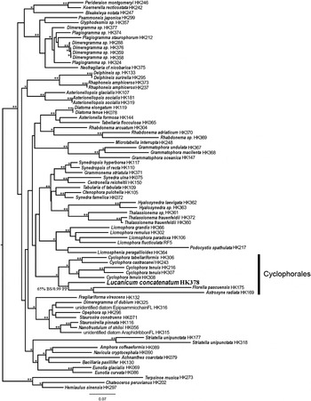
Phylogenetic analysis of Lucanicum was conducted using a three-gene (nuclear-encoded small subunit (SSU) rRNA, plastid-encoded rbcL and plastid-encoded psbC), 78-taxon dataset. The dataset was rooted with three non-pennate diatoms: Terpsinoë musica Ehrenberg; Chaetoceros peruvianus Brightwell; and Hemiaulus sinensis Greville. Sequence data were partitioned by gene and by codon position and run in RAxML ver. 7.4.2 (Stamatakis, Reference Stamatakis2006) for maximum likelihood (ML) analysis using the 6-substitution GTR + G model. To obtain a most likely tree, we ran 1,008 ML optimizations each starting from a parsimony tree. Clade support was obtained from 1,000 non-parametric bootstrap replicates using the rapid bootstrap algorithm in RAxML. The tree with the highest log likelihood score from 1,008 runs with bootstrap proportions is presented in Figure 4. All RAxML runs were completed on the LONESTAR supercomputer at the Texas Advanced Computing Center (TACC). Three Bayesian inference runs were also conducted with MRBAYES, using a 6-substitution model with partitioning by gene and by codon, for 30 million generations. All but the final million generations were discarded as ‘burn-in’ and the remaining trees were compared in Tracer ver. 1.5 (Rambaut & Drummond, Reference Rambaut and Drummond2007) for evidence of convergence. The final 10,000 trees from MRBAYES were used to generate a majority rule consensus tree and obtain posterior probabilities for nodes. Since the tree topology generated from MYBAYES was identical to the topology generated from the ML analysis, both the ML bootstrap values (bs) and the Bayesian posterior probabilities (pp) were plotted to nodes of the ML phylogram in Figure 4.
Terminology follows Anon (Reference Simonsen1975) and Ross et al. (Reference Ross, Cox, Karayeva, Mann, Paddock, Simonsen and Sims1979).
RESULTS
SYSTEMATICS
Order CYCLOPHORALES Round & Crawford, emend Lobban & Ashworth
Family CYCLOPHORACEAE Round & Crawford
Lucanicum Lobban & Ashworth, gen. nov.
(Figures 1–3)
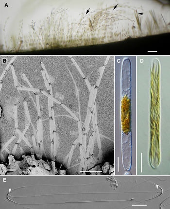
Lucanicum concatenatum sp. nov.: (A) numerous colonies (arrows), along with other diatoms (e.g. Toxarium hennedyanum (Gregory) Pelletan, arrowhead), attached to blue coral, as seen in dissection microscope with transmitted light (GU44AP); (B) whole mount of colonies in scanning electron microscopy showing the characteristic appearance of sausage links (GU44AO); (C) single live cell in chain from wild collection, showing plastids congregated around nucleus, DIC (GU44AO); (D) isolated cell from culture, plastids dispersed, their ribbon-like character clearly evident toward the apices; brightfield (GU44AI-3); (E) holotype, from culture, acid cleaned valve, showing rimoportulae (arrowheads) at each end of the sternum; DIC (GU44AI-3). Scale bars: A = ~250 µm; B = 100 µm; C, D = 25 µm; E = 10 µm.

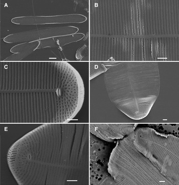
Lucanicum concatenatum sp. nov., scanning electron micrographs of culture (GU44AI-3), except (F) wild material: (A) acid cleaned valves, internal view, showing general shape and rimoportulae; (B) detail showing portion of parallel striae and sternum; (C) internal detail of apex showing rimoportula and radiating striae with some vimines; (D, E) external views of apices showing slit field; (F) whole mount showing copulae and mucilage pad (GU44AO). Scale bars: A = 100 µm; B–F = 1 µm.

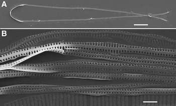
Lucanicum concatenatum sp. nov., scanning electron micrograph of girdle bands; culture (GU44AI-3): (A) entire band showing it is open at one end; (B) detail of bands and part of valve. Scale bars: A = 10 µm, B = 1 µm.

DIAGNOSIS
Cells linear with broadly rounded apices, joined apex to apex into nearly straight chains. Valves araphid with narrow sternum and apical slit field; finely striate, the striae comprising macroareolae without vimines, except in those radiating around the apex. Small rimoportula at each apex, with equal labia. Valvocopulae and copulae with two rows of slits.
ETYMOLOGY
From the Greek λουκάνικο (neuter noun), sausage, with reference to the shape of the cells and the colonies.
TYPE SPECIES
Lucanicum concatenatum sp. nov.
Lucanicum concatenatum Lobban & Ashworth, sp. nov.
TYPE MATERIAL
Holotype: GU44AI-3 (from culture) (Figure 1E), slide deposited at California Academy of Sciences, San Francisco, CA, accession number 627420, slide number 223037. GabGab reef, Apra Harbor, Guam, 13°26′33.63″N 144°38′34.25″E. GabGab reef, Apra Harbor, Guam, on blue coral in farmer fish territory at 10 m depth; collected by C. Lobban and M. Schefter 14 July 2011.
Syntype: GU44AO, SEM stub 270 (whole mount with wild sample) (Figure 1B), deposited at California Academy of Sciences, San Francisco, CA, accession number [pending]. As for holotype, collected by C. Lobban and M. Schefter 7 April 2012.
Additional material examined: GU44AL-3 and GU44AP.
DIAGNOSIS
With the characters of the genus. Cells 100–110 µm long (−140 in culture), 11 µm wide; plastids multiple, ribbon-like; striae 35–37 in 10 µm. Multiple copulae with two rows of areolae.
ETYMOLOGY
Latin: concatenatum meaning linked.
DESCRIPTION
Colonies (Figure 1A, B) were unbranched chains and comprised up to 15 cells (estimated from Figure 1A). Cells (Figures 1C–E, 2A) were sausage-shaped, approximately as deep as broad, with broadly rounded ends; multiple ribbon-like plastids were concentrated near the central nucleus or dispersed (presumably in response to irradiance) (Figure 1C, D). Frustules were not strongly silicified and presented few morphological characters. Valves had fine and uniformly parallel uniseriate striae closed by a velum (Figure 2B–E). Internally, interstriae were slightly thickened but there were no vimines and hence the areolae are single, macroareolae, except at the apices. The most distinctive feature of the valves was the lanceolate apical pore fields (Figure 2D, E), consisting of ~20 slits that started on the valve face and went down the mantle; in some images there appeared to be breaks across some slits (Figure 2E). There was a small rimoportula near each apex, on opposite sides of the sternum (Figures 1E, 2–C). Multiple, narrow, open, porate copulae were present (Figures 2F & 3A, B).
DNA PHYLOGENY
In three-gene phylogeny (Figure 4) this taxon is sister to Florella pascuensis Navarro and Astrosyne radiata Ashworth & Lobban, though not with a high level of support (65% bootstrap and 0.99 posterior probability). Although the placement of L. concatenatum within the clade is not certain, its inclusion within this clade is strongly supported (99% bootstrap and 1.00 posterior probability for the clade that includes L. concatenatum, Florella, Astrosyne and Cylophora). Lucanicum, Florella and Astrosyne all terminate long branches, but ML and Bayesian inference analyses are resistant to long-branch attraction problems, and it should be noted that other taxa on long branches (Podocystis spathulata (Shadbolt) Van Heurck and Striatella unipunctata (Lyngbye) Agardh) exist in this tree and are not found in the clade with L. concatenatum.
Tree constructed using aligned small subunit nuclear ribosomal RNA, chloroplast-encoded rbcL and psbC and rooted with non-pennate taxa Terpsinoë musica, Chaetoceros peruvianus and Hemiaulus sinensis. HK### refers to DNA and photograph vouchers for the specimens held at the Theriot Laboratory at UT Austin. Support for nodes illustrated by code: **, 100% maximum likelihood bootstrap (bs) and 1.00 Bayesian inference posterior probability (pp); *, ≥ 75% maximum likelihood bs and 0.95 Bayesian inference pp; exact values for nodal support shown at node bearing Lucanicum.

DISCUSSION
This species has been found several times, so far always on live blue coral (Heliopora coerulea Pallas, 1766) (notwithstanding the assertion by Lobban & Jordan (Reference Lobban, Jordan, Smol and Stoermer2010) that diatoms do not grow on live coral), in territories of Plectroglyphidodon lacrimatus (Quoy & Gaimard, 1825) at about 6–8 m depth, where it was associated with several other colonial diatoms including Stictocyclus stictodiscus (Mann) Round, Ardissonea fulgens var. gigantea (Lobarzewsky) De Toni and Toxarium J.W. Bailey spp. However, we do not suggest that this is its only habitat; the diatoms of the coral reefs are still too poorly sampled to make such an assertion, and the other species present on the coral are common on other substrata.
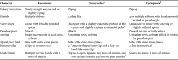
Lucanicum resembles Cyclophora and Neosynedra in chain-forming habit and general cell morphology. Both other genera have apical slit fields, a narrow sternum, and rounded to quadrate cribrate areolae (Round et al., Reference Round, Crawford and Mann1990; Ashworth et al., Reference Ashworth, Ruck, Lobban, Romanovicz and Theriot2012). Round et al. (Reference Round, Crawford and Mann1990) drew attention to the unusual character of the rimoportulae in Neosynedra in separating it from Cyclophora, and Cyclophora possesses a distinctive pseudoseptum, which is lacking in both Lucanicum and Neosynedra. Until recently Cyclophora was known only from C. tenuis, which has a pseudoseptum on only one valve and distinctively curved apical slit fields, but Ashworth et al. (Reference Ashworth, Ruck, Lobban, Romanovicz and Theriot2012) described several new species in which the apical slit field is straight, and thus similar to those in Neosynedra (especially N. tortosa) and Lucanicum. Moreover, the number of pseudosepta in the new species is variable both between species (C. castracanei has one on each valve) and within C. minor (which can have a pseudoseptum on one or both valves). However, a pseudoseptum is a defining character of Cyclophora (though not unique to it, also occurring in Astrosyne), so Lucanicum cannot be assigned to this genus.
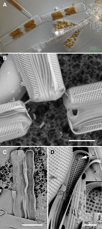
The differences between Lucanicum and Neosynedra are less distinct and a detailed comparison (Table 1) is necessary. Neosynedra Williams & Round (Reference Williams and Round1986) was separated from Synedra along with several other genera and ‘characterized by the compound, slit like apical pore-fields which resembles those of Cyclophora Castr., except that in Cyclophora the pore-field is divided into two parts by a plain central region’ (but we now know the latter is true only of C. tenuis). Round et al. (Reference Round, Crawford and Mann1990) also emphasized the rimoportula: ‘The genus is characterized by the slit-like pores of the apical pore field and the form of the rimoportula’, which has ‘a crescent-shaped inner lip and a flap- or knob-like outer lip’. For the sake of the comparison, and also to document the plastids, unknown when the foregoing descriptions were written, we show material of N. provincialis and N. tortosa from Guam samples in Figure 5 (see also Navarro & Lobban, Reference Navarro and Lobban2009, figures 46 & 47). The rimoportula in Figure 5D has the same characters as shown in Round et al. (Reference Round, Crawford and Mann1990, p. 375, figures i & j), in contrast to the nearly equal lips of the rimoportula in Lucanicum (Figure 2C). Neosynedra provincialis (Grunow) Williams & Round (Figure 5A) and N. tortosa (Grunow) Williams & Round have only four plate-like plastids, compared to multiple ribbon-like plastids in Lucanicum (also in Cyclophora). Striae are composed of individual areolae (as opposed to the undivided striae of L. concatenatum) (Figure 5B, C). The phylogenetic position of Neosynedra is unknown from DNA evidence, but it was left in Fragilariales: Fragilariaceae by Round et al. (Reference Round, Crawford and Mann1990), along with the other Synedra derivatives of Williams & Round (Reference Williams and Round1986). Although plastids are useful generic characters in some cases (e.g. some marine necklace-chain Fragilariaceae (Lobban et al., Reference Lobban, Ashworth, Arai, Jordan and Theriot2011)), difference in plastid type within genera is seen, for instance in Licmophora, where the norm is many small plastids but L. flabellata (Carmichael) Agardh has four large plates, and in Mastogloia and Achnanthes, in which most species have two ‘fore-and-aft’ plastids but one or two have many small plastids. Again with the difference in areolae, while the characters are often used to separate genera, continuous areola per striae can be seen within genera that normally have small areolae, e.g. Licmophora hyalina (Kützing) Grunow and L. comnavmaria Lobban & Schefter, which both have small areolae in the basal striae, and among species of Entomoneis (Round et al., Reference Round, Crawford and Mann1990) and Halamphora (Levkov, Reference Levkov and Lange-Bertalot2009). Thus each character individually is insufficient evidence to warrant a new genus. Likewise, the particular shape of the rimoportula is interesting but not a genus-defining character. DNA data are currently unavailable for Neosynedra, but we fully expect Neosynedra to be closely related to the Cyclophora/Lucanicum/Astrosyne clade based on the aforementioned morphological characters. Additionally, considering the great intraspecific genetic diversity seen in Cyclophora tenuis relative to other Cyclophora spp. (Figure 3), estimating genera based on genetic distance would be largely ambiguous. The choice is whether it is better to expand the generic concept of Neosynedra (which would presuppose that DNA data would show that the species now in this genus belong with Cyclophorales), or to erect a separate genus for concatenatum at the risk of having to sink it in future if expanded taxon sampling warrants. Although both species of Neosynedra occur in Guam (Lobban et al., Reference Lobban and Navarro2012), several attempts to isolate each species have been unsuccessful, and it is not a simple matter to supply the missing information. On the basis of the available evidence (Table 1) we propose the new genus Lucanicum, distinguishing it from Neosynedra on the basis of plastids and areolae.
Neosynedra spp.: (A) living chain of N. provincialis showing plate-like plastids (GU44AC-4); (B) whole mount of N. provincialis in scanning electron microscopy (SEM) showing apical pore fields and areolate striae (GU44AC-4); (C) internal view of acid-cleaned valve of N. tortosa (GU52N-7); (D) detail of internal apex of N. provincialis in SEM showing girdle bands and rimoportula (arrow) with inner crescent-shaped and outer knob-like lips; insert enlargement of rimoportula; (GU44P-B). Scale bars = A, C = 10 µm, B = 5 µm, D = 2 µm.

Comparison of characters of Lucanicum, Neosynedra and Cyclophora.

a, from Williams & Round (Reference Williams and Round1986) and Round et al. (Reference Round, Crawford and Mann1990) except colony and plastid characters from our own observations; b, from Ashworth et al. (Reference Ashworth, Ruck, Lobban, Romanovicz and Theriot2012).
Because Lucanicum is sister to a clade comprising Astrosyne and Florella, comparison with Florella is appropriate, even though the morphological similarity is obscure. Florella species have numerous septate girdle bands and were assigned by Navarro (Reference Navarro1996) to a new family within Striatellales; the genus bears more morphological resemblance to Striatellaceae (Striatella and Pseudostriatella) than to Cyclophora. The plastids are stellate. Florella has no apical pore field, although there is an unusual pattern of two longitudinal rows of elongate areolae in F. pascuensis Navarro. Although Navarro (Reference Navarro1996) emended the generic description to specify numerous scattered rimoportulae at the apices, he subsequently (Navarro, Reference Navarro2002) discovered F. pascuensis, which has only one rimoportula at each pole. Thus, both morphology and gene sequences show that Lucanicum cannot be placed in Florella, although they may be related.
It is becoming clear that the scope of the Cyclophorales must be broadened to include taxa without pseudosepta. When Round & Crawford (in Round et al., Reference Round, Crawford and Mann1990) erected Cyclophorales and Cyclophoraceae, they gave one description for both, based on the sole species Cyclophora tenuis. They thus included as a defining character, the presence of an annulus (pseudoseptum) on one valve. However, the discovery of new species of Cyclophora with pseudosepta present on one or both valves, and of the Astrosyne (Ashworth et al., Reference Ashworth, Ruck, Lobban, Romanovicz and Theriot2012) already indicated that these suprageneric taxa would need amending. It is now evident that the order must be amended to remove pseudosepta as a criterion, lest the other genera that are evidently closely related (particularly Lucanicum and Florella) are excluded by definition. Given the strong probability that other related genera will be identified in future, which might also result in additional families being identified, we presently restrict our amendment to deleting characters that are specific to Cyclophora or C. tenuis (i.e ‘Cellulae tabulares heterovalvares, una valva costa annulari centrali praedita’). The abbreviated description, after Round et al. (Reference Round, Crawford and Mann1990, p. 653), is thus:
Cyclophorales F.E. Round & R.M. Crawford, emend Lobban & Ashworth
Areae apicales porellorum elongatorum praesentes. Copulae multae simplices apertae. Plantae solum maris habitantes.
[Apical areas with elongate pores present. Multiple, simple, open copulae. Plants inhabiting the sea floor.]
Families included: Cyclophoraceae Round & Crawford, Florellaceae Navarro
This description would encompass Neosynedra should that genus prove to be related, as the ultrastructure of the linear pore fields suggests it might. Without reference to pseudosepta, the morphological definition of Cyclophorales is weak, and it will be desirable to establish additional characters as the order and family/families become better known; apical slits are present in other genera (Falcula and Asterionellopsis) and multiple open copulae are common. However, molecular data (Figure 4) suggest that the Cyclophorales, including Cyclophora, Astrosyne, Lucanicum and Florella, is monophyletic.
Until further taxon sampling is done, the families in Cyclophorales cannot be satisfactorily resolved. Round & Crawford's description of the Cyclophoraceae does not allow for the new species of Cyclophora that have pseudosepta on both valves, but we cannot revise it without knowing which other genera (if any) belong in it. Astrosyne, for example, has the distinctive pseudosepta but is circular and appears apart from Cyclophora in the phylogenetic tree. If Astrosyne is to be included in Cyclophoraceae, then Lucanicum and Florella must also be included. Florella appears most related to Astrosyne, but on long branches, and is presently in its own family, Florellaceae. Lucanicum is sister to the Florella/Astrosyne clade and the position of Neosynedra is not yet known. For the time being, therefore, we assign Lucanicum to Cyclophoraceae but make no amendment to the family description.
Supplementary material and methods
The supplementary material referred to in this article can be found online at journals.cambridge.org/mbdcup.