Case
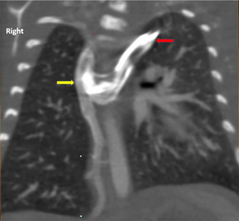
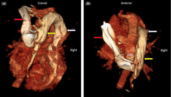
An infant with a concern for vascular ring underwent a CT that demonstrated a right aortic arch with aberrant left subclavian artery. There was an incidental finding of a retro-aortic left brachiocephalic vein coursing through the posterior mediastinum, draining into the azygous vein close to its connection with the right superior vena cava (Figs 1 and 2a–b).
Coronal CT image in venous phase. Contrast injection from the left hand delineating the LBV (red arrow) coursing rightward through the posterior mediastinum, draining into the azygous vein (yellow arrow).

(a and b) Volume-rendered 3D image of retro-aortic brachiocephalic vein in posteroanterior and superoinferior orientation. LBV (red arrow) courses posteriorly to the descending aorta to drain into the azygous vein (yellow arrow), which drains into the superior vena cava (white arrow).

Discussion
Retro-aortic LBV is seen in 0.5–1% of all CHD. Different LBV variants (including sub-aortic) have been described, but retro-aortic LBV with azygous continuation is a rare vascular variant. Embryologically, it is likely caused by the improper persistence of the inferior transverse venous plexus between the cardinal veins, which is preceded by an abnormal aortic arch formation that prevents the typical anastomosis between the anterior cardinal veins. Reference İkidağ1 Although there are no compressive effects, pre-procedural recognition is vital for surgeries involving the posterior mediastinum to avoid injury. Furthermore, identification is important prior to placement of central venous lines or transvenous pacemaker leads.
Acknowledgements
None.
Financial support
This research received no specific grant from any funding agency, commercial, or not-for-profit sectors.
Conflict of interest
None.
Ethical standards
The authors assert that all procedures contributing to this work comply with the ethical standards.