Introduction
Alzheimer's disease (AD) is the leading cause of late-onset dementia worldwide. Depressive symptoms are common in patients with AD, and they increase the caregiver burden (Akiyama et al., Reference Akiyama2008). Patient-rated depressive symptoms are an important independent predictor of subjective and objective quality of life (QOL) in mild AD (Naglie, Reference Naglie2011), while cognitive and functional statuses are not (Naglie, Reference Naglie2011).
Although the etiology and pathologic mechanism of depressive symptoms in AD patients remain unclear, there have been not a few studies on the relationship of objective depressive symptoms to regional cerebral blood flow (rCBF) or regional cerebral glucose metabolism in AD (Hirono et al., Reference Hirono1998; Liao et al., Reference Liao2003; Holthoff et al., Reference Holthoff2005; Lee et al., Reference Lee2006; Akiyama et al., Reference Akiyama2008; Levy-Cooperman et al., Reference Levy-Cooperman2008). Data from previous functional imaging studies have mainly supported the role of the dorsolateral prefrontal region in objective depressive symptoms in AD (Hirono et al., Reference Hirono1998; Holthoff et al., Reference Holthoff2005; Lee et al., Reference Lee2006; Akiyama et al., Reference Akiyama2008; Levy-Cooperman et al., Reference Levy-Cooperman2008). Associations with the anterior cingulate have been described inconsistently (Hirono et al., Reference Hirono1998; Liao et al., Reference Liao2003). However, there have been no studies focusing on the relationship between subjective depressive symptoms and rCBF in AD.
The correlation between objective and subjective depressive symptoms is significant but modest (r = 0.45), even in patients with normal cognitive function (Rane et al., Reference Rane, Fekadu, Wooderson, Poon, Markopoulou and Cleare2010). Significant differences were seen between self- and collateral-source reports of depression in mild cognitive impairment (MCI) and mild AD (Chopra et al., Reference Chopra, Sullivan, Feldman, Landes and Beck2008). Therefore, it is plausible that subjective and objective depressive symptoms may have somewhat different neural substrates in AD. In this study, we tried to identify the cerebral blood flow correlates of subjective depressive symptoms in AD. We performed this study on the hypothesis that there was a significant relationship between subjective depressive symptoms and rCBF in the dorsolateral prefrontal regions among AD patients.
Methods
Patients
Seventy-six consecutive patients with AD were recruited from the outpatient units of the Memory Clinic of Okayama University Hospital between September 2008 and April 2012 according to the following criteria. They all (i) underwent general physical and neurological examinations and extensive laboratory testing, including thyroid function tests, serum vitamin B12, and syphilis serology; (ii) took the revised Addenbrooke's Cognitive Examination (ACE-R; Yoshida et al., Reference Yoshida2012), the Mini-Mental State Examination (MMSE; Folstein et al., Reference Folstein, Folstein and McHugh1975), the Frontal Assessment Battery (FAB; Kugo et al., Reference Kugo2007), and the Geriatric Depression Scale (GDS; Yesavage and Blink, Reference Yesavage and Blink1983; Muraoka et al., Reference Muraoka, Ikuchi and Ihara1996); (iii) underwent single photon emission computed tomography (SPECT) with 99mTc-ethylcysteinate dimer of the brain as well as magnetic resonance imaging (MRI) of the head; (iv) were diagnosed with probable AD according to the criteria formulated by the National Institute of Neurological and Communicative Disorders and Stroke and the Alzheimer's Disease and Related Disorders Association (NINCDS-ADRDA) (McKhann et al., Reference McKhann, Drachman, Folstein, Katzman, Price and Stadlan1984); (v) had a dementia severity of 0.5 or 1 on the CDR (Hughes et al., Reference Hughes, Berg, Danziger, Coben and Martin1982); and (vi) were judged to have the ability to answer the GDS questions reliably by the chief clinician. The exclusion criteria were (i) complications from other neurological diseases or illnesses; (ii) history of mental illness or substance abuse prior to the onset of dementia; (iii) evidence of focal brain lesions on head MRI; (iv) treatment with cholinesterase inhibitors, memantine, antipsychotics, antidepressants, or anxiolytic drugs; and (v) left handedness or ambidexterity. The history of mental illness or substance abuse was ascertained by a family caregiver.
The profile of each patient (age, sex, months of disease duration, and years of education) was obtained. Scores of the Neuropsychiatric Inventory (NPI), Barthel Index of Activities of Daily Living (ADL), and Functional Assessment Questionnaire (FAQ) were calculated by a trained clinical psychologist, based on the information from family caregivers. The Clinical Dementia Rating (CDR; Hughes et al., Reference Hughes, Berg, Danziger, Coben and Martin1982) score was calculated by the chief clinician.
Instruments
The GDS, which has been extensively used in the fields of psychiatry and public health, was used to assess depression in the elderly (Yesavage and Blink, Reference Yesavage and Blink1983). The GDS has two different versions: a standard version with 30 items and a shortened version with 15 items. For the present study, the short 15-item version of the GDS was administered (Muraoka et al., Reference Muraoka, Ikuchi and Ihara1996). A higher score means more subjective depressive signs. The cut-off 5/6 is usually used (Muraoka et al., Reference Muraoka, Ikuchi and Ihara1996).
NPI is a valid and reliable instrument for measuring non-cognitive symptoms in dementia (Hirono et al., Reference Hirono1997). It is a caregiver-based tool that assesses ten different domains in dementia. NPI gives a composite score for each domain, which is the product of frequency by severity subscores: scores from 1 to 4 (with 4 being the most severe) for the frequency and from 1 to 3 (with 3 being the most severe) for the severity of each behavior (Akiyama et al., Reference Akiyama2008). The maximum attainable score is 12.
ACE-R was developed to provide a brief test sensitive to early stage dementia, and is capable of differentiating between dementia subtypes including AD, frontotemporal dementia, progressive supranuclear palsy, and other parkinsonian syndromes (Yoshida et al., Reference Yoshida2012). ACE-R includes MMSE, but extends it to encompass important areas not covered by MMSE, such as frontal-executive function and visuospatial skills. For this study, we used the Japanese version of ACE-R described by Yoshida et al. (Yoshida et al., Reference Yoshida2012).
The Barthel Index of ADL consists of ten items that measure a person's daily functioning, specifically the activities of daily living and mobility (Wade and Collin, Reference Wade and Collin1988). The ADL score ranges from 0 to 100. A higher score indicates a better performance. The FAQ measures functional activities of older adults using the patient's partner as an informant (Pfeffer et al., Reference Pfeffer, Kurosaki, Harrah, Chance and Filos1982). The FAQ consists of ten items and the score on each item ranges from 0 to 3. A higher score indicates more severe impairment.
Ethics
This study was approved by the Internal Ethical Committee of Okayama University Graduate School of Medicine, Dentistry and Pharmaceutical Sciences. After a complete description of the study to the patients and their relatives, written informed consent was obtained.
Brain perfusion SPECT imaging
All patients were examined by brain perfusion SPECT. Patients were examined in a comfortable supine position with their eyes closed in quiet surroundings. Ten minutes after intravenous administration of 99mTc-ethylcysteinate dimer (ECD, 600 MBq, Daiichi Radioisotope Laboratories Ltd., Tokyo, Japan), SPECT images were obtained using a triple-head, rotating gamma camera interfaced to a minicomputer (GCA9300A/DI; Toshiba, Tokyo, Japan), equipped with a fanbeam, low-energy, high-resolution collimator. Sixty projection images over a 360° angle in a 128 × 128 matrix were acquired. All images were reconstructed using ramp-filtered back-projection and then three-dimensionally smoothed with a Butterworth filter (order 8, cut-off 0.12 cycles/cm). The reconstructed images were corrected for gamma ray attenuation using the Chang method (μ = 0.09).
Data analysis
Spatial reprocessing and statistical analysis of images were performed on a voxel-by-voxel basis using Statistical Parametric Mapping 8 (SPM8, Wellcome Department of Imaging Neuroscience, University College, London, UK) running on MATLAB (The Mathworks, Inc., Natick, MA, USA). All SPECT images of each patient were normalized to the standard brain of the Montreal Neurological Institute (MNI), and spatial normalization was performed with 12-parameter affine and non-linear transformations (Friston et al., Reference Friston, Ashburner, Poline, Frith, Heather and Frackowiak1995). The voxel sizes of the reslice option were 2 mm × 2 mm × 2 mm. The non-linear parameters were set at 25-mm cut-off basis functions and 16 iterations. All the normalized SPECT images were then smoothed with an isotropic Gaussian kernel filter (12-mm full width at half maximum).
To examine the images for specific regions showing differences in perfusion, two sample t-tests were performed. Global normalization was performed by proportional scaling with the mean voxel value. Masking was applied using the threshold method (0.8 times the global value). The analysis used a threshold of p < 0.001 (uncorrected) at the voxel level and results were considered significant at 100 voxels at the cluster level.
Statistical analysis
Statistical analysis was performed using the SPSS 14.0J software program (SPSS Inc., Chicago, IL, USA). Comparisons between two groups were performed by independent sample t-tests. The correlation of NPI-depression and GDS scores to other clinical characteristics was analyzed using Pearson's correlation coefficient. A value of p < 0.05 was accepted as significant.
Statistical analysis of images was performed on a voxel-by-voxel basis using SPM8. The detailed method was described in the “Data analysis” section.
Results
Demographic characteristics
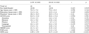
Demographic characteristics are shown in Table 1. Among the 76 AD patients, 44 were women and 32 were men. For dementia severity, 46 patients had CDR scores of 0.5 and 30 had CDR of 1. For GDS scores, 4 patients scored 0, 42 scored from 1 to 5, 27 patients scored from 6 to 10, and 3 patients scored 11 or over. On the NPI-depression scale, 50 patients had a score of 0, 6 patients scored 1, 10 patients scored 2, 9 patients scored 3, and only 1 patient showed a score of 6.
Clinical characteristics and neuropsychological tests
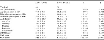
SD = standard deviation; ACE-R = revised Addenbrooke's Cognitive Examination; MMSE = Mini-Mental State Examination; FAB = Frontal Assessment Battery; GDS = Geriatric Depression Scale.aDuration – disease duration.
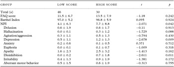
Comparison between high and low GDS scale groups showed that there were no significant differences in sex ratio, age, education, disease duration, cognition test scores, and ADL scores (Table 2). The patients with high GDS scores got high scores on the depression and disinhibition subscales of NPI (Table 2).
Instrumental ADL and BPSD
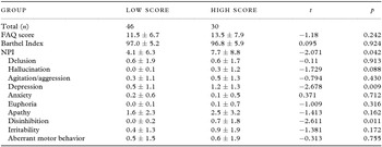
ADL = activities of daily living; BPSD = behavioral and psychological symptoms of dementia; FAQ = Functional Assessment Questionnaire; NPI = Neuropsychiatric Inventory.
rCBF
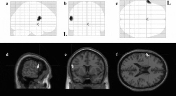
A group comparison between the SPM results of the high and low GDS score groups was performed (high GDS score group: GDS score ≥6; low GDS score group: GDS score ≤5). Specific voxels with significantly lower perfusion in the high GDS score group than in the low GDS score group are shown in Figure 1. No voxels with a significantly lower perfusion in the low GDS score group than in the high GDS score group were found.
(a)–(f) SPM (z) map of rCBF decrease in Alzheimer's disease (AD) patients with high GDS scores compared with AD patients with low scores. (a)–(c) Three-way glass view of the area of significant hypoperfusion. (d)–(f) Superimposed on head MRI T1 weighted images: (d) x = –62; (e) y = 8; and (f) z = 20; L – left.
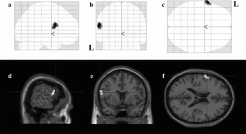
Figure 1 shows the z score for each voxel in this cluster, superimposed on a three-way glass brain view. The probability results of the SPM analysis indicated that there was a decreased uptake in the high GDS score group compared to the low GDS score group, in the left inferior frontal gyrus (number of voxels = 149; peak z score = 4.15; p < 0.001) (coordinates of MNI: x = –62, y = 8, z = 20).
Discussion
We found, patients with high scores showed significant hypoperfusion in the left inferior frontal region, compared to patients with low scores on GDS. There have been several published studies on rCBF or regional cerebral glucose metabolism in mild AD patients with objective depressive symptoms (Hirono et al., Reference Hirono1998; Holthoff et al., Reference Holthoff2005; Lee et al., Reference Lee2006; Akiyama et al., Reference Akiyama2008; Levy-Cooperman et al., Reference Levy-Cooperman2008). All studies showed that dorsolateral frontal regions are significantly related to objective depressive symptoms in AD. Most studies used the NPI-depression scale. In this study, we found hypoperfusion in the left inferior frontal region. The inconsistent results may reflect the fact that GDS scores and NPI-depression scores, in part, assess different dimensions of depression. The GDS captures somatic and behavioral dimensions as subjective scores, whereas NPI captures only the affective dimension as objective scores. Different depression subtypes show unique brain perfusion patterns (Fountoulakis et al., Reference Fountoulakis2004).
Of the published studies, only three provided detailed lesion localization with figures (Holthoff et al., Reference Holthoff2005; Lee et al., Reference Lee2006; Levy-Cooperman et al., Reference Levy-Cooperman2008). Lee et al. (Reference Lee2006) reported a significant decrease of rCBF in the right superior frontal region, and Levy-Cooperman et al. (Reference Levy-Cooperman2008) showed a decrease of rCBF in the right superior and middle prefrontal regions. In both studies, apathy and anxiety coexisting with depression were not considered. Meanwhile, Holthoff et al. (Reference Holthoff2005) reported the hypoperfusion of the area from the left superior to the inferior frontal gyri in the depressive AD patients, after discounting the effect of coexisting apathy. Hirono et al. (Reference Hirono1998) reported a significant decrease of rCBF in the bilateral superior frontal region without detailed figures. In the study of Akiyama et al. (Reference Akiyama2008), the two groups (depressive AD patients and not depressive AD patients) showed similar scores on apathy and anxiety scales, and a significant decrease of rCBF in the left superior frontal region was reported without detailed figures. In this study, we found hypoperfusion in the left inferior frontal region and there were no differences in NPI scores of apathy and anxiety between the two groups (high and low GDS score groups). The areas where rCBF was negatively correlated with apathy scores included the right temporal and right medial frontal gyri in AD (Kang et al., Reference Kang2012). In the studies of Lee et al. (Reference Lee2006) and Levy-Cooperman et al. (Reference Levy-Cooperman2008), coexisting apathy may cause the right-dominant laterality of hypoperfusion.
Among patients with post-stroke depression, inferior frontal lesions, irrespective of side, appeared to play a role as a risk factor (Singh et al., Reference Singh2000). In the study of Singh et al. (Reference Singh2000), the depressive symptoms were assessed by using both subjective and objective depression scales. Selective areas of hypometabolism in the inferior frontal lobe in depressed patients with Parkinson's disease were reported, though the depression in the study was evaluated by an objective depression scale (Mayberg et al., Reference Mayberg1990). Recently, the inferior frontal gyrus is thought to be important in emotional empathy (Shamay-Tsoory et al., Reference Shamay-Tsoory, Aharon-Peretz and Perry2009) and coping with distracting emotions (Dolcos et al., Reference Dolcos, Kragel, Wang and McCarthy2006). Moreover, inferior frontal regions have been observed to be crucial in emotional regulation (Ochsner et al., Reference Ochsner2004; Grimm et al., Reference Grimm2006). Therefore, it is plausible that hypoperfusion in the inferior frontal gyrus is related to subjective depressive symptoms. We suppose that the inferior frontal area is significantly related to subjective depressive symptoms in AD and that subjective depressive symptoms in AD are due to a specific pathogenesis rather than a reactive phenomenon (Liao et al., Reference Liao2003).
A few studies have reported a relationship between subjective depressive symptoms and rCBF or metabolism in patients with major depression without dementia. Dunn et al. (Reference Dunn2002) reported that psychomotor and anhedonia symptoms correlated with lower metabolism in the right insula, claustrum, anteroventral caudate/putamen, and temporal cortex in both unipolars and bipolars. In unipolars, the negative cognitions correlated with lower metabolism bilaterally in the frontal poles, right dorsolateral frontal cortex, and supracallosal cingulate (Dunn et al., Reference Dunn2002). Bench et al. (Reference Bench, Friston, Brown, Frackowiak and Dolan1993) reported that anxiety correlated positively with rCBF in the posterior cingulate cortex and inferior parietal lobule bilaterally. Psychomotor retardation and depressed mood correlated negatively with rCBF in the left dorsolateral prefrontal cortex and left angular gyrus (Bench et al., Reference Bench, Friston, Brown, Frackowiak and Dolan1993). Suh et al. (Reference Suh2009) observed a negative correlation between the Beck Depression Inventory (BDI) scores and relative rCBF in the bilateral ventrolateral prefrontal cortex and middle frontal gyri. The results are inconsistent and the interpretation of mutual differences is difficult. However, the presence or absence of dementing disorders may affect the neural substrates of subjective depressive symptoms.
Limitations
The results in this study should be interpreted with some caution. First, this study included outpatients at a single memory clinic. Therefore, selection bias cannot be excluded. Second, we used the short version of GDS in this study. GDS is a simple screening questionnaire for depression and the answer to each item is alternative (yes or no). Therefore, the detailed symptoms of depression could not be evaluated. Third, in this study patients with mild AD were scored using the GDS scale. The reliability of answers might be somewhat questionable. However, we selected only those patients, who the chief clinician judged, had the ability to answer the GDS reliably.
Conflict of interest
None.
Description of authors’ roles
Hajime Honda was involved in designation of the study and data analysis. He wrote the paper with Seishi Terada. Seishi Terada designed the study and analyzed the data with Satoshi Hayashi. Shuhei Sato was involved in design of the study and data analysis. Etsuko Oshima, Chikako Ikeda, Shigeto Nagao, and Osamu Yokota collected data. Yosuke Uchitomi supervised the study design, participated in data analysis, and assisted with writing the paper.
Acknowledgments
We sincerely thank Ms. Horiuchi, Ms. Imai, and Ms. Yabe for their skillful assistance. This work was supported by grants from the Japanese Ministry of Education, Culture, Sports, Science and Technology (21591517) and the Zikei Institute of Psychiatry.