INTRODUCTION
Recently, the morphology of spermatozoa and spermatophores have been studied extensively in the Decapoda (Latreille, 1803) and provide useful information on phylogenetic relationships and evolutionary divergence (see Bauer Reference Bauer1986, Reference Bauer, Bauer and Martin1991; Jamieson, Reference Jamieson1991; Jamieson & Tudge, Reference Jamieson, Tudge and Jamieson2000; Kronenberger et al. Reference Kronenberger, Brandis, Turkay and Storch2004; Tudge, Reference Tudge, Martin, Crandall and Felder2009). In particular, spermatophores and spermatozoa show a species-specific morphology, that has been successfully used in recent phylogenetic studies (Tudge Reference Tudge1997; Tirelli et al., Reference Tirelli, Pessani, Silvestro and Tudge2008, Reference Tirelli, Pessani, Silvestro and Tudge2010).
The male reproductive system of Decapoda consists of a pair of testes from which a pair of vasa deferentia (divisible into regions with different functions) emanate; ending via genital openings located on the coxa of the fifth pereopods. These openings may be simple gonopores, elevated papillae, or complex copulatory structures (McLaughlin, Reference McLaughlin and Mantel1983; Tudge & Lemaitre, Reference Tudge and Lemaitre2004, Reference Tudge and Lemaitre2006). The testes and the vasa deferentia are located dorsally or laterally to the gut and may be surrounded by the hepatopancreas (McLaughlin, Reference McLaughlin and Mantel1983). In the majority of the Decapoda they are located in the cephalothorax, while Paguroidea have the reproductive apparatus in the pleon (McLaughlin, Reference McLaughlin and Mantel1983). The testes produce spermatozoa, enveloped by spermatophores in which they wait to be transferred to females (Subramoniam, Reference Subramoniam1995; Hess & Bauer, Reference Hess and Bauer2002).
In Pagurus species described up to now, the male reproductive system shows the same patterns described for Decapoda and Paguroidea (McLaughlin, Reference McLaughlin1980, Reference McLaughlin and Mantel1983; Krol et al., Reference Krol, Hawkins, Overstreet and Harrison1992; Fantucci & Mantelatto, Reference Fantucci and Mantelatto2011), but they present a great difference in the gross morphology of the vasa deferentia. These last are relatively shorter and straighter (Scelzo et al., Reference Scelzo, Fantucci and Mantelatto2010; Fantucci & Mantelatto, Reference Fantucci and Mantelatto2011) than in described Diogenidae, in which ducts are proportionally longer and mostly helicoidal (Scelzo et al., Reference Scelzo, Mantelatto and Tudge2004; Tirelli et al., Reference Tirelli, Campantico, Pessani, Tudge and Asakura2006, Reference Tirelli, Campantico, Pessani and Tudge2007, Reference Tirelli, Pessani, Silvestro and Tudge2008; Amadio & Mantelatto, Reference Amadio and Mantelatto2009; Mantelatto et al., 2009b). This difference in length may be a consequence of the lack in Paguridae of the two regions responsible for lengthening of spermatophore stalk (Matthews, Reference Matthews1957; Greenwood, Reference Greenwood1972). The genital openings of the Pagurus species (Fantucci & Mantelatto, Reference Fantucci and Mantelatto2011) are simple gonopores, differing from hermit crabs species without sexual tubes in the large number of setae surrounding the gonopore itself. These setae have been supposed to have a sensory function during mating, to provide better adhesion to the female gonopore, and to act as sensors during the transfer of spermatophores to the partner (Manjón-Cabeza & García Raso, Reference Manjón-Cabeza and García Raso2000; Tudge & Lemaitre, Reference Tudge and Lemaitre2004). The spermatophores of decapods show a great morphological diversity, with different characteristics in different groups (Subramoniam, Reference Subramoniam, Blaxter and Southward1993). On the whole they show three different principal morphologies: spherical and small spermatophores, present in Brachyura; tubular in most Macrura (now Astacidea and Achelata); and pedunculate (or stalked) in Anomura (see Krol et al., Reference Krol, Hawkins, Overstreet and Harrison1992; Tudge, Reference Tudge, Martin, Crandall and Felder2009 for reviews). In Anomura in general, there is a lack of information regarding ampullar ultrastructure and in particular ampullar suture line morphology. Publications illustrated the lateral ridge as the point of dehiscence of the two ampulla halves in a variety of hermit crabs and other anomurans, including Paguridae (Tudge, Reference Tudge1999a). In particular, regarding the Paguridae, the ampulla wall and lateral ridge ultrastructure had been previously described only for Pagurus bernhardus (Linneus, 1758), Pagurus hirtimanus (White, 1847) and Pagurus prideaux (Leach, 1815) by Tudge (Reference Tudge1999a, Reference Tudgeb). More recently it had been described in Pagurus exilis (Scelzo et al., Reference Scelzo, Fantucci and Mantelatto2010) and in Pagurus criniticornis and Pagurus brevidactylus (Fantucci & Mantelatto, Reference Fantucci and Mantelatto2011). Also the spermatozoa show a great structural and morphological diversity (Subramoniam, Reference Subramoniam and Subramoniam1982; Felgenhauer & Abele, Reference Felgenhauer, Abele, Bauer and Martin1991; Tudge, Reference Tudge, Martin, Crandall and Felder2009). The spermatozoa are constructed of a large acrosomal vesicle with multiple concentric layers, a posterior, amorphous nucleus, an intermediate cytoplasm and three to many lateral arms. In Anomura, the acrosome is oblong (Jamieson, Reference Jamieson1991) or spherical (Tudge, Reference Tudge1992; Tudge & Scheltinga, Reference Tudge and Scheltinga2002) in shape and has a conspicuous apical operculum. In the area between the cytoplasm and nucleus three or more microtubular arms have their origin (Jamieson, Reference Jamieson1991). The association of the sperm morphology and ultrastructure with that of other parts of the reproductive system (e.g. testes, vasa deferentia and spermatophores) is important to better understand the reproductive biology of this taxon. Moreover, the great diversity found in recent studies of the morphology of spermatozoa and spermatophores, showing characteristics particular to members of different groups, allow us to conclude that these structures are taxonomically specific (see Tudge, Reference Tudge1991; Subramoniam, Reference Subramoniam, Blaxter and Southward1993; Santos & Mantelatto, Reference Santos and Mantelatto2011). Thus, together with other taxonomic criteria, they can be used to elucidate taxonomic and phylogenetic problems (Tirelli et al., Reference Tirelli, Pessani, Silvestro and Tudge2008, Reference Tirelli, Pessani, Silvestro and Tudge2010; Mantelatto et al., Reference Mantelatto, Scelzo and Tudge2009; Tudge, Reference Tudge, Martin, Crandall and Felder2009).
The hermit crab family Paguridae (Latreille, 1802) comprises 76 genera and approximately 542 species (De Grave et al., Reference De Grave, Pentcheff, Ahyong, Chan, Crandall, Dworschak, Felder, Feldmann, Fransen, Goulding, Lemaitre, Low, Martin, Ng, Schweitzer, Tan, Tshudy and Wetzer2009; McLaughlin et al., Reference McLaughlin, Komai, Lemaitre and Rahayu2010) and only for the genera Anapagurus Henderson, 1886, Cestopagurus Bouvier, 1897, Nematopagurus A. Milne-Edwards and Bouvier, 1892, Pagurus Fabricius, 1775, Solitariopagurus Tuerkay, 1986 and Xylopagurus A. Milne-Edwards, 1880 is the spermatophore and/or sperm morphology known (Jamieson & Tudge, Reference Jamieson, Tudge and Jamieson2000; Tudge, Reference Tudge1995a, Reference Tudge, Jamieson, Ausio and Justineb, Reference Tudge1999b; Scelzo et al., Reference Scelzo, Fantucci and Mantelatto2010; Fantucci & Mantelatto, Reference Fantucci and Mantelatto2011).
The genus Pagurus is a heterogeneous pagurid group consisting of more than 170 species worldwide (Lemaitre & Cruz Castano, Reference Lemaitre and Cruz Castaño2004). It is one of the most species-rich genera in Anomura (De Grave et al., Reference De Grave, Pentcheff, Ahyong, Chan, Crandall, Dworschak, Felder, Feldmann, Fransen, Goulding, Lemaitre, Low, Martin, Ng, Schweitzer, Tan, Tshudy and Wetzer2009) and is distributed in shallow waters such as littoral, subtidal and intertidal areas.
Pagurus excavatus (Herbst, 1791) is a common Mediterranean pagurid with a distribution in the Atlantic ranging from the Bay of Biscay, Spain and Portugal to Morocco, while in the Mediterranean Sea it ranges from Spain, the Balearic Islands, France and Italy to Cyprus (Ingle, Reference Ingle1993). It mainly lives at depths between 15 and 80 m (Ingle, Reference Ingle1993), but specimens have been sampled down to 450 m (Chintiroglou et al., Reference Chintiroglou, Doumenc and Koutsoubas1992).
Its symbiotic relationships with sea anemones (Pax & Muller, Reference Pax and Müller1962; Mainardi & Rossi, Reference Mainardi and Rossi1969; Ross, Reference Ross1979; Doumenc et al., Reference Doumenc, Chintiroglou and Koukouras1985; Chintiroglou et al., Reference Chintiroglou, Doumenc and Koutsoubas1992), reproductive behaviour under laboratory conditions (Bastock, Reference Bastock1967; Hazlett, Reference Hazlett1968), female sexual maturation and reproductive cycle (Mura & Cau, Reference Mura and Cau2002; Mura et al., Reference Mura, Orrù and Cau2006) are well known, but no exhaustive description of spermatophore and spermatozoa is available for this species, except a concise description of the external morphology of the spermatophore under light microscopy (Tudge, Reference Tudge1999b).
Therefore, the aim of this paper is to expand the knowledge about the reproductive morphology of P. excavatus by studying the spermatophore and sperm cell ultrastructure, and comparing them with similar structures in other members of the family Paguridae.
MATERIALS AND METHODS
Specimen
One sexually mature specimen (indicated by adult shield length and gonopores present) of Pagurus excavatus was collected by trawling during the Cruise ARSA 0395, organized by the Instituto Español de Oceanographia in 1994, in the Gulf of Cadiz, off south-west Spain, Mediterranean Sea. The sample was generously gifted by Professor Antonio Medina to one of the authors (C.T.). After collection, fragments of immediately dissected testes and reproductive tract were preserved in glutaraldehyde (2.5% in seawater) for 24 hours. Then the samples were prepared for transmission electron microscopy (TEM) observations according to standard protocols described in Tirelli et al. (Reference Tirelli, Campantico, Pessani, Tudge and Asakura2006, Reference Tirelli, Campantico, Pessani and Tudge2007).
Measurement of the different layers making up the spermatophore wall and the different regions of the spermatozoa were made from micrographs.
RESULTS
Spermatophore
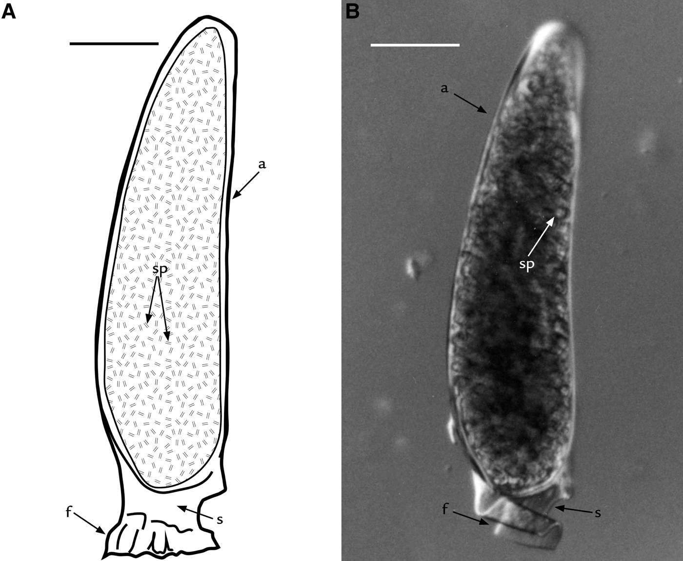
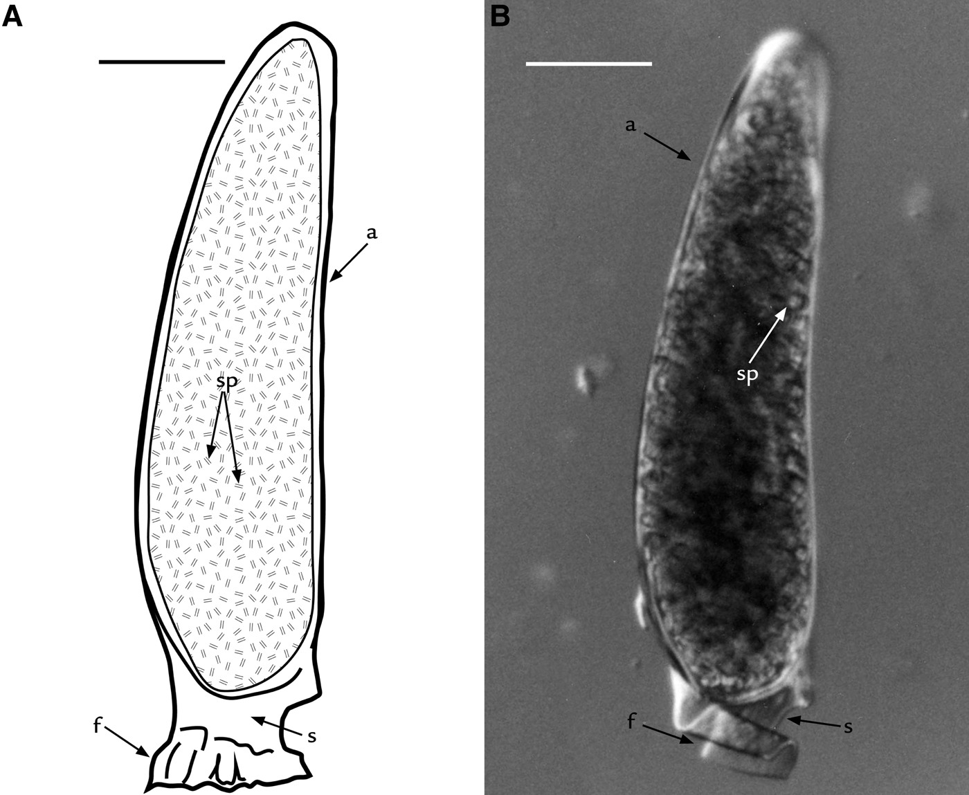
The tripartite spermatophore is composed of an ampulla raised on a short stalk with a pedestal at its base (Figure 1). The ampulla is divided into two halves (valves), separated by a line of dehiscence, the lateral ridge. See Tudge (Reference Tudge1991a, Reference Tudge, Jamieson, Ausio and Justine1995a, b) for a full description.
Pagurus excavatus (Herbst, 1791), spermatophore morphology: (A) drawing and (B) light microscopy of spermatophore showing the ampulla (a) filled with spermatozoa (sp), the stalk (s), and the foot (f). Scale bars: A,B, 50 μm.
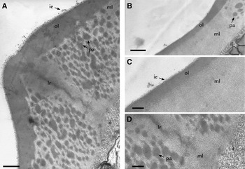
The whole ampulla wall is made up of three layers (Figure 2A–D). The first, the inner layer (il), is finely fibrillar and electron-dense. The outer layer, is made up of two strata (Figure 2C): both being finely granular, and homogeneous but with the external stratum being slightly more electron-dense and with an irregular exterior edge (ie) compared to the inner stratum beneath (ol). Between the inner and outer layers of the ampullar wall there is a homogeneous, finely granular middle layer (ml), showing some spherical electron-dense patches near the lateral ridge (see below). The inner and the outer layers of the spermatophore wall show a similar thickness, but the middle layer is between six and ten times as thick as the others.
Pagurus excavatus (Herbst, 1791), transmission electron microscopy wall ultrastructure at lateral ridge (lr) showing the outer electron-dense layer (ol) with irregular exterior edge (ie), the middle homogeneously granular layer (ml) with circular and/or elliptic patches (pa) and the inner fibrillar layer (il) with fibres with different orientations; (B) spermatophore wall ultrastructure far from the ridge showing the outer granular layer, the middle granular layer with electron-dense patches, and the inner fibrillar layer; (C) detail of the middle layer and of the outer granular layer; (D) detail at lateral ridge of the inner fibrillar layer and of the middle granular layer showing a large amount of electron-dense patches. Scale bars: A, 0.8 μm; B, 0.5 μm; C, 0.2 μm; D, 0.3 μm.

The lateral ridge is seen as a thickening of the ampullar wall where the two valves meet but they are separated by an irregular gap or discontinuity. At the TEM level (Figure 2A), in longitudinal section, the lateral ridge is conical in shape.
At the lateral ridge, the various layers thicken up to twice their depth in the ampullar wall (Figure 2A) and slightly modify their appearance. The outer layer appears to be more electron-dense, clearly distinguishing it from the middle layer and the exterior edge becomes more irregular. The middle granular homogeneous layer shows a large number of the electron-dense, circular and/or elliptic patches layer (Figure 2A). The inner fibrillar layer shows fibres with a different orientation (Figure 2A), not only parallel to the direction of cutting as seen in Figure 2D.
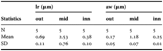
Table 1 shows the mean value and the standard deviation in the thickness of the different layers of the ampullar wall, including at the lateral ridge.
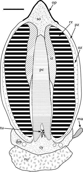
Pagurus excavatus (Herbst, 1791), drawing of longitudinal section of spermatozoa showing the acrosomal vesicle capped by the operulum (op), the subopercular zone (so), the perforatorial chamber (pc) surrounded by the inner acrosome zone (iz), the outer acrosome zone (oz), the reticulate zone (rz) and the peripheral acrosome zone (pz), the tubules inside the perforatorial chamber (tu), the cytoplasm (cy) with degenerated mithocondria and the bases of the microtubular arms (ma), and the nucleus (nu). Scale bar: 0.75 μm.

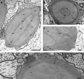
Pagurus excavatus (Herbst, 1791), transmission electron microscopy spermatozoa: (A) longitudinal section of spermatozoa showing the acrosomal vesicle capped by the operculum (op), the subopercular zone (so), the perforatorial chamber (pc) with smooth basal margin (sm) and indented middle and apical margin (im) surrounded by the inner acrosome zone (iz), the outer acrosome zone (oz), the reticulated zone (rz) and the peripheral acrosome zone (pz), the tubules inside the perforatoiral chamber (tu), the cytoplasm (cy), and the nucleus (nu); (B) transversal section of the spermatozoon showing the inner, outer, ray and peripheral zones, the perforatorial chamber, the cytoplasm with degenerating mitochondria (dm); (C) detail, in longitudinal section, of basal portion of the spermatozoa showing the nucleus, the connection between the cytoplasm and the perforatorial chamber with dense tubules, the outer and peripheral zones; (D) detail, in transversal section, of the spermatozoa showing the outer, ray and peripheral zones, and a microtubular arm (ma) in longitudinal section. Scale bars: A, 1.0 μm; B, 0.5 μm; C,D, 0.25 μm.

Thickness of ampulla wall and lateral ridge of Pagurus excavatus.
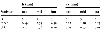
aw, ampulla wall; out, outer layer; mid, middle layer; inn, inner layer; lr, lateral ridge; N, number of lateral ridges or ampullae measured; SD standard deviation.
Spermatozoa
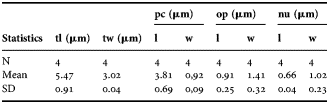
Spermatozoa (Figures 3 & 4A) are composed of an ovoid acrosomal vesicle capped by a conical, highly electron-dense operculum. The subopercular zone occupies the bulk of the space beneath, and within, the operculum, and is quite well developed and homogenously granular (Figures 3 & 4A). At the base of the acrosomal vesicle, there is the thin cytoplasm, the large nucleus and three arms positioned to form a 120° angle between each other. Pagurus excavatus total spermatozoal length is 5.5 µm, and total sperm width is 3.02 µm, (more detailed sperm cell dimensions are shown in Table 2).
Measurements of the various portions of the sperm cell of Pagurus excavatus.
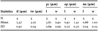
l, length; N, number of spermatozoa measured; nu, nucleus; op, operculum; pc, perforatorial chamber; SD, standard deviation; tl, total length; tw, total width; w, width.
The acrosome vesicle is posteriorly penetrated by the perforatorial chamber, which communicates with the adjacent cytoplasm directly (Figures 3, 4A, C) and extends anteriorly up to the subopercular zone. The perforatorial chamber has an overall cylindrical shape in longitudinal section, and exhibits a smooth margin in the basal portion and a minutely crenulated margin in the middle and distal portions (Figures 3 & 4A). The perforatorial chamber has fine longitudinal striations present in its entire length and often a series of obvious electron lucent tubules or vesicles (also orientated longitudinally) in the basal portion (Figures 3 & 4A, C).
A slightly granular layer, more electron-dense than the subopercular zone and the perforatorial chamber, thinly surrounds the whole chamber. It is thinnest in the basal portion and doubles in thickness more anteriorly. This layer constitutes the inner acrosomal zone (Figure 4A, B). Immediately exterior to this is a reticulated acrosome zone (Figure 4A, B, D), showing an elongate kidney shape in longitudinal sections, but obviously a thick, open-ended cylinder in three dimensions. In both longitudinal and transverse sections this zone has the appearance of densely radiating bands of alternating dark and light electron-density (Figure 4A–D).
Exterior to the reticulated acrosome zone there is the outer acrosomal layer (Figures 3 & 4A, B, D). It is thin, homogenously granular, of moderate electron-density and extends peripherally around the acrosome from the posterior end of the acrosomal vesicle to the posterior edge of the operculum.
Exterior to the outer acrosome zone there is the peripheral acrosome zone (Figures 3 & 4A, B, D), extending around the periphery of the acrosome vescicle, from the operculum to the base of the perforatorial chamber. This is an homogeneous, finely granular and moderately electron-dense zone. The cytoplasmic material surrounds the base of the acrosomal vesicle and a portion protrudes into the posterior opening of the perforatorial chamber (Figures 3 & 4A, C). It is slightly granular and homogeneous. Some cytoplasmic bodies are present including spherical, acristate or degenerating mitochondria (Figures 3 & 4D), and the longitudinally arranged bundles of microtubules, which are the bases of the microtubular arms (Figures 3 & 4A). The electron-lucent nucleus (Figures 3 & 4A, C) is irregular in shape and contains fibrous material and chromatin filaments; in longitudinal section, it is as wide as the upper cytoplasmic region.
DISCUSSION
Spermatophore
As previously mentioned, the spermatophores of Pagurus excavatus have the classic tripartite structure typical of anomurans and their general morphology was previously described by Tudge (Reference Tudge1995a, Reference Tudge, Jamieson, Ausio and Justineb, Reference Tudge1999b). According to Tudge (Reference Tudge1991, Reference Tudge1999a), the spermatophores produced by species belonging to the genus Pagurus are morphologically very different from any other Paguroidea Latreille, 1802. The main ampulla constitutes the largest part of the spermatophore and is cylindrical in shape and attached individually, or in multiples, to a pedestal (Mouchet, Reference Mouchet1931; Greenwood, Reference Greenwood1972; Tudge, Reference Tudge1999a). In addition, an accessory ampulla is present. The accessory ampulla is a much smaller pocket (ampulla), sometimes containing one to a few sperm cells, adjacent to where the main ampulla and stalk meet and is a typical element of the mature spermatophores of pagurids, parapagurids and lithodids (Tudge Reference Tudge1991, Reference Tudge1999a; Tudge et al., Reference Tudge, Jamieson, Sandberg and Ersèus1998a); all taxa in which the spermatophores do not undergo significant stalk elongation. A plausible mechanism for accessory ampulla formation was first suggested by Mouchet (Reference Mouchet1931) and later reiterated and illustrated by Tudge (Reference Tudge1999b).
In the present study it is evident that the spermatophores show a distinct lateral ridge where the ampullar halves meet, as first described by Mouchet (Reference Mouchet1930, Reference Mouchet1931) and later by Tudge (Reference Tudge1991, Reference Tudge1999a) and Tirelli et al. (Reference Tirelli, Campantico, Pessani, Tudge and Asakura2006, Reference Tirelli, Campantico, Pessani and Tudge2007, Reference Tirelli, Pessani, Silvestro and Tudge2008, Reference Tirelli, Pessani, Silvestro and Tudge2010) for some coenobitids, pagurids and diogenids.
The suture line is an unelevated line around the ampulla and it is more evident in the distal portion, where it becomes a crest. It is assumed, as with other anomurans, that the ampulla in P. excavatus opens along the lateral ridge with a zip-like mechanism, as previously described by Tudge (Reference Tudge1991) and Tirelli et al. (Reference Tirelli, Campantico, Pessani, Tudge and Asakura2006, Reference Tirelli, Pessani, Silvestro and Tudge2008, Reference Tirelli, Pessani, Silvestro and Tudge2010).
The lateral ridge and the spermatophore wall ultrastructure are predominantly homogeneously granular. An homogeneously granular spermatophore wall ultrastructure is considered typical of the Paguridae and has been previously reported for Pagurus hirtimanus (Miers, 1880), Pagurus prideaux, and Solitariopagurus tuerkayi (as Porcellanopagurus Filhol, 1885) (see Tudge, Reference Tudge1999a). A similar condition is also recorded for the diogenid Paguristes eremita (Linnaeus, 1767) (see Tirelli et al., Reference Tirelli, Pessani, Silvestro and Tudge2010), the parapagurid Sympagurus (Smith, 1883), the lomisid Lomis hirta (Lamarck, 1818), the galatheids Allogalathea elegans (Adams & White, 1948) (see Tudge, Reference Tudge1995a, Reference Tudge1997) and Pleuroncodes planipes (Stimpson, 1860) (see Hinsch, Reference Hinsch1991) and the porcellanid Aliaporcellana suluensis (Dana, 1852) (see Tudge, Reference Tudge1995a; Tudge & Jamieson, Reference Tudge and Jamieson1996). The presence of the homogeneously granular spermatophore wall in all this diversity of Anomura, outside of the Paguridae, indicates that it could be the plesiomorphic condition for this trait.
The ampullar wall ultrastructure of P. excavatus though, does show some similarities with some Diogenidae Ortmann, 1892 including the electron-dense patches near the ridge, the irregular edge of the outer layer and the thickness of the ridge (Tudge, Reference Tudge1991, Reference Tudge, Jamieson, Ausio and Justine1995b, Reference Tudge1999a; Tirelli et al., Reference Tirelli, Pessani, Silvestro and Tudge2008, Reference Tirelli, Pessani, Silvestro and Tudge2010).
All the above-mentioned details regarding spermatophore wall and lateral ridge ultrastructure seem to be important characters to improve our knowledge about Paguroidea reproductive products, therefore they should be carefully studied.
Spermatozoa
The spermatozoa of Pagurus excavatus show the typical morphology of anomurans: (1) an ovoid to elongate acrosomal vesicle organized into concentric zones; (2) an electron-dense operculum capping the acrosomal vesicle; (3) a perforatorial chamber partially or wholly penetrating the acrosomal vesicle; (4) cytoplasm with degenerate mitochondria; (5) a generally diffuse posterior nucleus; and (6) three or more microtubular arms emanating from the cytoplasm (see Jamieson, Reference Jamieson1991; Jamieson & Tudge, Reference Jamieson, Tudge and Jamieson2000; Tudge, Reference Tudge, Martin, Crandall and Felder2009 for reviews).
The typical acrosome vesicle shape recorded for the Anomura, varies from spherical to cylindrical, with a length:width ratio of approximately one or more, as reported for all anomurans studied to date (see Tudge et al., Reference Tudge, Scheltinga and Jamieson2001), among them, e.g. Clibanarius erythtopus (Tudge & Justine, Reference Tudge and Justine1994), Lithodes maja (Tudge et al., Reference Tudge, Jamieson, Sandberg and Ersèus1998a), Paguristes eremita (Tirelli et al., Reference Tirelli, Pessani, Silvestro and Tudge2010), Calcinus tubularis (Tirelli et al., Reference Tirelli, Campantico, Pessani, Tudge and Asakura2006), Calcinus minutus (Tudge, Reference Tudge, Jamieson, Ausio and Justine1995b) and Loxopagurus loxochelis (Scelzo et al., Reference Scelzo, Medina, Tudge and Asakura2006). An exception to this ratio is the symmetrical hermit crab Pylocheles A. Milne Edwards, 1880 (see Tudge et al., Reference Tudge, Scheltinga and Jamieson2001). The acrosome of P. excavatus, is ovoidal, quite large, constitutes almost the entire sperm volume and has a length:width ratio of approximately 1.75, being in agreement with data reported in the literature (Tudge, Reference Tudge, Jamieson, Ausio and Justine1995b, Reference Tudge1997, Reference Tudge1999a, Reference Tudgeb).
In particular, the sperm cells of P. excavatus are characterized by not having microvillar projections in the perforatorial chamber, an absence similarly reported for other Paguridae (especially belonging to the genus Pagurus), the Parapaguridae and Lithodidae (Tudge & Justine Reference Tudge and Justine1994; Tudge, Reference Tudge1995a, Reference Tudge, Jamieson, Ausio and Justineb, Reference Tudge1999a, Reference Tudgeb; Jamieson & Tudge, Reference Jamieson, Tudge and Jamieson2000), and the diogenids Loxopagurus loxochelis (Scelzo et al., Reference Scelzo, Medina, Tudge and Asakura2006), Cancellus H. Milne-Edwards, 1836 (see Tudge, Reference Tudge1995a, Reference Tudge, Jamieson, Ausio and Justineb) and Paguristes eremita (Tirelli et al., Reference Tirelli, Pessani, Silvestro and Tudge2010). Furthermore, in P. excavatus the perforatorial chamber shows conspicuous longitudinal tubules and vesicles, especially basally. Similar tubules have already been reported for the diogenid hermit crab P. eremita (Tirelli et al., Reference Tirelli, Pessani, Silvestro and Tudge2010) but are more commonly observed in the Brachyura (e.g. Jamieson, Reference Jamieson1991; Tudge et al., Reference Tudge, Grellier and Justine1994, Reference Tudge, Jamieson, Segonzac and Guinot1998b; Jamieson & Tudge, Reference Jamieson, Tudge and Jamieson2000).
Pagurus excavatus, as with other members of the genus Pagurus already described, is characterized by the presence of one or more reticulated acrosome zones (also possibly present in the parapagurid, Sympagurus) (Tudge, Reference Tudge1995a, Reference Tudge, Jamieson, Ausio and Justineb, Reference Tudge1997; Jamieson & Tudge, Reference Jamieson, Tudge and Jamieson2000) between the inner and the outer acrosome zones. Furthermore, the perforatorial chamber shows a conical shape in longitudinal section and penetrates to the subopercular zone. It shares this deep penetration with other members of the genus Pagurus, Lithodes, and the diogenids Calcinus and Clibanarius, in contrast to much shorter perforatorial chambers in Diogenes, Dardanus and Coenobita (Tudge, Reference Tudge1992, Reference Tudge1995a, Reference Tudge, Jamieson, Ausio and Justineb, Reference Tudge1997).
Another important characteristic present in the spermatozoa of Pagurus excavatus is the presence of the peripheral acrosome zone. This external-most acrosome zone has been previously described in many other hermit crabs, including pagurids, parapagurids, and diogenids. For a detailed list of the species studied up to 1995 and showing this zone see Tudge (Reference Tudge1995a, Reference Tudge, Jamieson, Ausio and Justineb). After that date, only diogenids were reported to show a peripheral acrosome zone. Among them, Paguristes eremita (Tirelli et al., Reference Tirelli, Pessani, Silvestro and Tudge2010), Loxopagurus loxochelis (Scelzo et al., Reference Scelzo, Medina, Tudge and Asakura2006), and Strigopagurus boreonotus (Tudge, Reference Tudge1996). Moreover, the peripheral acrosome zone was observed in other anomurans including the hippids Emerita talpoida (Say, 1817) and Hippa pacifica (Dana, 1852) (see Tudge, Reference Tudge1995a; Tudge et al., Reference Tudge, Scheltinga and Jamieson1999), and even in some brachyurans like Cancer pagurus Linnaeus, 1758 (see Tudge et al., Reference Tudge, Grellier and Justine1994), Segonzacia mesatlantica (Williams, 1988) and Austinograea alayseae (Guinot, 1990) (see Tudge et al., Reference Tudge, Jamieson, Segonzac and Guinot1998b), for example. The minimal cytoplasmic region enveloping the base of the acrosomal vesicle, with only a small portion connected with the perforatorial chamber; more or less degenerate spherical mitochondria, the characteristic ‘triad’ pattern of the base of the three microtubular arms; and, finally, an electron-lucent and fibrillar nucleus are all fairly ubiquitous throughout the Anomura.
The overall dimensions of the spermatozoa, of P. excavatus (5.5 µm long by 3.0 µm wide) are much smaller than many hermit crabs described to date (e.g. Birgus, Coenobita and Diogenes) but larger than others (e.g. Calcinus and Clibanarius) (Tudge, Reference Tudge1992, Reference Tudge1997). In particular, spermatozoa of Anomura, and Decapoda in general, have been shown to be consistently sized within an ejaculate of a species and most sperm dimensions are measured from dozens of individual cells (usually via TEM) but there is no reference to support the idea that spermatozoa within a species are all the same size. Although measurements from dozens of published papers on sperm morphology show this relationship (see references in the reviews by Jamieson, Reference Jamieson1991; Jamieson & Tudge, Reference Jamieson, Tudge and Jamieson2000; Tudge, Reference Tudge, Martin, Crandall and Felder2009), no direct comparison has been made between species of the same genus. Some information on sperm cell sizes and ratios of their acrosome vesicles can be found in Tudge (Reference Tudge1992, Reference Tudge1997) and Tudge et al. (Reference Tudge, Scheltinga and Jamieson2001).
In conclusion, as reported above, both spermatophores and spermatozoa produced by P. excavatus show the general characteristics already described for other Paguroidea and in particular show some similarities with spermatophores and spermatozoa produced by other representatives of the genus. This may be due to the fact that, being Paguridae a very diverse and speciose taxon comprising 76 genera (De Grave et al., Reference De Grave, Pentcheff, Ahyong, Chan, Crandall, Dworschak, Felder, Feldmann, Fransen, Goulding, Lemaitre, Low, Martin, Ng, Schweitzer, Tan, Tshudy and Wetzer2009) with approximately 176 species in the genus Pagurus alone (De Grave et al., Reference De Grave, Pentcheff, Ahyong, Chan, Crandall, Dworschak, Felder, Feldmann, Fransen, Goulding, Lemaitre, Low, Martin, Ng, Schweitzer, Tan, Tshudy and Wetzer2009), our knowledge about spermatophore and sperm morphology of this genus is at the very beginning. This means that available knowledge on the group may just represent a small part of the great variety of morphologies and ultratructures potentially developed in the taxon. Therefore, it is of primary importance to enlarge the study of the ultrastructure of pagurid spermatophores and spermatozoa, in order to increase our knowledge about their reproductive biology, and to perform a phylogeny of this family, as already done for Diogenidae (Tirelli et al., Reference Tirelli, Pessani, Silvestro and Tudge2010). This is an essential step to enlarge our understanding of such a complex taxon as the Paguridae, and its relationship to other Paguroidea and Anomura.
ACKNOWLEDGEMENTS
The authors thank Professor Antonio Medina (University of Cádiz, Department of Biology) for supplying the Pagurus excavatus specimen and Professor Ezio Campantico (Life Sciences and Systems Biology Department, University of Torino) for assistance with transmission electron microscopy.