INTRODUCTION
Some parasites absorb heavy metals in much greater concentrations than their hosts (Zimmermann et al. Reference Zimmermann, von Bohlen, Messerschmidt and Sures2005; Sures et al. Reference Sures, Thielen, Baska, Messerschmidt and von Bohlen2005; Malek et al. Reference Malek, Haseli, Mobedi, Ganjali and MacKenzie2007). They can therefore be excellent early-warning indicators of changes in environmental conditions and ecosystem health (Marcogliese, Reference Marcogliese2004). In fish populations, parasitic species have both a temporal and a spatial sensitivity to pollutants and other water quality parameters that can be measured readily (Landsberg et al. Reference Landsberg, Blakesley, Reese, Mcrae and Forstchen1998). Furthermore, parasites can cause environmental changes by influencing host physiological and behavioural characteristics (Sures, Reference Sures2004). All organisms have parasites which may be trophically transmitted to other organisms (Marcogliese, Reference Marcogliese2005). To date, studies comparing heavy metal concentrations in host organisms and their parasites have focused mainly on cestodes and acanthocephalans (Szefer et al. Reference Szefer, Rokicki, Frelek, Skóra and Malinga1998; Zimmermann et al. Reference Zimmermann, Sures and Taraschewski1999; Barus et al. Reference Barus, Tenora, Kráčmar and Prokeš2001). Although free-living nematodes have been recognized as good bioindicators (Bongers and Ferris, Reference Bongers and Ferris1999), there have been few comparative studies on heavy metal concentrations in parasitic nematodes and their hosts.
Fish species potentially useful in pollution studies should have the following characteristics: they should be abundant and easily obtainable in large numbers and, since contaminants are frequently associated with sediments, demersal fish have more potential as indicators than pelagic species (Vethaak and Rheinallt, Reference Vethaak and Rheinallt1992). Trichiurus lepturus L. (Teleostei: Trichiuridae) satisfies all of these criteria. This species, with the common name cutlassfish or large head hairtail, is a commercially important fish living in tropical and temperate marine habitats on continental shelves down to 350 m. It is a shoaling bentho-pelagic species, with daily vertical and seasonal horizontal migrations, which feeds on organisms such as crustaceans, mollusks and worms and can also be piscivorous (Nakamura and Parin, Reference Nakamura and Parin1993). The only previous study on heavy metal accumulation in T. lepturus was on mercury (Hg) concentrations in the tissues of one specimen from the Sea of Japan (Suzuki et al. Reference Suzuki, Miyama and Toyama1973).
Nematodes of the genus Hysterothylacium Ward et Magath, 1917 (Nematoda: Anisakidae) are parasites, as both larvae and adults, of the body cavities, mesenteries and intestines of marine fishes and are also found in anadromous fishes in fresh water (Moravec, Reference Moravec1994). The aim of the present study was to investigate the potential of Hysterothylacium sp. as bioindicators of heavy metal pollution by comparing lead and cadmium concentrations in the nematodes, in the tissues of infected and uninfected T. lepturus, and in locally sampled sea water.
MATERIALS AND METHODS
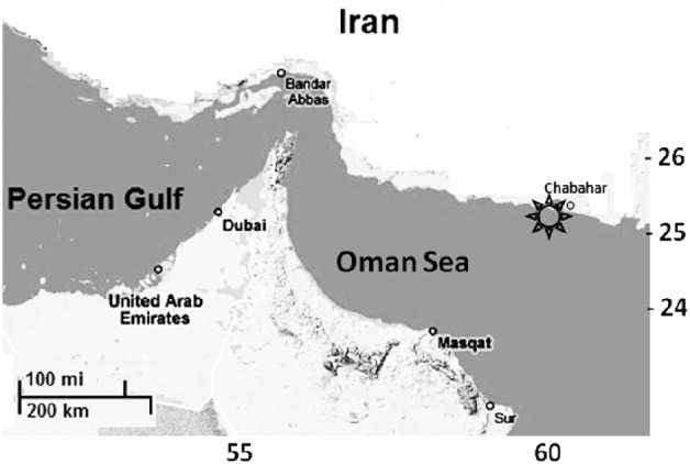
Two samples of sea water and 20 specimens of T. lepturus from the same location were examined. All fish were caught by the trawler Ferdows I in October 2008 off the Iranian coast of the Gulf of Oman at 60° 12′ to 60°17′E by 25°12′ to 25°14′N (Fig. 1), at a depth of 50 m. This region is a busy highway for petroleum ships from the Persian Gulf and adjacent seas and is thus likely to show high levels of pollution. Fish were dissected and samples of liver, muscle, gonad and intestinal tissues and of nematodes were placed in microtubes in a nitrogen tank, in which they were transferred to the laboratory. Nematodes were collected from the intestinal lumen attached to the intestinal wall, and in the mesenteries of the host fish.
Sampling location.

In the laboratory the samples were prepared according to the protocol described by Malek et al. (Reference Malek, Haseli, Mobedi, Ganjali and MacKenzie2007). They were weighed to the nearest 0·01 mg (wet weight) and then completely dissolved in a solution of 2 ml of 65% nitric acid and 2 ml of 75% perchloric acid. To speed up digestion, samples were heated to 50°C. The solutions were mixed with 4 ml of chloroform to extract lipids and diluted to 10 ml with distilled water. They were then ready for ICP-OES simultaneous set analysis (Inductive Coupling Plasma Optical Emission Spectrometry, Model: VISTA-PRO, Detector type: CCD) to measure their heavy metal contents. Two heavy metals commonly used in environmental assessment studies, lead (Pb) and cadmium (Cd), were measured in the samples, with the detection limits set at 0·06 ppm and 0·007 ppm respectively. All solutions were obtained from Merck Germany Darmstadt. Statistical analyses, including Kolmogrov-Smirnov, paired sample t-tests and graphs were carried out using SPSS 13.0 package.
RESULTS
Nine fish were infected with larvae of a nematode which was identified as Hysterothylacium sp. type MB of Deardorff and Overstreet (Reference Deardorff and Overstreet1981).
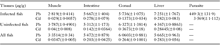
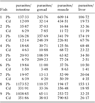
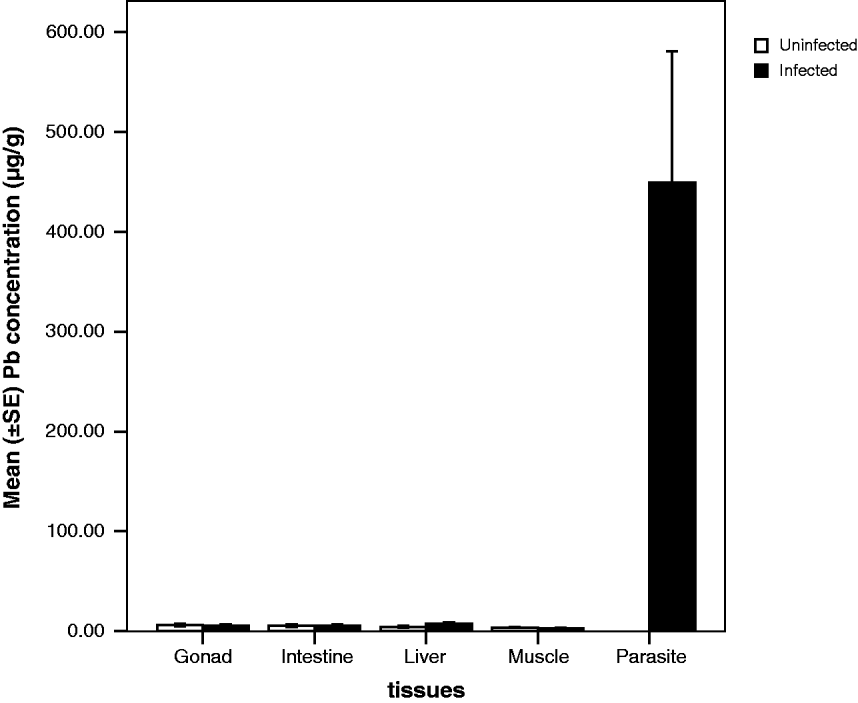
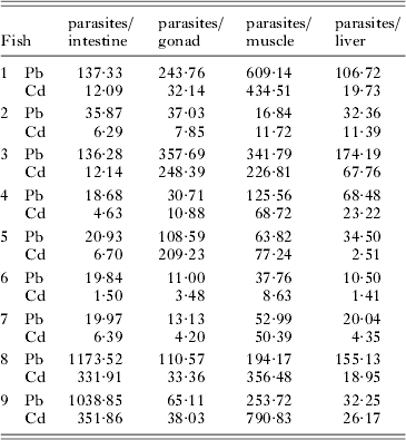
Concentrations of lead and cadmium in every nematode sampled were much higher than in infected host tissues, but with a wide range of variation in individual fish (Tables 1 and 2). Lead and cadmium concentrations in sea water were 0·0603 (±0·005) and 0·011 (±0·003), respectively. Mean concentrations of lead and cadmium in nematodes were 289·03 and 81·5 times higher than in host intestine, 188·4 and 225 times higher than in host muscle, 108·6 and 65·3 times higher than in host gonads, 70·5 and 19·5 times higher than in host liver and 3351 and 148 times higher than in sea water. The Kolmogorov–Smirnov normality test was applied to test the normal distribution of data. As the data were normally distributed, paired sample t-tests showed significant differences between cadmium concentrations in parasites and host gonads (t=−2·666 and P=0·008), liver (t= 2·928 and P=0·019), muscle (t=3·178 and P=0·013) and intestine (t=3·006 and P=0·017). For lead, parasites also showed much higher concentrations than in host gonads (t=−3·359 and P=0·010), liver (t=−3·369 and P=0·010), muscle (t=−3·381 and P=0·010) and intestine (t=−3·361 and P=0·010). Cadmium concentrations in the muscle of infected hosts were significantly lower than in both the intestine (t=2·898 and P=0·016) and liver (t=−3·038 and P=0·013). In uninfected hosts, cadmium concentrations in muscle were lower than in the intestine (t=3·245 and P=0·012), gonad (t=−2·223 and P=0·026) and liver (t=−3·036 and P=0·016).
Lead and cadmium mean concentrations (±S.E.) in host tissues and parasites

Ratio of Pb and Cd concentrations between host tissues and parasites in individual fish
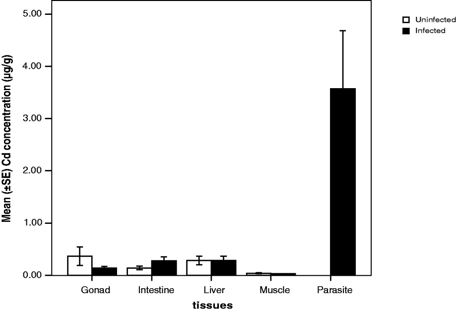
There were no significant differences in lead and cadmium concentrations between infected and uninfected hosts (Figs 2 and 3). In both infected and uninfected hosts, cadmium concentrations in muscle were significantly lower than in intestine (t=4·018 and P=0·001), liver (t=−4·395 and P<0·0005) and gonad (t=−3·248 and P=0·001).
Comparison of mean cadmium concentrations in infected and uninfected hosts.

Comparison of mean lead concentrations in infected and uninfected hosts.

Metal concentrations in sea water were significantly lower than in all fish organs and parasites.
DISCUSSION
According to the results of the present study, Hysterothylacium sp. type MB can act as a bioindicator in the marine habitiat. Petter and Sey (Reference Petter and Sey1997) described Hysterothylacium sp. type KE larvae from 7 species of fish, including T. lepturus, from Kuwaiti waters. They commented that their larvae were conspecific with Hysterothylacium sp. type MB larvae described by Kardousha (Reference Kardousha1992) from 4 other species of fish from the Persian Gulf. Hysterothylacium sp. type MB was originally described by Deardorff and Overstreet (Reference Deardorff and Overstreet1981) from 9 species of fish, including T. lepturus, from the Gulf of Mexico. We have therefore identified our nematodes as Hysterothylacium sp. type MB Deardorff and Overstreet, Reference Deardorff and Overstreet1981.
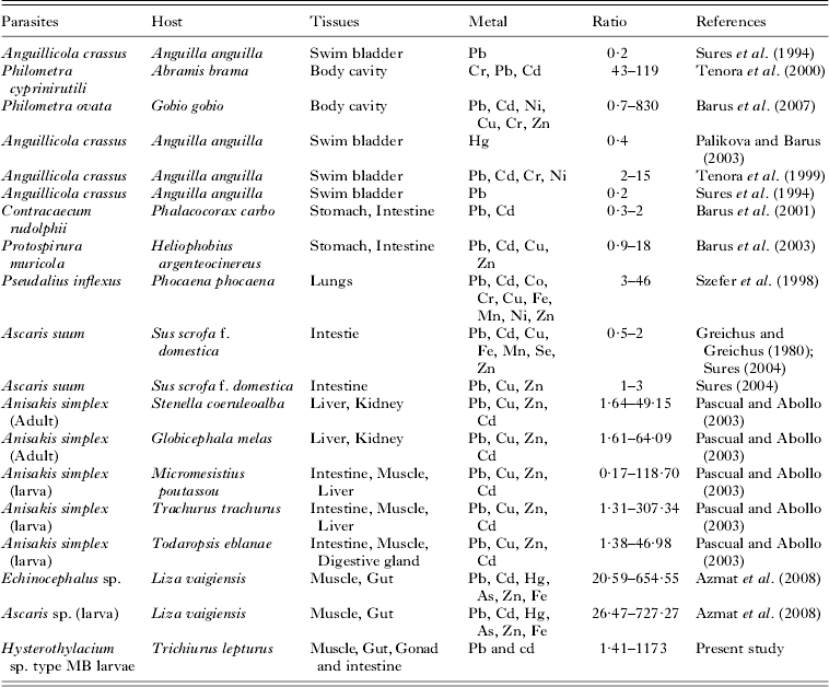
There are few studies on heavy metal accumulation in nematodes. These are summarized in Table 3. Studies by Sures et al. (Reference Sures, Taraschewski and Jackwerth1994) and Palikova and Barus (Reference Palikova and Barus2003) revealed that the lead concentration in Angullicola crassus from the swim bladder of Anguilla anguilla was significantly lower than in the host muscle. The failure of this parasite to absorb metal could be related to its microhabitat. According to Sures et al. (Reference Sures, Siddall and Taraschewski1999) the host intestine is the most likely site for metal uptake by parasites.
Reported ratios of metal concentrations between nematode parasites and host tissues

The higher concentration of heavy metals in some parasites compared with their hosts is probably due to the inability of such parasites to produce certain organic compounds, including organo-metallic compounds that have heavy metals in their structure. This has been shown for acanthocephalans (Sures, Reference Sures2001) and the results of the present study suggest it may also be true for intestinal nematodes. Whether or not this is the reason for the higher concentrations of lead and cadmium in these nematodes, it is clear that they can be useful indicators of heavy metal pollution. Because T. lepturus is a cosmopolitan fish and Hysterothylacium sp. type MB is one of its common parasites, we suggest that similar studies in other regions could provide useful data for comparisons between different regions and for the effects of contaminants on parasite abundance and prevalence in their fish hosts.
When Sures (Reference Sures2004) compared metal accumulations in different endoparasitic helminths with that in their hosts, he concluded that, as a general pattern, nematodes appear not to be suitable as sentinel species. Table 3 summarizes the results of studies on heavy metal accumulation of nematodes in different host-parasite systems. Based on these results it appears that some nematodes may serve as bioindicators. These include Philometra cyprinirutili in Abramis brama, Philometra ovata in Gobio gobio: the adult stage of Anisakis simplex in Stenella coeruleoalba and Globicephala melas and the larval stage of Anisakis simplex in Micromesistius poutassou and Trachurus trachurus.
Studies of heavy metal absorption by parasites have 2 main applications: in environmental studies and in food hygiene. In the present study no significant differences in the concentrations of cadmium and lead between tissues of infected and uninfected hosts were observed. This finding was also recorded by Brotheridge et al. (Reference Brotheridge, Newton and Evans1998) for the parasitic nematode Eustrongylides sp. in brown trout. This is in contrast to other studies such as those of Sures and Siddall (Reference Sures and Siddall1999) and Bergey et al. (Reference Bergey, Weis and Weis2002), in which uninfected hosts had significantly higher heavy metal concentrations than infected hosts. Azmat et al. (Reference Azmat, Fayyaz, Kazi, Mahmood and Uddin2008) also showed that metal accumulation in the muscle and gut of Liza veigiensis parasitized with the nematodes Echinocephalus sp. and Ascaris sp. was lower than in uninfected fish. In the present study it may be that the small numbers of Hysterothylacium in each host or high heavy metal concentrations in the environment and in host tissues were the reasons that a reduction in concentration in infected hosts was not observed. In the present study host muscle showed lower cadmium concentrations than other tissues, which from the food hygiene point of view is important because fish muscle is used more as human food than the other tissues tested.
ACKNOWLEDGEMENTS
Special thanks to Dr Valinasab and Mr Azhir from Iran Fishery Research Institute for providing facilities for sampling, and to Mr Hassani from the Analytical Chemistry Department, Tarbiat Modarres University for helping with the analytical parts of the study. We also appreciate the help of Mr Mirmasumy of the Plant Physiology Laboratory. The project was financed by the Research Affairs Department of The University of Tehran.