Introduction
Toxoplasmosis, which stands out as a major public health problem and a serious disorder during pregnancy, can cause abortion, fetal death, impaired fetal growth and congenital infection, with serious consequences to the neonate (Tenter et al., Reference Tenter, Heckerotha and Weissb2000; Pappas et al., Reference Pappas, Roussos and Falagas2009). Although vertical transmission of toxocariasis has been recorded only once (Maffrand et al., Reference Maffrand, Avila-Vazquez, Princich and Alasia2006), its importance in the transmission of Toxocara canis has been widely proven, as has the potential risk of complications it poses during pregnancy (abortion and impaired fetal growth), according to studies involving experimental models (Schoenardie et al., Reference Schoenardie, Scaini, Pepe, Borsuk, Avila, Villela, McBride, Borsuk and Berne2013a; Aguiar et al., Reference Aguiar, Farias, Avila, Telmo, Martins, Berne, Almeida and Scaini2015; Telmo et al., Reference Telmo, Avila, Santos, Aguiar, Martins, Berne and Scaini2015). Moreover, the seroprevalence of Toxocara spp. in pregnant women was registered as 6.4% in Brazil, which indicates that this population is also exposed to the infection (Santos et al., Reference Santos, Lehmann, Lorenzi, Hirsch, Telmo, Mattos, Cadore, Klafke, Berne, Gonçalves and Scaini2015).
The aetiological agents of toxoplasmosis and human toxocariasis are different; however, the risk factors for infection, related to social, economic, cultural and dietary habits, are the same for both of these diseases (Campos et al., Reference Campos, Elefant, Silva, Gandolfi, Jacob, Tofeti and Pratesi2003; Tenter, Reference Tenter2009).
In toxoplasmosis, it has been proven that the main mode of transmission is through the consumption of sausages or raw or undercooked meat that contains intermediate hosts of the parasite Toxoplasma gondii and tissue cysts (Sukthana, Reference Sukthana2006). However, in toxocariasis this type of transmission is still neglected, even though there have been reports of toxocariasis associated with the consumption of raw or undercooked meat or viscera of paratenic hosts of the helminth Toxocara spp., which contains infective encapsulated larvae (Hoffmeister et al., Reference Hoffmeister, Glaeser, Flick, Pornschlegel, Suttorp and Bergmann2007; Choi et al., Reference Choi, Lim, Choi, Lee, Paik, Kim, Choi and Huh2012). Another common mode of infection by these parasites is through the accidental ingestion of contaminated soil. Sporulated oocysts of T. gondii (Tenter et al., Reference Tenter, Heckerotha and Weissb2000) are transmitted through soil contaminated with cat faeces, while embryonated eggs of Toxocara spp. (Despommier, Reference Despommier2003) are transmitted through soil contaminated with dog faeces. Studies show that the prevalence of co-infection with T. gondii and Toxocara spp. in children ranges from 3% (Marchioro et al., Reference Marchioro, Colli, Ferreira, Viol, Araujo and Falavigna-Guilherme2015) to 12% (Franscisco et al., Reference Franscisco, Souza, Gennari, Pinheiro, Muradian and Soares2006). In addition, experimental and human studies regarding the co-infection with both parasites have been conducted to investigate behavioural changes in individuals (Corrêa et al., Reference Corrêa, Chieffi, Lescano and Santos2014; Khademvatan et al., Reference Khademvatan, Khajeddin, Izadi and Yousefi2014). However, research on the implications of co-infection during pregnancy is rare. The objective of this study was to determine the co-infection with T. gondii and Toxocara spp. in pregnant women at a university hospital in southern Brazil and to identify the risk factors associated with infection by both parasites.
Materials and methods
Population study
This was a cross-sectional study of 280 pregnant women treated at the University Hospital of the Federal University of Rio Grande (FURG), Rio Grande do Sul, Brazil. The sample size was calculated using the Epi Info 3.5.2 software (CDC, Atlanta, Georgia, USA) and an expected co-infection rate of 3.2% (Marchioro et al., Reference Marchioro, Colli, Ferreira, Viol, Araujo and Falavigna-Guilherme2015) with a confidence interval of 95% and 10% losses. This study was performed from May 2013 to April 2014. The prevalence of IgG antibodies to Toxocara spp. in this population was 6.4% (Santos et al., Reference Santos, Lehmann, Lorenzi, Hirsch, Telmo, Mattos, Cadore, Klafke, Berne, Gonçalves and Scaini2015).
Pregnant women were invited to participate in the study and those who agreed signed an informed consent form. Participants were required to allow researchers to analyse blood samples collected during routine prenatal care and access medical records, and to answer an epidemiological questionnaire. For pregnant women under the age of 18 years, a legal guardian was required to sign the informed consent form.
Serology
Serology tests for the detection of anti-T. gondii antibodies were performed during prenatal care using indirect immunofluorescence assays in the clinical analysis laboratory at the University Hospital, Rio Grande, Rio Grande do Sul. To conduct research on the IgG anti-Toxocara spp. antibodies, indirect enzyme-linked immunosorbent assays (ELISAs) were performed using the antigen excretion and secretion of T. canis. Serum samples were pre-adsorbed with the Ascaris suum antigen (Santos et al., Reference Santos, Lehmann, Lorenzi, Hirsch, Telmo, Mattos, Cadore, Klafke, Berne, Gonçalves and Scaini2015).
Data analysis
Two trained researchers used a structured questionnaire to survey participants after parturition in the maternity ward of University Hospital. This questionnaire covered the obstetric history (a history of abortions, premature births, number of pregnancies and number of births), general aspects (contact with pets, type of food), and social and economic characteristics (social, economic and level of education) of the participants. The questionnaires were double-entered using the Epidata 3.1 program (http://www.epidata.dk).
To verify the association of infection with T. gondii and with Toxocara spp. and the association of co-infection with socio-demographic characteristics, obstetric history, eating habits and contact with animals, we performed an analysis using the Chi-square test with a categorical comparison between variables. The prevalence ratio (RP) was calculated for each variable with a significant difference of P < 0.05 and confidence interval (CI) of 95%. Multivariate analysis was performed via logistic regression, followed by the construction of a hierarchical linear model, which incorporated variables with P ≤ 0.20 in the crude analysis. The first level was composed of demographic and socio-economic variables (family income and place of residence), while the second level was composed of the risk factors for infection with Toxocara spp. (contact with a domesticated dog, contact with a domesticated cat and vegetable consumption). All analyses were performed using SPSS (SPSS Inc., Chicago, Illinois, USA) and Epi-info 3.5.2 programs.
Results
The prevalence of IgG antibodies to Toxocara spp. was 6.4% in the population under study (Santos et al., Reference Santos, Lehmann, Lorenzi, Hirsch, Telmo, Mattos, Cadore, Klafke, Berne, Gonçalves and Scaini2015). Serology tests for toxoplasmosis were performed prenatally in 71.4% of women, and 28.6% were not tested for toxoplasmosis. From the group of pregnant women who were tested for toxoplasmosis, 62.5% (125) were positive for IgG antibodies to T. gondii. According to these results, of all the women who participated in our study, 200 were tested for both toxoplasmosis and human toxocariasis.
In this study, a co-infection rate of 8% was observed in the total population of pregnant women and, among women who were seropositive for T. gondii, the co-infection rate was found to be 12.8%. In addition, seropositivity for Toxocara spp. increased the risk of infection by T. gondii (P = 0.019) and seropositivity for T. gondii increased the risk of infection by Toxocara spp. (RP: 5.35; 95% confidence interval (CI): 1.19–24.0; P = 0.015).
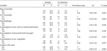
There was no association between the socio-demographic characteristics of the study population and co-infection with T. gondii and Toxocara spp., nor between risk factors and co-infection (table 1).
The proportion (%) of risk factors associated with co-infection with Toxocara spp. and T. gondii in pregnant women treated in a hospital in Rio Grande – Rio Grande do Sul. N, Number of samples examined; CI, 95% confidence intervals; the prevalence ratio denotes comparisons between variables using the Chi-square test.

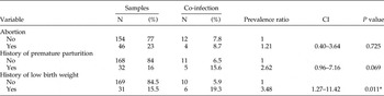
In the multivariate analysis of the obstetric history of pregnant women who tested positive for IgG antibodies to T. gondii and Toxocara spp., there was no significant relationship between abortions and premature birth and co-infection; however, we observed that co-infection increases the chances of an infant being born with a low birth weight (P = 0.011) (table 2).
The proportion (%) of obstetric disorders associated with co-infection by Toxocara spp. and T. gondii in pregnant women treated at a hospital in Rio Grande – Rio Grande do Sul. N, Number of samples examined; CI, 95% confidence intervals; the prevalence ratio denotes comparisons between variables using the Chi-square test.

*Level of significance with P < 0.05.
Discussion
Helminth infections during pregnancy have been associated with low birth weights in newborns, neonatal mortality (Friedman et al., Reference Friedman, Mital, Kanzaria, Olds and Kurtis2007; Imhoff-Kunsch & Briggs, Reference Imhoff-Kunsch and Briggs2012) and metabolic disorders in pregnancy (Tweyongyere et al., Reference Tweyongyere, Mawa, Kihembo, Jones, Webb, Cose, Dunne, Vennervald and Elliott2011). Among helminth infections, toxocariasis is a parasitic zoonosis with worldwide distribution (Smith et al., Reference Smith, Holland, Taylor, Magnaval, Schantz and Maizels2009) and, according to other studies, its impact on children's health has been observed, with a registered prevalence rate of 50% (Schoenardie et al., Reference Schoenardie, Scaini, Brod, Pepe, Villela, McBride, Borsuk and Berne2013b). However, only a few studies have been conducted on pregnant women, even though the registered seroprevalence of T. canis in Brazil is 6.4%, which shows that this population is also exposed to infection (Santos et al., Reference Santos, Lehmann, Lorenzi, Hirsch, Telmo, Mattos, Cadore, Klafke, Berne, Gonçalves and Scaini2015). Furthermore, the intracellular parasite T. gondii is responsible for disorders during pregnancy and complications during fetal development (Hill et al., Reference Hill, Chirukandoth and Dubey2005). In a serological study of pregnant women in Paraná, the prevalence of IgG antibodies to T. gondii was found to be 59% (Ferezin et al., Reference Ferezin, Bertolini and Demarchi2013). Few studies have evaluated co-infection with T. gondii and Toxocara spp. and its potential for harm during pregnancy and fetal development. However, the present study showed that 88.9% of the pregnant women who were seropositive for T. canis were also seropositive for T. gondii.
During helminthiasis an increased T-helper 2 (Th2) immune response occurs (Hernández et al., Reference Hernández, Escobedo, Guzman, Ibarra Coronado, López Griego and Morales-Montor2010), and the same occurs during pregnancy, which negatively affects the immune response to intracellular pathogens, as these pathogens promote a Th1 immune response (Spellberg & Edwards, Reference Spellberg and Edwards2001).
In this study, the total population presented a co-infection rate of 8%, which is lower than the 12.4% rate found in a study of 339 children from São Paulo (Franscisco et al., Reference Franscisco, Souza, Gennari, Pinheiro, Muradian and Soares2006) and higher than the 3.2% rate found in a study on children from Paraná, Brazil. However, both of the studies on children showed that positivity for Toxocara spp. increases the risk of infection by T. gondii in children (Franscisco et al., Reference Franscisco, Souza, Gennari, Pinheiro, Muradian and Soares2006; Marchioro et al., Reference Marchioro, Colli, Ferreira, Viol, Araujo and Falavigna-Guilherme2015). This association was also observed in the population of pregnant women in this study. However, we also observed that seropositivity for T. gondii increases the risk of infection by Toxocara spp., which was not observed by Marchioro et al. (Reference Marchioro, Colli, Ferreira, Viol, Araujo and Falavigna-Guilherme2015).
The similar mode of transmission of both parasites, which includes the consumption of raw or undercooked meat and the ingestion of embryonated eggs of Toxocara spp. (Smith et al., Reference Smith, Holland, Taylor, Magnaval, Schantz and Maizels2009) and oocysts of T. gondii (Montoya & Remington, Reference Montoya and Remington2008 ), may explain the co-infection rate observed in our study. Thus, it is important to emphasize that co-infection with these parasites may occur because the intermediate hosts of T. gondii and the paratenic hosts of Toxocara spp. include the same species of animals, such as cattle, sheep and pigs (Sukthana, Reference Sukthana2006; Hoffmeister et al., Reference Hoffmeister, Glaeser, Flick, Pornschlegel, Suttorp and Bergmann2007; Choi et al., Reference Choi, Lim, Choi, Lee, Paik, Kim, Choi and Huh2012). Therefore, these animals may be considered sources of infection as they may carry both tissue cysts containing bradyzoites of T. gondii (Sukthana, Reference Sukthana2006) and infective larvae of Toxocara spp., which are found in the animal's muscle tissue or viscera (Choi et al., Reference Choi, Lim, Choi, Lee, Paik, Kim, Choi and Huh2012).
Another common mode of infection of these parasites is the accidental ingestion of sporulated oocysts of T. gondii (Montoya & Remington, Reference Montoya and Remington2008) and embryonated eggs of Toxocara spp. (Smith et al., Reference Smith, Holland, Taylor, Magnaval, Schantz and Maizels2009), both of which are developmental forms of the parasites that may be found in soil, on fomites and on poorly cleaned vegetables. In toxoplasmosis, even though the ingestion of oocysts is a well-known mode of infection, it is not the most significant mode in the epidemiology of the disease (Dubey, Reference Dubey2004), while in human toxocariasis, ingestion of embryonated eggs is considered to be the main mode of infection of Toxocara spp. (Smith et al., Reference Smith, Holland, Taylor, Magnaval, Schantz and Maizels2009).
In addition to factors related to the similar modes of transmission of these parasites that may facilitate co-infection, helminthiasis can increase the severity and incidence of infections by intracellular parasites as it causes an increased Th2 response and decreased Th1 response. Such immunomodulation is also observed in pregnancy, and Hernández et al. (Reference Hernández, Escobedo, Guzman, Ibarra Coronado, López Griego and Morales-Montor2010) discuss the possibility of a synergistic immune response to helminthiasis during pregnancy. A previous study showed that patients with a co-infection with helminths are at increased risk for malaria, which may increase the risk of complications during pregnancy and harm to the fetus (Gallagher et al., Reference Gallagher, Malhotra, Mungai, Wamachi, Kioko, Ouma, Muchiri and King2005).
Epidemiological studies on helminth infections often relate infection to low birth weights in newborns (Imhoff-Kunsch & Briggs, Reference Imhoff-Kunsch and Briggs2012), which was confirmed by experimental studies on these infections (Anderson, Reference Anderson1996). The only study that considered a relationship between seropositivity for Toxocara spp. and low birth weights of newborns found no association between these variables (Santos et al., Reference Santos, Lehmann, Lorenzi, Hirsch, Telmo, Mattos, Cadore, Klafke, Berne, Gonçalves and Scaini2015). However, in the population under study, we found a significant association between co-infection with T. gondii and Toxocara spp. and low birth weights in newborns.
The association between co-infection with T. gondii and Toxocara spp. in pregnant women and low birth weights in neonates demonstrates the importance of serological diagnosis during prenatal care. It also demonstrates the importance of further research in this area to identify risk factors associated with co-infection, and the possible implications of co-infection during pregnancy and on the health of the newborn.
Acknowledgements
We thank the Post-graduate Program in Public Health, Coordenação de Aperfeiçoamento de Pessoal de Nível Superior (CAPES), and professionals from the Center of Obstetrics and the clinical laboratory of University Hospital, Rio Grande (FURG) for their significant contribution in the development of this study.
Financial support
This research received no specific grant from any funding agency, commercial or not-for-profit sectors.
Conflict of interest
None.
Ethical standards
The authors assert that all procedures contributing to this work comply with the ethical standards of the relevant national and institutional committees on human experimentation. This study was approved by the Ethics Committee on Research in Health, FURG (CEPAS no. 33/2011).