Introduction
This article continues a series of papers on the mineralogy and crystal chemistry of new mineral species containing rare-earth elements (REE) as species-defining cations (henceforth ‘REE minerals’) discovered at the Mochalin Log deposit, Chelyabinsk Oblast, South Urals, Russia (55°48′42″N, 60°33′46″E). A general description and mineralogical data for this deposit were given in the first paper which also contained the characterisation of two new minerals ferriperbøeite-(La) and perbøeite-(La) (Kasatkin et al., Reference Kasatkin, Zubkova, Pekov, Chukanov, Škoda, Polekhovsky, Agakhanov, Belakovskiy, Kuznetsov, Britvin and Pushcharovsky2020a). In the second article we described radekškodaite-(La) and radekškodaite-(Ce), two members of the epidote–törnebohmite polysomatic series with a novel-type structure including one epidote and two törnebohmite modules (ET2) (Kasatkin et al., Reference Kasatkin, Zubkova, Pekov, Chukanov, Ksenofontov, Agakhanov, Belakovskiy, Polekhovsky, Kuznetsov, Britvin, Pushcharovsky and Nestola2020b).
This paper reports data on a new REE silicate from the Mochalin Log deposit that is named as an analogue of percleveite-(Ce), ideally Ce2Si2O7 (Holtstam et al., Reference Holtstam, Norrestam and Andersson2003), with La predominance among REE. Both the new mineral and its name have been approved by the Commission on New Minerals, Nomenclature and Classification of the International Mineralogical Association (IMA2019–037, Kasatkin et al., Reference Kasatkin, Zubkova, Pekov, Chukanov, Škoda, Agakhanov, Belakovskiy, Plášil and Kuznetsov2019a). The holotype specimen is deposited in the systematic collection of the Fersman Mineralogical Museum of the Russian Academy of Sciences, Moscow with the catalogue number 96697.
Occurrence and general appearance
Percleveite-(La) was found during systematic scanning electron microscope (SEM) energy-dispersive spectroscopy examination of polymineralic REE-bearing nodules collected in 1980s by local collectors at the historical dump no. 2 situated in the Mochalin Log river valley and, since then, deposited in the collection of one of the authors (AMK).
As reported by Kasatkin et al. (Reference Kasatkin, Zubkova, Pekov, Chukanov, Škoda, Polekhovsky, Agakhanov, Belakovskiy, Kuznetsov, Britvin and Pushcharovsky2020a), the gross Ce:La ratio in many such nodules is often close to 1:1 and both Ce- and La-dominant members of the same series of REE minerals may occur in the same specimen. Thus, the absence of a particular Levinson suffix below in the text means the presence of Ce- and La-dominant mineral species with the same rootname.
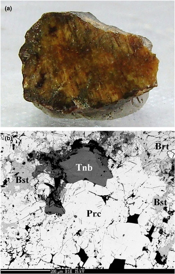
Percleveite-(La) forms isolated anhedral grains commonly up to 0.2 mm × 0.4 mm and very rarely up to 1 mm × 1 mm in polymineralic nodules composed mainly by allanite/ferriallanite, bastnäsite/hydroxylbastnäsite, fluorbritholite-(Ce), ferriperbøeite/perbøeite and törnebohmite. The new mineral is typically intimately intergrown with törnebohmite and/or bastnäsite or is overgrown by them (Figs 1a,b). The very rich REE mineral assemblage with percleveite-(La) also includes alexkuznetsovite-(La) (Kasatkin et al., Reference Kasatkin, Zubkova, Pekov, Chukanov, Škoda, Agakhanov, Belakovskiy and Pushcharovsky2019b), alexkuznetsovite-(Ce) (Kasatkin et al., Reference Kasatkin, Zubkova, Pekov, Chukanov, Škoda, Agakhanov and Belakovskiy2020c), biraite-(Ce), biraite-(La) (Kasatkin et al., Reference Kasatkin, Zubkova, Pekov, Chukanov, Škoda, Agakhanov, Belakovskiy, Kuznetsov and Pushcharovsky2020d), lanthanite-(La), percleveite-(Ce), perrierite, radekškodaite-(La), stetindite-(Ce) and stillwellite-(Ce). Other associated minerals are annite, calcite, hollandite, potassic feldspar, quartz, thorite and thorianite.
(a) Cut and polished section of nodule showing zones of light yellow percleveite-(La) intergrown with orange-brown bastnäsite/hydroxylbastnäsite and fluorbritholite-(Ce), black allanite/ferriallanite and greenish törnebohmite. Size of sample: 0.6 cm × 0.4 cm, specimen no. ML 89-2; (b) fragment of (a): percleveite-(La) (Prc) in association with bastnäsite (Bst), törnebohmite-(La) (Tnb) and fluorbritholite-(Ce) (Brt). Black grains are quartz. Polished section. SEM (back-scattered electron) image. Scale bar = 200μm.

According to the distribution scale of RΕΕ minerals found at the Mochalin Log deposit (Kasatkin et al., Reference Kasatkin, Zubkova, Pekov, Chukanov, Škoda, Polekhovsky, Agakhanov, Belakovskiy, Kuznetsov, Britvin and Pushcharovsky2020a), percleveite-(La) should be considered as rare: among 300 nodules with REE-bearing minerals investigated by us, it was found in only 12 of them, in minor amounts.
Physical properties and optical data
The new mineral is very pale yellow to colourless in thin fragments to light yellow in aggregates, transparent, with white streak and greasy lustre. It does not fluoresce under ultraviolet light. The imperfect cleavage on {001} is observed and the fracture is uneven (observed under the SEM). Percleveite-(La) is brittle. The Mohs’ hardness is ca. 6, based on scratch tests. Density could not be measured due to lack of pure material in grains suitable for measurements and the absence of necessary heavy liquids. A density value calculated using the empirical formula and the unit-cell parameters from single-crystal X-ray diffraction (XRD) data is 5.094 g cm–3. In transmitted plane-polarised light percleveite-(La) is colourless and non-pleochroic. Optically it is uniaxial (+), with ω = 1.825(10) and ɛ = 1.835(10) (589 nm).
Raman spectroscopy
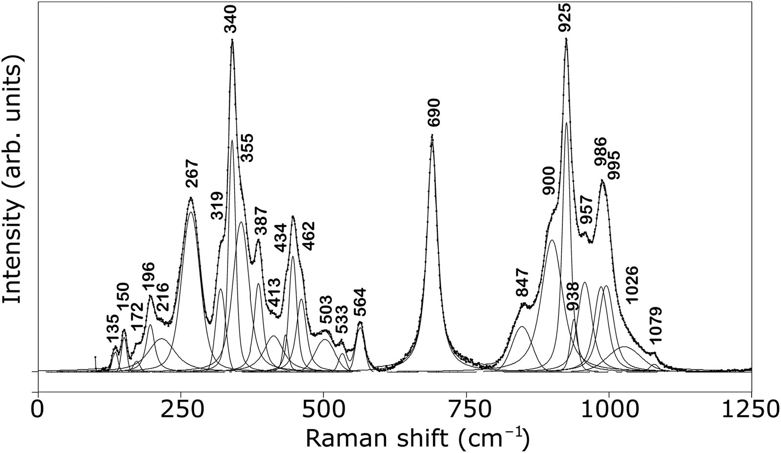
The Raman spectrum of percleveite-(La) (Fig. 2) was obtained from a polished section by means of a Horiba Labram HR Evolution spectrometer. This dispersive, edge-filter-based system is equipped with an Olympus BX 41 optical microscope, a diffraction grating with 600 grooves per millimetre and a Peltier-cooled, Si-based charge-coupled device (CCD) detector. After careful tests with different lasers (473, 532 and 633 nm), the 633 nm He–Ne laser with the beam power of 10 mW at the sample surface was selected for spectra acquisition to minimise analytical artefacts. The Raman signal was collected in the range of 100–4000 cm–1 with a 100× objective (NA 0.90) and the system operated in the confocal mode, beam diameter was ~1 μm and the lateral resolution ~2 μm. No visual damage of the analysed surface was observed at these conditions after the excitation. Wavenumber calibration was done using the Rayleigh line and low-pressure Ne-discharge lamp emissions. The wavenumber accuracy was ~0.5 cm–1 and the spectral resolution was ~2 cm–1. Band fitting was done after appropriate background correction, assuming combined Lorentzian–Gaussian band shapes using the Voight function (PeakFit; Jandel Scientific Software).
Raman spectrum of percleveite-(La) excited by a 633 nm laser. The measured spectrum is shown by dots. The curve matched to dots is a result of spectral fit as a sum of individual Voigt peaks shown below the spectral envelope.

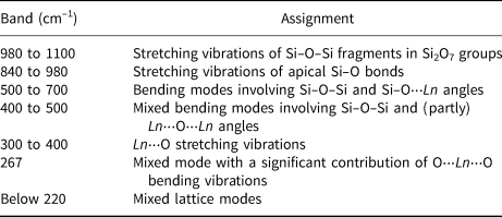
Based on the results of quantum chemical calculations for Y2Si2O7 polymorphs (Kaindl et al., Reference Kaindl, Többens and Kahlenberg2010) and taking into account Raman spectroscopic data for synthetic keiviite-(Yb) Yb2Si2O7 (Bretheau-Raynal et al., Reference Bretheau-Raynal, Dalbiez and Drifford1979) and melilite-type compounds (Kaminskii et al., Reference Kaminskii, Rhee, Lux, Eichler, Bohatý, Becker, Liebertz, Ueda, Shirakawa, Koltashev, Hanuza, Dong and Stavrovskii2011), the Raman bands of percleveite-(La) have been assigned according to Table 1. No bands are observed in the Raman spectrum of percleveite-(La) above 1100 cm–1 which indicates the absence of H2O, OH–, CO32– and BO33– groups.
Raman bands and mode assignments for percleveite-(La).

‘⋅⋅⋅’ denotes non-covalent (essentially ionic) bonds.
Chemical data
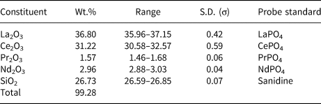
Chemical data (eight spot analyses) were obtained using a Cameca SX-100 electron microprobe (wavelength dispersive spectroscopy mode, acceleration voltage of 15 kV, a beam current of 20 nA and a 3 μm beam diameter). Both the crystal structure and Raman spectroscopy data confirm the absence of H2O, OH–, CO32– and BO33– groups in the new species. Analytical data are given in Table 2. Contents of other elements with atomic numbers higher than that of carbon are below detection limits.
Chemical composition of percleveite-(La).

S.D. – standard deviation
Percleveite-(La) grains are chemically homogeneous and the WDS analyses reveal the uniform chemical composition with only minimal inter-REE chemical variation. Lanthanum significantly prevails among REE in all analyses in the type specimen. The empirical formula calculated on the basis of 7 O atoms per formula unit is (La1.02Ce0.86Nd0.08Pr0.04)Σ2.00Si2.00O7. The idealised, end-member formula is La2Si2O7 which requires La2O3 73.00, SiO2 27.00, total 100 wt.%.
The Gladstone–Dale compatibility index (1 – K p/K c) calculated for percleveite-(La) using its empirical formula and the unit-cell parameters determined from single-crystal XRD data is –0.001, rated as superior (Mandarino, Reference Mandarino1981).
Percleveite-(La) does not react with either cold hydrochloric or nitric acids.
X-ray crystallography and crystal structure
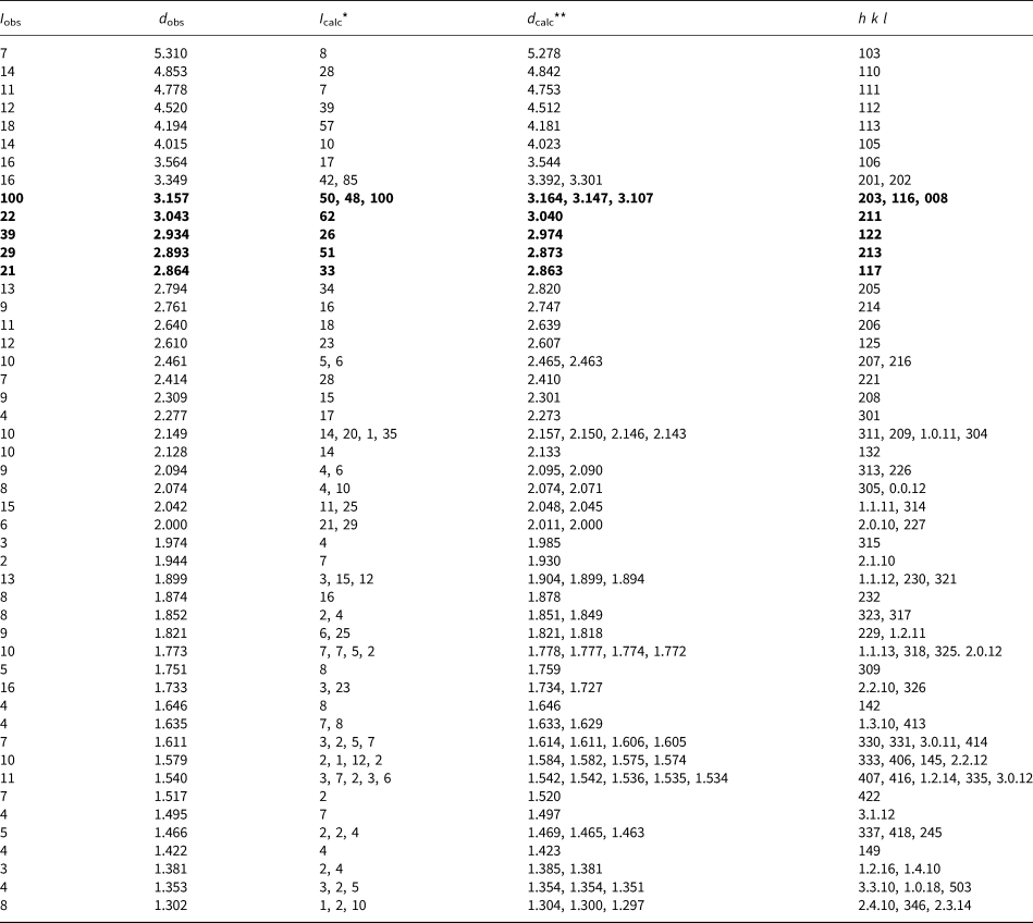
Powder XRD data for percleveite-(La) (Table 3) were collected with a Rigaku R-AXIS Rapid II single-crystal diffractometer equipped with a cylindrical image plate detector (radius 127.4 mm) using Debye-Scherrer geometry, CoKα radiation (rotating anode with VariMAX microfocus optics), 40 kV, 15 mA and exposure = 15 min. Angular resolution of the detector is 0.045°2θ (pixel size is 0.1 mm). The data were integrated using the software package osc2tab (Britvin et al., Reference Britvin, Dolivo-Dobrovolsky and Krzhizhanovskaya2017). Parameters of the tetragonal unit cell refined from the powder XRD data are: a = 6.8472(12), c = 24.848(14) Å and V = 1165.0(11) Å3.
Powder X-ray diffraction data (d in Å) of percleveite-(La).

*For the calculated pattern, only reflections with intensities ≥1 are given; **for the unit-cell parameters calculated from single-crystal data. The strongest lines are given in bold.
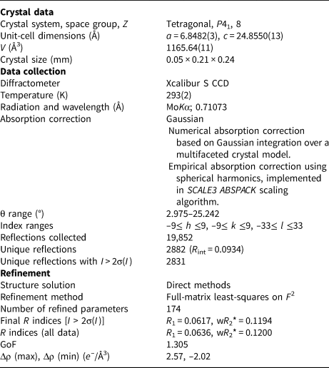
For the single-crystal XRD study, a grain of percleveite-(La), 0.05 mm × 0.21 mm × 0.24 mm in size, extracted from the polished section and analysed using electron microprobe, was mounted on a glass fibre and examined with an Xcalibur S single-crystal diffractometer equipped with a CCD detector. A full sphere of three-dimensional data was collected. Data reduction was performed using CrysAlisPro software Version 1.171.37.35 (Agilent Technologies, 2014). The data were corrected for Lorentz factor and polarisation effect.
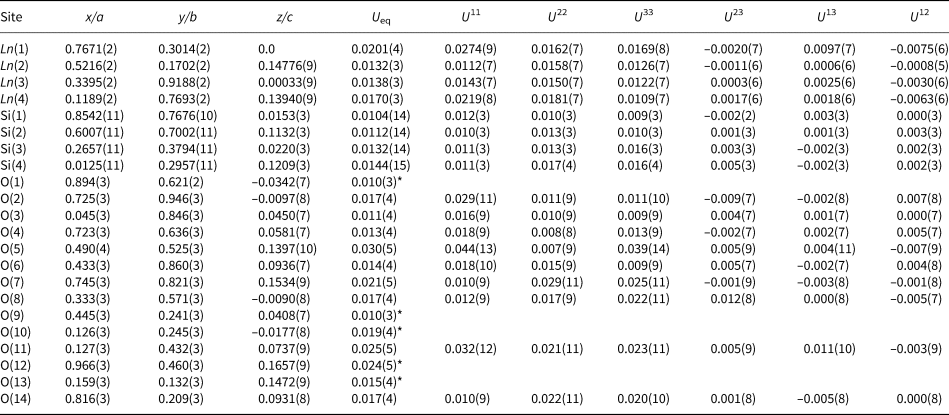
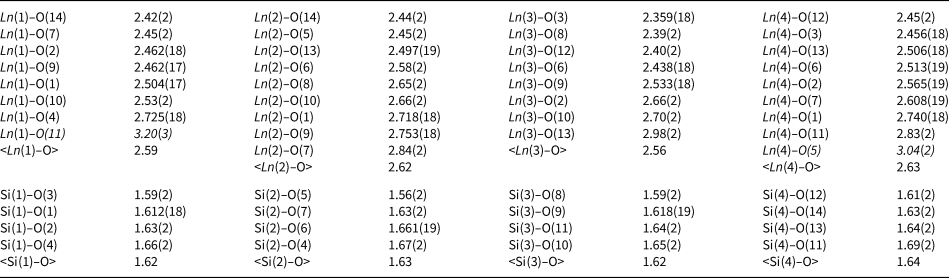
The crystal structure of percleveite-(La) was solved by direct methods and refined with the use of SHELX-97 software (Sheldrick, Reference Sheldrick2008) to R = 0.0617 for 2831 unique reflections with I > 2σ(I). The crystal was an enantiomeric twin; the Flack parameter (Flack, Reference Flack1983) was refined to 0.46(7). For the refinement of the site occupancies of four crystallographically non-equivalent lanthanide sites, the scattering curves of Ce were used according to the averaged value of 57.6 electrons derived from the chemical data. The refinement resulted in the following site occupancy factors: 0.984(10), 1.009(11), 1.017(11) and 0.993(11) for Ln(1), Ln(2), Ln(3) and Ln(4), respectively. At the last cycles of the refinement these values were fixed at 1.00. The highest peak at difference-Fourier synthesis is at the distance of 1.03 Å from the Ln(1) site and the deepest hole is at the distance of 1.36 Å from the same atom. The crystal data, data collection information and structure refinement details are given in Table 4, atomic coordinates and thermal displacement parameters of atoms in Tables 5 and selected interatomic distances in Table 6. The crystallographic information file has been deposited with the Principal Editor of Mineralogical Magazine and is available as Supplementary material (see below).
Crystal data, data collection information and structure refinement details for percleveite-(La).

*w = 1/[σ2(F o2) + (0.0361P)2 + 31.9202P]; P = ([max of (0 or F o2)] + 2F c2)/3
Atom coordinates, anisotropic and equivalent thermal displacement parameters (U eq, in Å2) for percleveite-(La).

*U iso.
Selected interatomic distances (Å) in the structure of percleveite-(La)*.

* Elongated Ln–O distances are italicised.
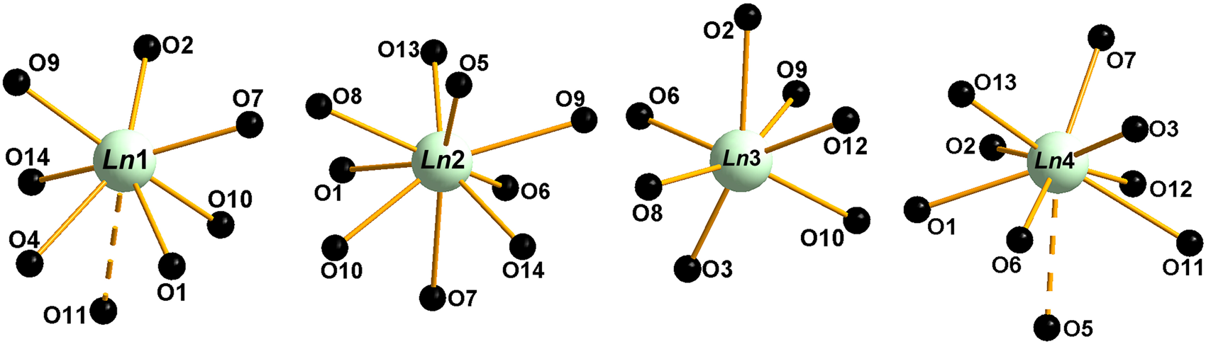
The crystal structure of percleveite-(La) (Fig. 3) contains [Si2O7]6– dimers connected via Ln-centred polyhedra. The arrangement of [Si2O7]6– disilicate groups could be described as the (001) ‘layers’ with Ln cations embedded in them and connecting adjacent ‘layers’. Four crystallographically non-equivalent Ln cations occupy 7- [Ln(1)], 9- [Ln(2)] and 8-fold [Ln(3,4)] polyhedra taking the limit of 3.0 Å for Ln–O distances. Additional elongated Ln–O distances were found for Ln(1) [Ln(1)–O(11) of 3.20(3) Å] and Ln(4) [Ln(4)–O(5) of 3.04(2) Å]. The coordination of Ln cations is shown in Fig. 4.
The crystal structure of percleveite-(La). SiO4 tetrahedra are red. The unit cell is outlined.

Coordination environment of Ln 3+ cations in the structure of percleveite-(La). Elongated Ln–O bonds (longer than 3 Å) are shown as dashed lines.

Discussion
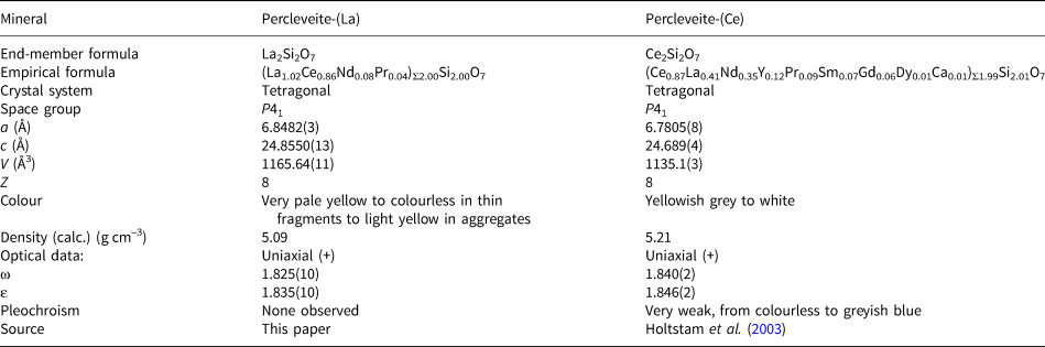
Percleveite-(La) is the La-dominant analogue of percleveite-(Ce) (Holtstam et al., Reference Holtstam, Norrestam and Andersson2003). For comparison of the two minerals see Table 7. Both species are also isostructural with a number of synthetic light rare earth elements, disilicates with the general formula Ln 2Si2O7 [Ln = La, Ce, Pr, Nd and Sm] (Deng and Ibers, Reference Deng and Ibers2005 and references therein) which crystallise in space group P41 and, according to the classification of Felsche (Reference Felsche1973), have a Type A structure with basic structure type of the high-temperature pyrophosphate β-Ca2P2O7 (Boudin et al., Reference Boudin, Grandin, Borel, Leclaire and Raveau1993). Strontium diarsenate Sr2As2O7 (Weil et al., Reference Weil, Djordjević, Lengauer and Kolitsch2009; Edhokkar et al., Reference Edhokkar, Hadricha, Graiab and Mhiria2012) and high-temperature strontium divanadate β-Sr2V2O7 (Baglio and Dann, Reference Baglio and Dann1972) also belong to this structure type.
Comparative data for percleveite-(La) from Mochalin Log and percleveite-(Ce) from Bastnäs, Sweden.

Several polymorphs of REE silicates have been synthesised to date (Smolin and Shepelev, Reference Smolin and Shepelev1970; Felsche, Reference Felsche1973; Fleet and Liu, Reference Fleet and Liu2003; Deng and Ibers, Reference Deng and Ibers2005). Individual structural types in this family of compounds are designated by Latin capital letters [following the terminology of Felsche (Reference Felsche1973)] or by Greek symbols. These structure types differ from each other in coordination number of REE 3+, reflecting their variable ionic radius and the arrangement of SiO4 tetrahedra. Each type is characterised by specific kinds of silicate anionic groups: [Si2O7]6– for the types A, C, D to H and K; linear triple tetrahedral group of [Si3O10]8– combined with isolated [SiO4]4– tetrahedron for the type B; catena-tetrasilicate [Si4O13]10– combined with two [SiO4]4– tetrahedra for the type I.
Naturally occurring phases i.e. sorosilicate minerals of trivalent rare-earth elements and scandium containing disilicate groups, include tetragonal percleveite-(Ce) (type A), monoclinic keiviite and thortveitite [(Y,HREE)2Si2O7 and Sc2Si2O7, respectively; type C, also termed type-β] and yttrialite (Y2Si2O7; type D, also termed type-γ) [where HREE and LREE are heavy and light REE respectively]. The type A structure shows affinity to large LREE ions, whereas the smaller HREE, Y and Sc are favoured by types D and C. Triclinic type B (also termed type-α), incorporating medium-sized Ln, has not been found in nature to date. Despite a simple chemical composition, these REE disilicate minerals are rare to very rare in nature. Yttrialite, keiviite and thortveitite either crystalise as late-magmatic phases in the final stages of solidification of some highly evolved NYF (Niobium–Yttrium–Fluorine) pegmatites (terms from Černý and Ercit, Reference Černý and Ercit2005) or formed from hydrothermal fluids rich in REE and/or Sc in different environments (e.g. Bjørlykke, Reference Bjørlykke1937; Voloshin et al., Reference Voloshin, Pakhomovskii and Tyusheva1985; Spandler et al., Reference Spandler, Hermann and Rubatto2004; Guastoni et al., Reference Guastoni, Nestola, Ferraris and Parodi2012). Percleveite-(Ce) has been reported from very few occurrences worldwide: its type locality at the Nya Bastnäs deposit, Riddarhyttan ore field, Sweden (Holtstam et al., Reference Holtstam, Norrestam and Andersson2003), El Seboah peralkaline rocks in Egypt (Soliman Abu Elatta, Reference Soliman Abu Elatta2018) and alkali pegmatites of the Konder Pluton, Khabarovsk Krai, Russia (Osipov et al., Reference Osipov, Antonov, Panikorovskii and Zolotarev2018). However, identification of the mineral from Egypt is based on the energy-dispersive spectroscopy analysis alone while the report from Konder is only mentioned amongst other minerals of the area with no analytical data given.
The Nya Bastnäs deposit bears a specific skarn-hosted magnetite-REE mineralisation formed ~1.9 Ga by interaction of felsic magmatic-related fluids with limestones at temperatures above 400°C (Holtstam et al., Reference Holtstam, Andersson, Broman and Mansfeld2014; Sahlström et al., Reference Sahlström, Jonsson, Högdahl, Troll, Harris, Jolis and Weis2019). At Bastnäs, percleveite-(Ce) is a primary mineral, mainly associated with cerite-(Ce), bastnäsite-(Ce) and delhuyarite-(Ce). At Mochalin Log, percleveite-(Ce) and percleveite-(La) were probably formed during a high-temperature metasomatic replacement of primary bastnäsite. They usually occur in central parts of the REE-rich nodules in association with bastnäsite, allanite, törnebohmite, ferriperbøeite, perbøeite and fluorbritholite-(Ce). Regardless of some genetic differences, both REE deposits have several common geochemical and mineralogical features. (1) Percleveite is associated closely with silicates and carbonates with very high nominal REE contents (~60–75 wt.% LREE 2O3). (2) There is a scarcity to almost complete absence of P, As, Nb, Ta and Ti in the environment. Though P is an exception, as monazite is absent at Bastnäs (Holtstam and Andersson, Reference Holtstam and Andersson2007) but quite abundant at Mochalin Log, however, it was never observed in the association with percleveite-(La) and percleveite-(Ce). (3) The REE mineralisation is depleted in actinides, U and Th which prevents REE minerals from radiation-induced amorphisation and subsequent alteration. However, primary bastnäsite from Mochalin Log contains a distinct Th admixture (up to 0.9 wt.% ThO2), and during metasomatic processes thorium, released from bastnäsite, concentrates in forms of thorianite and thorite while monazite and coexisting REE silicates are Th-poor and, thus, crystalline but not metamict.
Formation of percleveite-(La) and percleveite-(Ce) requires a LREE- and Si-rich environment, concurrently with very low activities of P, As, Nb, Ta, Ti and alkali elements. Otherwise, an elevated activity of these elements favours a crystallisation of REE phosphates, arsenates, complex Nb–Ti–Ta oxides and alkali-REE silicates. Hence, the rarity of percleveite-(La) and percleveite-(Ce) in Nature might be caused by specific geochemical conditions at which A-type Ln 2Si2O7 become stable phases with respect to other REE minerals. Moreover, low Ca supresses the formation of britholite and along with Mg supresses the formation of cerite. In summary, we can infer from both above-mentioned occurrences, that percleveite-(La) and percleveite-(Ce) were formed during a high-temperature fluid-assisted process and the scarcity of these minerals at both localities indicates that such conditions were achieved only locally.
Acknowledgements
We thank Associate Editor Elena Zhitova, Referees Dan Holtstam and Alessandro Guastoni, Structures Editor Daniel Atencio and Principal Editor Stuart Mills for constructive comments that improved the manuscript. This study was supported by the Russian Foundation for Basic Research, grant no. 18-05-00332 (in part of XRD studies and crystal chemistry elaboration). This work was partly performed in accordance with the state task, state registration no. ААА-А19-119092390076-7 for NVC. JP acknowledges support by the Ministry of Education, Youth and Sports National sustainability program I of the Czech Republic (project No. LO1603). The technical support by the SPbSU X-Ray Diffraction Resource Center in the powder XRD study is acknowledged.
Supplementary material
To view supplementary material for this article, please visit: https://doi.org/10.1180/mgm.2020.81.