Introduction
The mammalian eye has a dynamic range spanning approximately 9 log units, and it uses multiple mechanisms in adaptation to varied ambient illumination. These include changing the size of the pupil, shifting between cone- and rod-mediated visual functions and also probably relatively complex neuronal adaptations (Reuter, Reference Reuter2011). However, it has also been claimed that there is a longer-term adaptive mechanism known as photostasis where rod photoreceptors can regulate photon capture in response to varied lighting conditions (Penn & Williams, Reference Penn and Williams1986). This is based on the observation that rodent photoreceptor outer segments (OSs) lengthen and their rhodopsin concentration increases when they experience prolonged periods of darkness or reduced illumination (Penn & Williams, Reference Penn and Williams1986; Penn & Anderson, Reference Penn and Anderson1987; Williams & Thistle, Reference Williams and Thistle1998; Daly et al., Reference Daly, DiLeonardo, Balkema and Balkema2004). Although this will increase the probability of photon capture, the mechanism for this type of adaptation is unclear but may reside in reduced patterns of phagocytosis by the retinal pigment epithelium (RPE). These observations are based on earlier studies showing significant increases in rhabdomeric structure in larval mosquitoes (White & Lord, Reference White and Lord1975), spiders (Blest, Reference Blest1978), and Limulus eyes (Behrens & Krebs, Reference Behrens and Krebs1976) in darkness compared with light environments. These were detailed, often ultrastructural studies (Behrens & Krebs, Reference Behrens and Krebs1976; Blest, Reference Blest1978), and in the case of White and Lord (Reference White and Lord1975), very tightly controlled with rhabdomeric volume measured not only during progressive periods of light and darkness but also at progressive energy levels.
Mammalian photoreceptor OS undergo periodic changes. Rods lose their apical tips, generally in the morning, and new discs are synthesized at their base (Young, Reference Young1967, Reference Young1971). The consequence of this is that discs are constantly migrating toward the RPE with all being replaced over about 10 days, which is approximately the time taken for a newly produced disk to reach the apical end of the OS. A range of experiments have claimed that there is marked lengthening of the segments when animals are kept in restricted illumination. The reported extent of this can be relatively dramatic, with some studies claiming 90% lengthening between light and dark environments in rodents (Penn & Anderson, Reference Penn and Anderson1987), whereas others have revealed 78% (Penn & Williams, Reference Penn and Williams1986) or just 25% or 30% extension (Battelle & LaVail, Reference Battelle and LaVail1978; Schremser & Williams, Reference Schremser and Williams1995).
Although photostasis has become widely accepted as a long-term mechanism of adaptation in mammals since first proposed by Penn and Williams (Reference Penn and Williams1986), there are concerns. All of the experiments except one (Daly et al., Reference Daly, DiLeonardo, Balkema and Balkema2004) have been confined to albino rats (Battelle & LaVail, Reference Battelle and LaVail1978; Penn & Williams, Reference Penn and Williams1986; Penn & Anderson, Reference Penn and Anderson1987; Schremser & Williams, Reference Schremser and Williams1995), which by definition have mutations of their RPE with no melanin synthesis. Further, albino RPE cells have progressive morphological abnormalities (Adams et al., Reference Adams, Shahabi, Hoh-Kam and Jeffery2010). Second, results between labs have been highly varied in the reported magnitude of OS extension. Third, the methods used have often been unclear, with limited sampling and no statistical analysis ever undertaken. Also, in the only study to address the issue in both pigmented and albino mice, there appears to be some limited impact in albinos but very little in pigmented mice (Daly et al., Reference Daly, DiLeonardo, Balkema and Balkema2004). However, again no statistics or raw data were offered. The above concerns gain further traction by a recent report that shows that RPE/OS interactions are very different between the two pigmentation phenotypes, with that in albinos being significantly disrupted (Shahabi et al., Reference Shahabi, Golding, Lenassi and Jeffery2011).
Because the previously published data show such variability and lack clarity, we have gone back and asked whether the mechanism of photostasis actually exists in normally pigmented and albino mice in terms of changes in OS length. Further, we have extended the study to ask this in young and old pigmented animals to determine if it is only a feature of the young retina because in aged rats OSs start to shorten, implying that they may be less plastic (Cunea & Jeffery, Reference Cunea and Jeffery2007). We also sampled a much larger population than previously undertaken in any study. After measuring approximately 1300 individual OSs, we find no statistically significant evidence for changes of OS length in our mice as a function of varied environmental light at any age or in either pigmentation phenotype.
Methods
All animals were used with University College London ethics committee approval and under a UK Home Office animal project license. All animal procedures conformed to the United Kingdom Animals Scientific Procedures Act, 1986.
Twenty C57BL/6J mice of two age groups, 6-week old and 1-year old as well as ten Balb/c mice, 6-week old, were used. All mice were raised in a normal 12:12 light/dark (L/D) cycle. The intensity of the in-cage illumination was 50–100 lux, depending on the position. They were group-caged on lower racks, where there was no direct illumination. The mice were divided equally into those kept in constant darkness (D/D), in a commercial unit for dark rodent maintenance (Scantainer, Scanbur, Denmark) for 21 days and those that continued to be maintained on a 12:12 L/D cycle. The 21-day duration period of darkness was chosen because it should correspond to two phases of complete OS renewal (Young, Reference Young1967). Animals maintained in the dark were checked every third day under a deep red indirect light which had a spectral peak of 765 nm with a half power width of −660 to +820 nm. This produced approximately 1.2 × 10⁻² Scotopic cd/m2.
All mice were killed by cervical dislocation at midday, and their eyes were immediately enucleated. Animals maintained in the dark were killed immediately upon removal from the dark in indirect light. The average time between removal from the dark and eyes being placed in fix was approximately 1 min. Eyes were placed in a mixture of 2% glutaraldehyde and 2% paraformaldehyde in phosphate buffer (PBS, pH 7.2) for 24 h. The anterior chamber and lens were dissected free, and the eye cups were rinsed in PBS before being placed in 1% osmium tetroxide for 2 h. Eye cups were then washed and dehydrated through a graded series of alcohols and embedded in historesin (Technovit, Aldermaston, UK). Sections were cut with a glass knife at 1.5 μm, mounted on glass slides and stained with toluidine blue.
Analysis was confined to central sections adjacent to, but not including the optic nerve head, within the central third of the eye cup. Images from sections were digitally captured at a magnification of 1000× (Nikon DXM 1200 camera, Japan). Measurements of individual OS lengths were undertaken in Adobe Photoshop CS5 extended at a magnification such that individual OS were approximately 15-cm long on the screen. One eye was examined from each animal. Measurements were made over a maximum of 5–6 central sections per eye, where there was no ambiguity about tracing the full length of the OS. Commonly, measurements of length were made within 150–250 µm of the posterior pole of the retina. In each group, 140–324 OS were measured. For statistical analysis, the mean OS lengths for each eye in each group were calculated. Hence comparisons of OS length were made with five eyes in each group.
To gain a profile of the outer retina, in each group additional measurements were undertaken. In each retina examined, the thickness of the outer nuclear layer (ONL) was measured at central locations at a magnification of 1000× on digitally stored images, which came from similar regions in which OS lengths were measured. These measurements were made in regions where the thickness of this layer was uniform. Two measurements in two sections were taken from each animal in each group. Estimates of cell number in the ONL were also undertaken. The number of nuclei encountered along a strip orthogonally to the outer limiting membrane was counted at the same locations as measurements of ONL thickness. In the same regions and at the same magnification, the number of OS was counted along strips approximately 45-µm wide. A total of 3118 OSs were counted. An ANOVA test was used in the statistical analysis followed by post hoc testing with a t-test when sample sizes were large and a Mann–Whitney U Test when they were small.
Results
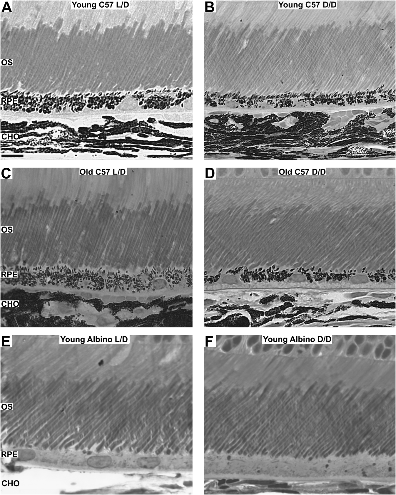
Outer retinal structure was clear in each group (Fig. 1). The OS could be clearly identified as they stand out following processing with osmium tetroxide. In each eye, the majority of central regions contained OS that were aligned and could be traced from their base to their tip at the RPE within a single cutting plane. Although the location of OS tips did not vary, in a small proportion of cells, the junctions between the OS and the inner segment varied with OS extending deeply toward ONL. It has been argued that these may be cone photoreceptors (Carter-Dawson & LaVail, Reference Carter-Dawson and LaVail1979). No attempt was made to differentiate between these, rather all were included in the analysis, but as the sample sizes were large, this element of variability was minimal. The number of OSs counted per unit length did not differ significantly between the groups (ANOVA: P = 0.659, df = 8). Likewise, there were no statistically significant differences in the thickness of the ONL between aged matched pigmented and albino phenotypes (Mann–Whitney U: P = 0.273), although it was thinner in the albinos than the age matched pigmented mice. However, the albino mice had significantly fewer cells in their ONL compared with the pigmented mice (Mann–Whitney U: P = 0.025). This may reflect the fewer number of photoreceptors found in albinos (Jeffery et al., Reference Jeffery, Brem and Montoliu1997; Grant et al., Reference Grant, Patel, Philp, Grey, Lucas, Foster, Bowmaker and Jeffery2001), although it is not clear why this is not reflected in the number of OSs.
Outer retina in young pigmented (A and B: 6-week-old), old pigmented (C and D: 1-year-old), and young albino (E and F: 6-week-old) mice maintained in cyclic illuminance (12:12 L/D) and dark (12:12 D/D) environment. No significant difference in OS length could be found between any of the groups. See text for statistical values. RPE = retinal pigment epithelium at the bottom. OS = outer segment. CHO = choroid. Scale bar = 103 m. 213 × 267 mm (300 × 300 DPI).

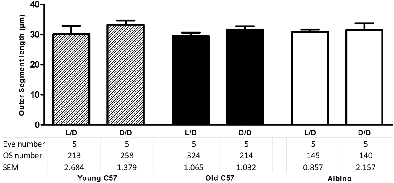
Measurements of OS length failed to reveal any significant difference between mice exposed to the two light environments in each of the young pigmented, old pigmented, or in the albino groups (ANOVA: P = 0.175, df = 29). However, in spite of this, in each group, mean OS lengths were slightly longer in D/D mice than in 12:12 L/D, but this never came close to being statistically significant in any of the groups (t-test, df = 4: Young C57 L/D vs. D/D: P = 0.180; Old C57 L/D vs. D/D: P = 0.112; Albino L/D vs. D/D: P = 0.374; Fig. 2).
Bar charts of photoreceptor OS length in young pigmented (6 weeks), old pigmented (1 year), and young albino (6 weeks) mice maintained in cyclic illuminance (12:12 L/D) (50 lux) and dark (12:12 D/D) environment. No significant differences in OS length were being found between the groups. The number of individual OSs and eyes per animal group is given at the bottom. Error bars = SEM. 170 × 83mm (300 × 300 DPI).
Discussion
These results clearly demonstrate that there is no statistically significant lengthening of photoreceptor OSs in either normal C57BL/6J mice or in albino Balb/c mice when maintained in darkness compared with those maintained on a normal light dark cycle. However, within each of the three groups, there appeared to be a small trend in favor of longer OS in animals maintained in the dark. But this was minimal, particularly when one takes into account the extensive sampling undertaken in this study and the relatively small variability in the data.
These results stand in stark contrast to an extensive literature claiming that there is a marked extension of OSs in reduced light by as much as 90% (Penn & Anderson, Reference Penn and Anderson1987), although other studies have been lower at 78% (Penn & Williams, Reference Penn and Williams1986), and others have been as low as 25% (Battelle & LaVail, Reference Battelle and LaVail1978) and 30% (Schremser & Williams, Reference Schremser and Williams1995), while at best we found an approximate 9% nonsignificant increase in the mean OS length, but in only one group, the young C57 mice. Further, in all previous studies, it was very difficult to assess the number of OS measured or the methods employed. Some studies used animals in reduced light for up to 88 days (Battelle & LaVail, Reference Battelle and LaVail1978), whereas others did so for only 16 h (Penn & Williams, Reference Penn and Williams1986). Battelle and LaVail (Reference Battelle and LaVail1978) seem to have examined approximately 150 OS, others less, and some fail to give any clear indication of numbers at all. Commonly, there is no statistical analysis of data or even error bars on graphs revealing variability. This is in contrast to our study where approximately 1300 OS were measured in 30 mice. The period of darkness employed here was similar to that in some previous studies and is consistent with complete OS renewal, which takes approximately 10 days (Young, Reference Young1967).
One possible explanation for the results we have obtained is that photostasis is in fact a significant adaptive mechanism for long-term retinal adaptation but that somehow our mice were exposed to light during the 21 days when we thought they were in complete darkness. However, there are compelling reasons for this being unlikely. First, the units in which the mice were housed were large-purpose built, commercially constructed, and capable of housing many cages. They were in a dedicated room. They have a single compartment for housing. When purchased, they were tested for light penetration with both a light meter and by placing a member of staff in the cabinet and allowing them to dark adapt. Neither the light meter nor the subject detected any light. While our mice were housed in these, other experiments were also taking place in them and the results of these were consistent with the animals having been in complete darkness. These included a mouse model of retinal degeneration that was triggered by light. No degeneration was noted or shortening of OSs. Second, photostasis has been claimed to be an adaptive mechanism even when animals were exposed to low levels of illumination (Battelle & LaVail, Reference Battelle and LaVail1978; Penn & Williams, Reference Penn and Williams1986; Penn & Anderson, Reference Penn and Anderson1987; Schremser & Williams, Reference Schremser and Williams1995). Hence, had there been a low level of light contamination, one might have still expected an effect on OS length, which was not there. Finally, it could be argued that there may have been brief inadvertent light exposure that only affected our mice. Given the design of the containers, this would not be possible. However, the albino mice were maintained in darkness at a different time to the pigmented animals. Hence, one might then be forced to argue that the two groups were both inadvertently exposed to light independently without affecting other experiments, which seems very unlikely.
An additional potential problem is that mice were removed from the boxes and killed directly. During this brief time, they were exposed to indirect light. However, the time between removal from the dark box and killing was of the order of 1 min maximum, and in many cases, it was shorter. Eyes were removed at the point of death and placed directly into fixative. It is highly unlikely that OSs changed significantly during this time window. The process of OS removal and tip ingestion by the RPE probably takes tens of minutes if not more, although it has never been directly measured. Further, if in this period there had been a significant shortening of OSs, this would have been reflected in some type of change at the RPE/OS tip junction in the D/D groups detected at high magnification at which we viewed our images. This was not the case. If such light exposure had a role to play, one might also expect variability in the D/D groups to be consistently larger than in the L/D groups, but again this was not the case. Hence, we do not think that this very brief exposure had any impact on our results.
Other studies have used minimal lighting perhaps for the purpose of animal husbandry, but no rationale has been offered for the varied low level lighting or their durations employed by different studies. However, Daly et al. (Reference Daly, DiLeonardo, Balkema and Balkema2004) used very dim light (2 × 10⁻⁴ cd/m2) and appear to have strikingly similar results to those shown here using exactly the same strain of pigmented mice, showing no difference between L/D and D/D reared animals. Their study was the only one to include pigmented animals. The similarity of the data between this study and ours implies that neither dim light nor its absence drives photostasis in pigmented mice.
We initially hypothesized that the use of albino mice might represent the reason why studies had reported excess OS lengthening in the dark. There were three reasons for this. First, albinos have a mutation that targets the RPE, which is responsible for OS phagocytosis. Second, Daly et al. (Reference Daly, DiLeonardo, Balkema and Balkema2004) appear to show stasis in albino but not pigmented mice, but offered no analysis. Third, it has been shown that albino RPE is highly polyploid (Adams et al., Reference Adams, Shahabi, Hoh-Kam and Jeffery2010), and there is evidence that this impacts on their function (Shahabi et al., Reference Shahabi, Golding, Lenassi and Jeffery2011). In spite of these compelling reasons, we could find no significant evidence for photostasis in the albino mice. On the contrary, while all groups showed a very limited nonsignificant extension of OS in the dark, this was least obvious in albinos.
Photostasis is widely accepted as a long-term mechanism of visual adaptation in mammals, both in terms of changes in OS length, which we have examined here, and rhodopsin content, which we have not addressed. However, Daly et al. (Reference Daly, DiLeonardo, Balkema and Balkema2004) did start to question the validity of the concept. They found that there was evidence for abnormal rhodopsin regulation in albino mice and claimed that these animals had an improper input into the photostasis mechanism. However, overall their data are incomplete and difficult to understand. It is not stated how many OSs were measured, the graphs for relative OS length in pigmented animals in normal light and dim light are similar to those presented here, but means and statistics are not provided for differing light condition (Daly et al., Reference Daly, DiLeonardo, Balkema and Balkema2004, Figure 7A). Relative differences in OS length are apparent for albinos under their different lighting conditions, but again no analysis is provided. Part of the reason for this is that the focus of the study was more on rhodopsin concentrations rather than a large detailed study of OS lengths. Daly et al. (Reference Daly, DiLeonardo, Balkema and Balkema2004) did show that rhodopsin levels were greater in animals maintained in darkness than light, and overall there was less rhodopsin in albinos than in pigmented animals. This reflects an earlier study by Grant et al. (Reference Grant, Patel, Philp, Grey, Lucas, Foster, Bowmaker and Jeffery2001) showing reduced rhodopsin in albinos compared with pigmented phenotypes.
The other key problems associated with albinos, which have been so widely used in photostasis, are that there is growing evidence that they may have faulty phagocytosis mechanisms as their OSs become enlarged and compressed consistent with their tips not being removed appropriately (Shahabi et al., Reference Shahabi, Golding, Lenassi and Jeffery2011). Also the morphology of their RPE cells becomes progressively abnormal due to the development of polyploidy (Adams et al., Reference Adams, Shahabi, Hoh-Kam and Jeffery2010). Interestingly, in light of this, Penn and Williams (Reference Penn and Williams1986) stated that “The albino rat has been questioned as a model for retinal research in the past…” Also they concluded that “It is possible, perhaps, that photostasis may be an anomalous phenomenon, applicable only to the albino rat.” Our data do not support this notion, as we failed to find significant evidence of photostasis in either pigmentation phenotype.
Given these problems, it is surprising how resilient the concept of photostasis has remained. It is known that sensory receptors of the olfactory (Wang et al., Reference Wang, Okutani, Murata, Taniguchi, Namba and Kaba2012) and the auditory systems (Goutman et al., Reference Goutman, Fuchs and Glowatzki2005; Rabbitt & Brownell, Reference Rabbitt and Brownell2011) are modulated in response to changes in sensory stimulation. In the cochlea, outer hair cell electromotility and stiffness are directly regulated through efferent neurons modulating their function (Dallos et al., Reference Dallos, He, Lin, Sziklai, Mehta and Evans1997; Castellano-Munoz et al., Reference Castellano-Munoz, Israel and Hudspeth2010). Further, it is clear that rhabdomer size is modulated by light in some invertebrates (White & Lord, Reference White and Lord1975; Behrens & Krebs, Reference Behrens and Krebs1976; Blest, Reference Blest1978). Hence, it is not surprising that similar mechanisms have been sought in mammalian photoreceptors. However, previous mammalian studies on photostasis have been highly variable in their approach and outcome. We hope that by standardizing some of these variables with animals kept in complete darkness for a minimum of two phases of complete OS renewal and examining large OS numbers statistically that we have established a baseline where we can clearly say that significant photostasis does not occur. It is possible that under different circumstances, OS lengthening may take place. However, to date, our data significantly question the notion of photostasis in mammals.
Acknowledgment
We are grateful to Anne-Kathrin Wesselmann and Jaimie Hoh Kam for critical reading and valuable comments and to Chris Hogg for technical assistance. This research was supported by The Rosetrees Trust.