Introduction
A catheter ablation procedure is a treatment option currently selected for children following diagnostic electrophysiology studies because of its high success rate and low complication rate. Success rates of>95% and a complication rate of 3–5% have been reported for catheter ablation in cases with supraventricular tachycardia. Reference Sawan, Eitel, Thiele and Tilz1,Reference Swissa, Birk and Dagan2
Radiofrequency (RF) catheter ablation of the atrioventricular accessory pathways has become the first choice in the treatment of paediatric tachycardia. However, in performing safe and effective radiofrequency ablation in critical regions of the septum (close to the atrioventricular node or close to the His bundle), there continue to be difficulties in ablation of the anteroseptal and midseptal accessory pathways, which carry the risk of complete heart block. Therefore, cryoablation of supraventricular arrhythmias has emerged as an alternative to radiofrequency ablation in children. It is an especially appropriate option when ablation is required in a location close to normal signalling tissue because of the advantages of cryothermal energy, such as increasing catheter stability and the ability to reverse lesions during cryomapping. Reference Ergul, Tola, Kiplapinar, Akdeniz, Saygi and Tuzcu3
While electrophysiological studies and catheter ablation are generally performed under fluoroscopy, the non-use of fluoroscopy or decreasing the usage time has become the most important target with the increasing use of 3-dimensional electroanatomic mapping systems.
The aim of this study was to evaluate the outcomes of patients who underwent electrophysiology studies and a catheter ablation procedure in our centre.
Patients and method
A retrospective screening was made of paediatric patients who underwent catheter ablation and were followed up in the Arrhythmia Clinic of the Paediatric Cardiology Department of Umraniye Training and Research Hospital between November 2017 and December 2024, or who were referred for ablation from another centre. The demographic data, clinical characteristics, electrocardiographic and Holter findings, echocardiographic examination results, and data related to the ablation procedure and follow-up were retrieved from the patient files. Patients who had an intracardiac defibrillator, pacemaker, or ventricular arrhythmia were excluded from the study. Approval for the study was granted by the hospital Ethics Committee.
EPS and catheter ablation procedure
As standard procedure for all the patients, appropriate sheaths were placed on the right and left femoral veins under general anaesthesia. A laryngeal mask was used during the procedure. The inducibility of tachycardia was evaluated by increasing the good atrial signals obtained and programmed atrial pacing. Using a catheter placed in the right atrium under the guidance of a 3-dimensional electroanatomic mapping system (3D EAM; EnSite Velocity system, St. Jude Medical), the right atrium anatomy was established. Under the guidance of the EnSite Velocity system, catheters were placed in the right atrium, coronary sinus, and right ventricle without the use of fluoroscopy, and right atrium mapping was performed. Ventricular and atrial stimulation protocols were used to evaluate the signalling characteristics and determine the tachycardia mechanism. Basic measurements were taken.
When the left-side arrhythmogenic focus was determined in the patient, it was first investigated whether or not there was a patent foramen ovale. When the foramen ovale was present, the left atrium was reached via the patent foramen ovale and ablation was performed. When there was no foramen ovale, trans-septal punction was made and passed through for ablation to be performed on the left atrium. Cryoablation was used in atrioventricular nodal re-entry tachycardia and in right anterolateral, anteroseptal, and midseptal accessory pathway ablations. Cryocatheters were selected as 7F 6 mm or 9F 8 mm tips (Medtronic, Minneapolis, USA) according to the patient’s weight, type of arrhythmia, and the operator’s preference. The radiofrequency ablation catheters were selected as a 7F 4 mm tip ablation catheter (Medtronic) or a 7F with irrigation radiofrequency ablation catheter (St Jude Medical). All the ablations were performed by the same paediatric electrophysiologist (M.K.). Following successful ablation, a period of 20 mins was waited to evaluate recurrence. After this period, the ablation success and inducibility of tachycardia were evaluated with programmed and atrial burst stimulations under basal conditions and intravenous isoprenaline infusion. In the presence of an accessory pathway, ablation was defined as successful with antegrade and retrograde signal block with adenosine.
On the day of the procedure, the patients were monitored with 12-derivation electrocardiography (ECG). At 24 hours after the procedure, ECG and echocardiography follow-up were performed and patients were then discharged. Routine follow-up examinations were made at 15 days, 4 months, and 1 year after the procedure. At each follow-up examination, the patients were asked about symptoms, ECG and echocardiography were performed, and if there were complaints of palpitations, the patient was evaluated with Holter or an event recorder with respect to recurrence.
Complications
Adverse events related to the applied procedure were defined as major and minor complications. A major complication was defined as an adverse event requiring intervention for treatment. Events such as catheter-related perforation, pericardial effusion, and embolism requiring an additional invasive procedure such as pericardiocentesis, vascular surgery, or blood transfusion were considered major complications. A minor complication was defined as an adverse event that did not require intervention and could be managed on an outpatient basis. Atrioventricular block and thrombus formation in the vessel were considered minor complications.
Statistical analysis
Data obtained in the study were analysed statistically using SPSS for Windows vn. 25.0 software (IBM Corpn., Armonk, NY, USA). Continuous variables with normal distribution were stated as mean ± standard deviation (SD) values and those not showing normal distribution as median (range) values. Categorical variables were stated as number (n) and percentage (%). For the comparisons between groups, the Student’s t-test, Chi-square test, and Mann–Whitney U-test were used as appropriate. Correlations between variables were determined using the Spearman correlation coefficient. A value of p < 0.05 was accepted as the level of statistical significance.
Results
The data were screened from a total of 586 patients (M/F = 300/286), aged 3–20 years, who underwent an ablation procedure in our centre between 2017 and 2024. All but two of the patients were between the ages of 3 and 18 years (a 19-year-old patient with Ebstein anomaly and a 20-year-old patient with an operated atrial septal defect and tricuspid valve anomaly).
In 5 (0.85%) patients who presented with complaints of palpitations and no documented supraventricular tachycardia, diagnostic electrophysiology studies were performed, and as tachycardia could not be stimulated, the ablation procedure was not performed.
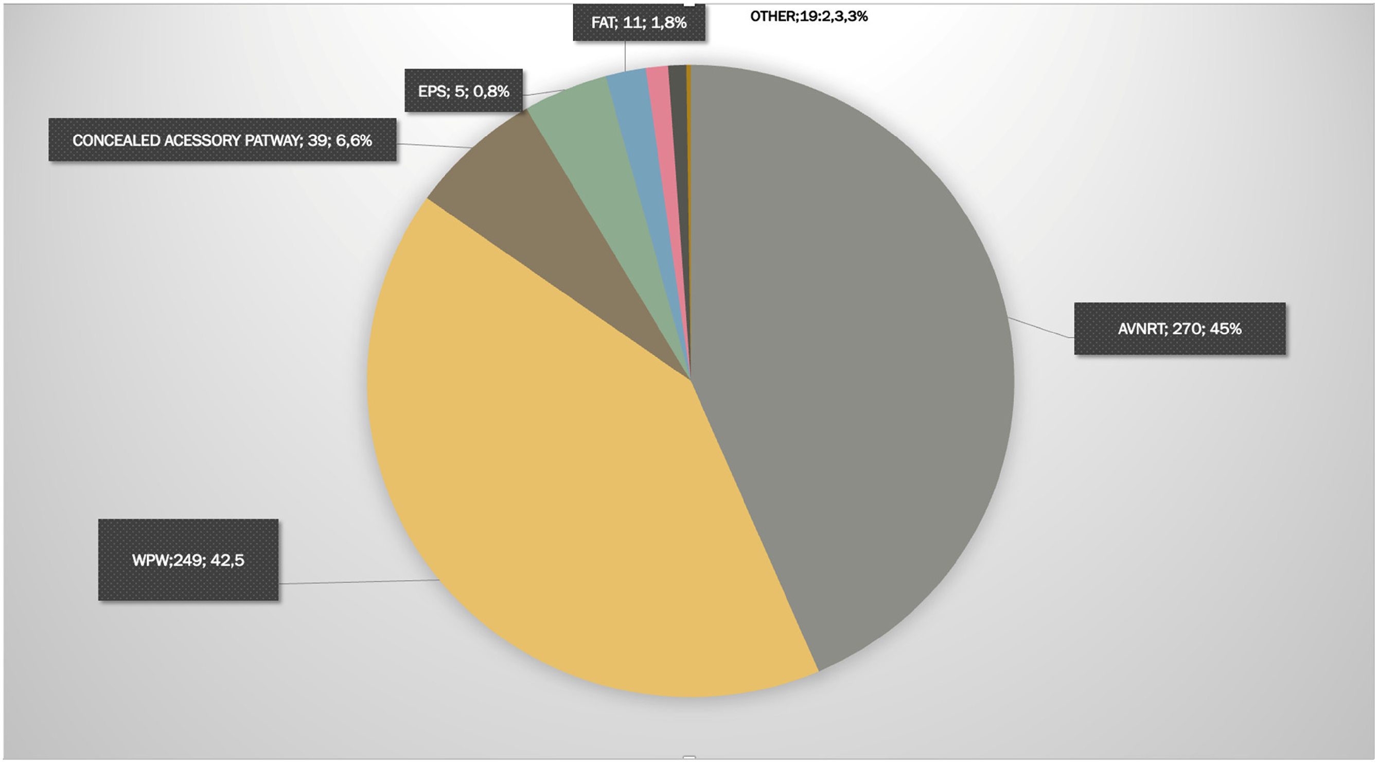
The procedure was applied to 288 (49 %) patients with atrioventricular re-entry tachycardia (Wolf Parkinson White 86.4%-n:249; occult accessory pathway 13.9%- n:39), and to 270 (45%) with atrioventricular nodal re-entry tachycardia. Eleven (1.8%) patients with focal atrial tachycardia, 11 (1.8%) with frequent extrasystole or ventricular tachycardia, 6 (1%) with supraventricular premature beat (SVE), and 2 patients with a Mahaim diagnosis underwent the procedure (Figure 1).
Diagnoses of patients who underwent the procedure.
Fluoroscopy was used during the procedure in 75 (12.8 %) patients, and the mean duration of fluoroscopy was 5.2 ± 3.2 mins (range 0.2–19.6 mins). Of these 75 patients, trans-septal punction was performed on the left side for the procedure to be applied in 71 patients. Fluoroscopy was used to guide the catheters during the procedure in 4 patients. With the exception of 2 patients in whom the procedure was very prolonged, the duration of fluoroscopy was shorter than 10 mins.
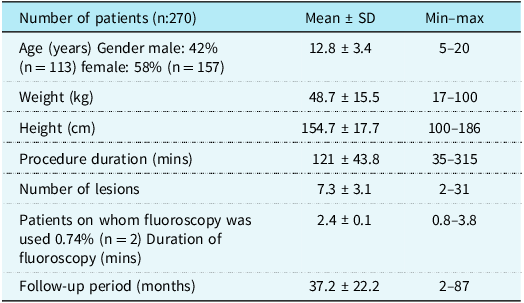
The patient group diagnosed with AVNRT
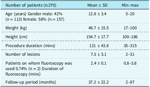
Ablation was performed because of atrioventricular nodal re-entry tachycardia in 270 patients. The characteristics of these patients are shown in Table 1. In 248 (92%) of these patients, the cryoablation procedure alone was performed with 6 mm or 8 mm cryoablation catheters. In the other 22 (8%) patients, radiofrequency catheters were used for ablation. In 9 of these patients, radiofrequency catheters were placed in the lesions after cryoablation, and a radiofrequency procedure was performed when tachycardia stimulus reoccurred. Since 2024, our centre has performed ablations using only radiofrequency catheters in atrioventricular nodal re-entry tachycardia patients with appropriate age and weight. The other 13 patients were patients who underwent ablation only with radiofrequency catheters. In 2 of the patients who underwent ablation because of atrioventricular nodal re-entry tachycardia, trans-septal punction was made on the left side, and radiofrequency ablation was performed. One of these patients also had a left accessory hidden pathway, so ablation was performed on the left side. The other patient was a patient with atypical atrioventricular nodal re-entry tachycardia who underwent ablation only on the right side in the first procedure. Due to recurrence, ablation was performed on the left side in the second procedure. Fluoroscopy was not used except for these two patients.
The characteristics of patients with atrioventricular nodal re-entry tachycardia
The procedure was successful in 97.7% of the patients who underwent ablation because of atrioventricular nodal re-entry tachycardia. Partial success was obtained in 2 (0.7%) patients, and failure was determined in 4 (1.5%). No permanent complications due to the procedure were observed. The procedure was terminated as a complete atrioventricular block developed during the procedure in 1 patient, and this block recovered 5 days after the procedure.
Recurrence was seen after the procedure in 12 (4.4%) patients. The time to the development of recurrence was determined to be a mean 11.9 ± 10.1 months (range, 1–36 months). No statistically significant correlation was determined between recurrence and the number of lesions (p > 0.05) and duration of the procedure (p > 0.05).
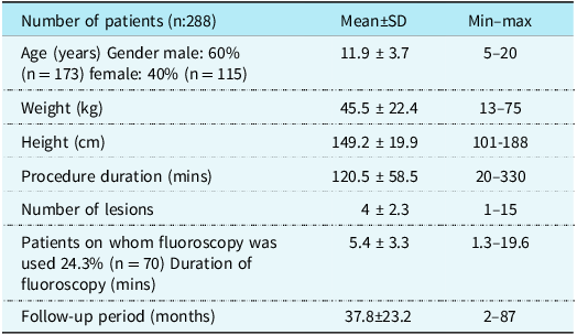
The patient group diagnosed with atrioventricular re-entry tachycardia (AVRT)
The ablation procedure was performed on 288 (49%) patients with a diagnosis of atrioventricular re-entry tachycardia, comprising 249 (86.4%) with Wolf Parkinson White, and 39 (13.9%) with occult accessory pathway. The characteristics of these patients are shown in Table 2. In these 45(15.6%) patients with wolff parkinson white (WPW), the ablation procedure was not performed, as tachycardia could not be stimulated during electrophysiology studies, and there was not a high risk of accessory pathway sudden death. Of the patients who underwent the ablation procedure, radiofrequency ablation was applied with 4F radiofrequency and/or radiofrequency catheters with irrigation to 205 (71%), and cryoablation was performed on 83 (29%) patients. In this group, the procedure was applied on the left side by performing trans-septal punction in 68 patients. Fluoroscopy was used in a total of 70 (24.3%) patients, including the 68 with trans-septal punction. In one patient, the left side was accessed through the foramen ovale and short-term fluoroscopy was performed for catheter control. In one patient, short-term fluoroscopy was used for catheter manipulation. The duration of fluoroscopy was determined to be mean 5.4 ± 3.3 mins.
The characteristics of patients with atrioventricular re-entry tachycardia
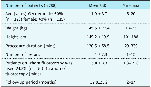
An accessory pathway was determined on the right most often in a posteroseptal localisation (29%) and on the left in a lateral localisation (28.2%).
Following the procedure, the acute success rate was 96.2%, partial success was 2.4%, and the procedure was accepted as a failure in 6 (2%) patients. No permanent complications due to the procedure were observed.
Recurrence after the procedure was seen in 17 (5.9%) patients. The duration of the procedure was determined to be significantly shorter in the patients with recurrence (p = 0.008). No statistically significant correlation was determined between recurrence and lesion localisation (p > 0.05).
The patient group diagnosed with focal atrial tachycardia
The ablation procedure was performed on 11 patients with a diagnosis of focal atrial tachycardia. The procedure was performed with radiofrequency ablation catheters. Fluoroscopy was used in 2 patients, and the mean duration of fluoroscopy was 2.4±0.14 mins. One of these patients underwent trans-septal puncture, and the other patient underwent short-term fluoroscopy for catheter manipulation. The procedure was successful in all the patients, and no recurrence was observed in any patient.
Discussion
Catheter ablation of supraventricular tachycardia has become the standard treatment procedure in recent years. The success rates of ablation treatment are very high for most types of supraventricular tachycardia. Generally, catheter ablation is more cost-effective than long-term drug treatment. In supraventricular tachycardia, with the exception of atrial fibrillation, the complication rate of catheter ablations is 3–5%, and the serious complication rate is<1%. Reference Sawan, Eitel, Thiele and Tilz1,Reference Karacan, Çelik, Akdeniz and Tuzcu4 The EUROPA study, a prospective collection of catheter ablation data from 5 European paediatric arrhythmia centres, demonstrated the overall high efficacy and safety of catheter ablation of various tachycardia substrates in paediatric patients. Reference Krause, Paul and Bella5
In the treatment of atrioventricular nodal re-entry tachycardia, radiofrequency ablation is the preferred treatment method in the adult age group because of high success rates and low recurrence rates. However, there is a risk of complete atrioventricular block requiring pacemaker implantation at the rate of 0.5–2%. Reference Sawan, Eitel, Thiele and Tilz1,Reference Swissa, Birk and Dagan2,Reference Karacan, Çelik, Akdeniz and Tuzcu4 Therefore, cryoablation has become an alternative treatment method to radiofrequency in recent years, especially in children and young people. Many studies have compared the two methods. Reference Swissa, Birk and Dagan2,Reference Karacan, Çelik, Akdeniz and Tuzcu4,Reference Rodriguez-Entem and Exposito6–Reference Jabarkhyl, Marwat and Haider10 The success rates of cryoablation have been shown to be increasing, with success of the procedure determined at rates of 83–100%. There has also been a significant decrease (4–6.5%) in recurrence rates that were previously reported to be high (22–27%). Reference Swissa, Birk and Dagan2,Reference Karacan, Çelik, Akdeniz and Tuzcu4,Reference Reents, Springer and Ammar8 Cryoablation is applied in our centre as the first option in the treatment of atrioventricular nodal re-entry tachycardia. The procedure was performed to 92% of the current study patients with 6 mm (30%) and 8 mm (62%) tip cryoablation catheters. Ablation was delivered to mean 7.3 lesions during the procedure with cryocatheters. In 22 (8%) patients, where success could not be obtained with cryoablation, radiofrequency ablation was performed with a 4 mm radiofrequency catheter during the procedure. Acute success of the procedure was found to be 97.7%, and partial success was 0.7%. In the follow-up period of a mean 37.5 months (range, 2–90 months), the recurrence rate was determined to be 4.4%, with the mean time to recurrence of 12 months. The results obtained in this study were consistent with the high success rates and low recurrence rates in the literature.
In previous studies, the decrease in the recurrence rate in cryoablation has been attributed to the use of catheters with a larger tip compared to radiofrequency catheters, and the number of lesions was greater. Reference Karacan, Çelik, Akdeniz and Tuzcu4,Reference Drago, Russo, Silvetti, De Santis, Iodice and Naso Onofrio11 In the current study, no statistically significant correlation was determined between recurrence and the number of lesions and duration of the procedure.
In the treatment of atrioventricular re-entry tachycardia in children, radiofrequency ablation is performed as the first option, as it is for the adult age group. The success of the procedure is lower in children than in adults. However, with an increase in the number of centres and operators and 3-D mapping systems coming into widespread use, the radiofrequency ablation success rates have increased, and complication rates have decreased. While paediatric radiofrequency ablation success rates have been reported to be 90–92%, these success rates increase to 95% with the use of 3-D systems. Reference Krause, Paul and Bella5,Reference Kugler, Dandford, Houston and Felix12,Reference Lee, Hwang, Chen, Tai, Chen and Chiang13,Reference He, Yi and Meiting14 Recurrence rates have been reported to have decreased from 10% to 5.3%. Reference Krause, Paul and Bella5,Reference Van Hare, Javitz and Carmelli15,Reference Liu, Jacobsen, Pehrson and Chen16 In our centre, radiofrequency ablation was performed more (71%) for the patients diagnosed with atrioventricular re-entry tachycardia. The acute success rate was found to be 96%, and the recurrence rate 5.9%. The most important reason for the high recurrence rate in the paediatric patient group may be due to the high frequency of multiple accessory pathways. The frequency of multiple accessory pathways has been reported to be 3–22% in adults, and this rate is much higher in children. Reference Kugler, Dandford, Houston and Felix12 The majority of recurrences after a successful procedure in the current study patients were observed to be because of multiple accessory pathways.
Although focal atrial tachycardia is one of the more rarely seen (5–17%) causes of supraventricular tachycardia, it is seen more often in children than in adults. Reference Liu, Jacobsen, Pehrson and Chen16,Reference Kistler, Chieng and Tonchev17 Catheter ablation is the first-stage treatment because of the weak effect of short-term and long-term pharmacological treatment. In focal atrial tachycardia, the radiofrequency ablation procedure is based on accurate mapping of the region. Acute success rates have been reported in the range of 69– 100% in ablation series. Reference Krause, Paul and Bella5,Reference Liu, Jacobsen, Pehrson and Chen16,Reference Roberts-Thomson, Kistler and Kalman18 Recurrence rates have fallen significantly with the detailed mapping of the atrium with 3-D mapping technology. Previously reported recurrence rates of up to 33% have decreased to 7%. Reference He, Yi and Meiting14–Reference Liu, Jacobsen, Pehrson and Chen16 The patient group with focal atrial tachycardia constituted the smallest group (11 patients, 1.8%) of the current study patients. The procedure success was 100%, and no recurrence was observed in this group that underwent radiofrequency.
In catheter ablation treatment in the paediatric patient group, it is very important to reduce the duration of fluoroscopy during the procedure to as short a time as possible. Fluoroscopy is traditionally used for catheter placement and guidance of the trans-septal procedure. The mean fluoroscopy duration during the ablation procedure has been reported to be 16–27 mins, some publications have stated it to be 18–38 mins, and it has also been reported to exceed 50 mins in 20% of paediatric cases. Reference Sawan, Eitel, Thiele and Tilz1,Reference Kistler, Chieng and Tonchev17 As children have a longer life expectancy and are more sensitive to radiation, it is important that the patient is protected against the short and long-term side-effects of radiation. With the more widespread use of 3-D electroanatomic mapping systems, fluoroscopy durations have significantly decreased. Reference Kistler, Chieng and Tonchev17–Reference Papagiannis, Avramidis, Alexopoulos and Kirvassilis20 The results of previous long-term studies of procedures performed using very little fluoroscopy have shown the long-term results to be very effective and reliable. Reference Karacan, Çelik, Akdeniz and Tuzcu4,Reference Krause, Paul and Bella5,Reference Jan, Yazici and Kalinšek21 It is currently aimed to perform the procedure without using any fluoroscopy, and with this aim, studies have been conducted with the use of intracardiac echocardiography during the procedure. Reference Tuzcu22 Technological developments in the last few years, especially in the fields of electroanatomic mapping and intracardiac echocardiography, have enabled this target to be reached without any sacrifice in reliability, efficacy, and procedure duration. Reference Luani, Zrenner and Basho23–Reference Sánchez, Yanics, Wilson, Doshi, Kurian and Pieper27 In studies that have compared the procedure outcomes using traditional fluoroscopy and without any use of fluoroscopy have determined no difference in procedure success and complication rates. Reference Yazici, Lakič, Prolič Kalinšek, Žižek and Ažman Juvan28–Reference Razminia, Manankil and Eryazici30
Procedures applied in our centre are performed with the principle of limited fluoroscopy use. Fluoroscopy was not used during the procedure in 98% of the patients diagnosed with atrioventricular nodal re-entry tachycardia, 73% of those with atrioventricular re-entry tachycardia, and 82% of those with focal atrial tachycardia. Fluoroscopy was used for a very short time, only for catheter manipulations and trans-septal punction procedures. The reason for the high mean fluoroscopy time was the durations of 13 and 19.6 mins in the two patients diagnosed with WPW. The presence of multiple accessory pathways, performing trans-septal punction, and having to use different catheters prolong both the duration of fluoroscopy and the procedure. In the patients with atrioventricular re-entry tachycardia, the mean fluoroscopy duration was 5.4 mins, and of the 77 patients in this group in whom fluoroscopy was used, trans-septal punction was applied to 65. Especially in the atrioventricular nodal re-entry tachycardia patient group, the target was to perform the procedure without the use of fluoroscopy, and it was used for a mean of 2.4 mins in only 5 patients.
Conclusion
The results of the catheter ablation study performed in our centre demonstrated that catheter ablation using 3D EAM systems on arrhythmia substrates in paediatric patients with normal cardiac anatomy can be performed with limited use of fluoroscopy, low complications, and acute success rates. No major or irreversible minor complications due to the procedure were observed in any patient. As the number of procedures performed with the goal of limited or even zero fluoroscopy increases and the systems used are further developed, the procedures continue with the hope that their success rates will increase and the recurrence rate will further decrease.
Author contribution
Ozlem Sarisoy: Conception, design, data collection and/or processing, analysis and/or interpretation, literature review, and writer.
Ramazan Arı: Data collection and/or processing.
Mehmet Karacan: Conception, design, supervision, and critical review.
Financial support
This research received no specific grant from any funding agency, commercial, or not-for-profit sectors.
Competing interests
The authors have no conflicts of interest to declare.
Ethical standard
The study was approved by the local ethics committee of Istanbul Health Sciences University, Umraniye Training And Research Hospital Scientific Research Ethics Committee (No: B.10.1.TKH.4.34.H.GP.0.01/148, Date: 16.5.2024).