Introduction
The development of a tracheo-arterial fistula is an unusual but severe complication of persistent tracheostomy, with high morbidity and mortality rates. The survival rate following tracheo-arterial fistula bleeding has been reported as 14.3 per cent.Reference Wood and Mathisen1
An aberrant right subclavian artery, termed an arteria lusoria, is the most common aortic arch abnormality and is usually asymptomatic.
Herein, we describe a case of major bleeding from an arteria lusoria aneurysm via a tracheotomy wall fistula, in a patient with muscular dystrophy disease. This unique occurrence emphasises the need for long term vigilance in the care of tracheostomised patients, particularly those with significance comorbidity.
Case report
A 30-year-old man presented to the emergency department with bleeding from his tracheotomy. He had previously received long term treatment for Duchenne muscular dystrophy, and had required invasive assisted ventilation for chronic respiratory failure since the age of 10 years. Hitherto, this treatment had been well tolerated. The patient's mother performed tracheostomy tube placements every week. The last change had been performed using a low pressure cuff tube, without difficulty. Prior to presentation, the patient had developed an abrupt tracheotomy haemorrhage after an acute coughing episode.
On examination, there was no active bleeding, but a bulky blood clot around the tracheostomy tube was noticed. Flexible fibre-optic endoscopic examination of the tracheal tube was unremarkable, with no active bleeding or mucosal erosion. However, removal of the tracheostomy tube induced massive bleeding; this was stopped by over-inflation of the cannula cuff.
Surgical exploration under general anaesthesia was immediately undertaken. The brachiocephalic trunk and carotid arteries were exposed. No vascular injury was seen. Through the tracheotomy, bleeding was identified emerging from an erosion of the posterior tracheal wall. A diagnosis of posterior tracheo-arterial fistula was suggested. A new cuffed tracheostomy tube was placed, and the cuff over-inflated.
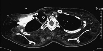
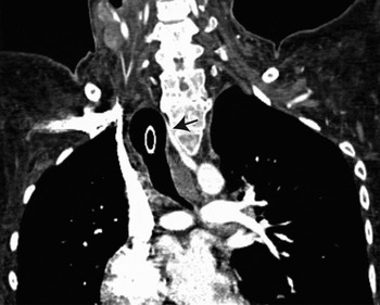
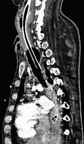
A thoracic computed tomography (CT) scan with contrast injection showed an aberrant right subclavian artery (arteria lusoria) adherent to the damaged tracheal posterior wall. Tracheostomy cuff inflation had achieved emergency haemostasis by crushing the aneurysm against the adjacent dorsal vertebra (Figures 1 and 2). A scoliosis deformity was also observed, causing unusual contact between the trachea, the arteria lusoria anomaly and the spine (Figure 3). As a result of these radiological features, a diagnosis of tracheo-arterial fistula from an aberrant right subclavian artery was highly suspected.
Axial, contrast-enhanced computed tomography scan of the thorax (mediastinal window setting). The aberrant right subclavian artery (arrow) is located lateral to the trachea and behind the oesophagus, next to the spine.

Coronal, contrast-enhanced computed tomography scan of the thorax (mediastinal window setting). The arteria lusoria (arrow) arises from the aortic arch and is pushed back onto the adjacent dorsal vertebra by the tracheostomy cuff projecting into the trachea.

Sagittal, contrast-enhanced computed tomography scan of the thorax (mediastinal window setting), showing a curved indentation of the posterior trachea wall caused by the tracheostomy cuff, allowing haemostasis of the arteria lusoria anomaly (arrow).

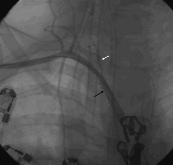
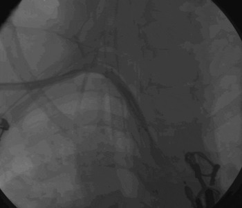
A surgical mediastinal approach was not considered in the first instance, due to the risks of the procedure and the patient's weak general condition. Instead, a vascular interventional radiology procedure was performed via a femoral approach. Initial angiography confirmed the presence of an arteria lusoria aneurysm, with extravasation of contrast material (Figure 4). The aneurysm was ablated by percutaneous implantation of a 7 × 40 mm Fluency Plus® stent graft (Bard Peripheral Vascular, Tempe, Arizona, USA). The self-expanding, covered stent (7 mm in diameter and 40 mm in length) was advanced along a guide wire and positioned at the aneurysm site. Thereafter, no residual bleeding was seen. All adjacent vessels remained permeable, with good patency of the right aberrant subclavian artery (Figure 5).
Pre-treatment arteriography of the supra-aortic branches. Opacification indicates the aberrant right subclavian artery arising from the left. The eroded aneurysm (black arrow) causes active bleeding (white arrow).

Post-treatment arteriography of the right subclavian artery. The self-expanding, covered stent is positioned at the aneurysm site, with good patency of the right subclavian artery and distal branches.

The patient's post-procedure course was uneventful, with no recurrent bleeding or right arm ischaemia. He was admitted to the intensive care unit for medical supervision.
The patient was discharged six weeks later, and the previous home management of his respiratory failure was resumed.
Discussion
Post-tracheotomy bleeding from a tracheo-arterial fistula is unusual, but is one of the most devastating long term complications of tracheotomy. The incidence of tracheo-arterial fistula ranges from 0 to 1 per cent, with a mean of 0.3 per cent.Reference Allan and Wright2–Reference Wright4 The brachiocephalic (innominate) arteries and the carotid arteries are often involved.
An aberrant right subclavian artery, termed an arteria lusoria, is the most common aortic arch abnormality, occurring in 0.5 to 2.5 per cent of the general population.Reference Myers, Fasel, Kalangos and Gailloud5 In this vascular malformation, four vessels arise sequentially from the aortic arch; the arteria lusoria is the last one, after the left subclavian artery. The aberrant right subclavian artery courses alone from the left side to the right side of the chest, usually behind the oesophagus (in 80 per cent of cases), as in our patient. In the remaining cases, it crosses between the oesophagus and the trachea (15 per cent), or in front of the trachea (5 per cent).Reference Myers, Fasel, Kalangos and Gailloud5
This vascular malformation is usually asymptomatic, occurring as an isolated anomaly. However, in rare cases it can be associated with vascular or cardiac abnormalities, with clinical consequences. Atherosclerosis can induce ‘dysphagia lusoria’, with oesophageal compression.Reference Levitt and Richter6 Dyspnoea or chronic coughing can occur in cases of tracheal compression. Finally, aneurysmal dilation of this artery with oesophageal fistulisation has been described.Reference Myers, Fasel, Kalangos and Gailloud5, Reference Glock, Chaffai, Tasrini, Meusberger, Marre and Portales7 There have been no previous reports of arteria lusoria fistulisation into the trachea after previous tracheotomy. Thus, an aberrant right subclavian artery should be kept in mind as a potential cause of tracheotomy bleeding, similar to brachiocephalic and carotid arteries.
Patients with Duchenne muscular dystrophy and a tracheostomy seem to have a higher incidence of tracheo-arterial fistula. In these patients, nearly 10 per cent of post-tracheotomy haemorrhages are induced by a tracheo-arterial fistula.Reference Saito, Sawabata, Matsumura, Nozaki, Fujimura and Shinno8 Some reports indicate that, in this patient population, tracheo-arterial fistula may be associated with the frequent occurrence of spinal deformity.Reference Saito, Sawabata, Matsumura, Nozaki, Fujimura and Shinno8 Scoliosis often constricts the chest cavity of such patients, resulting in unusual anatomical relations of the trachea and the brachiocephalic arteries. In our patient, a combination of a retro-oesophageal arteria lusoria and spinal scoliosis appeared to result in increased mechanical forces pressing the artery against the tracheostomy cannula.
The spinal deformities associated with Duchenne muscular dystrophy are gradually progressive; thus, so is the resulting vascular compression. This fact probably explains the long delay (10 years) between our patient's tracheotomy procedure and his bleeding event; this is one of the longest such delays described, and is in agreement with an aetiology of progressive spinal distortion and aneurysm formation.Reference Schaefer and Irwin3, Reference Saito, Sawabata, Matsumura, Nozaki, Fujimura and Shinno8, Reference Epstein9 Thus, a post-tracheotomy haemorrhage can appear several months after the surgical procedure. A tracheo-arterial fistula must be always suspected in cases of tracheostomy bleeding, especially in Duchenne muscular dystrophy patients.
• Tracheo-arterial fistulae usually involve the brachiocephalic (innominate) or carotid arteries
• An aberrant right subclavian artery, termed an arteria lusoria, is the most common aortic arch abnormality
• Aneurysmal disruption of an arteria lusoria in patients with spinal deformities should be kept in mind as a potential cause of tracheotomy bleeding in patients requiring long term follow up
Several precautions have been advised to prevent the development of tracheo-arterial fistula. The tracheotomy should not be performed under the third tracheal ring.Reference Wood and Mathisen1, Reference Saito, Sawabata, Matsumura, Nozaki, Fujimura and Shinno8, Reference Epstein9 Tracheostomy cuff pressure should be limited to 20 mmHg.Reference Wood and Mathisen1, Reference Wright4, Reference Saito, Sawabata, Matsumura, Nozaki, Fujimura and Shinno8 In the intensive care unit, neck hyperextension and repetitive pulling on the cannula should be avoided.Reference Wright4, Reference Saito, Sawabata, Matsumura, Nozaki, Fujimura and Shinno8–Reference Gelman, Aro and Weiss10 Tracheotomy infections must be treated adequately to avoid tissue necrosis.Reference Gelman, Aro and Weiss10 In patients with myopathy, it is suggested to perform a thoracic CT scan before the tracheotomy in order to clearly establish the anatomical location of blood vessels. The follow up of such patients should include repeated CT scanning; however, no clear scheduling procedure has been described.Reference Epstein9
Conclusion
Post-tracheotomy bleeding from a tracheo-arterial fistula is unusual. It is generally caused by a brachiocephalic trunk or carotid aneurysm. However, the presence of an aberrant right subclavian artery, termed an arteria lusoria, is not rare, and surgeons should be aware of this potential aetiology for tracheo-arterial fistula. Moreover, the development of tracheo-arterial fistula is probably influenced by spinal deformities such as those found in Duchenne muscular dystrophy patients, which may result in unusual anatomical relations of the mediastinal vessels. Such distortion is gradually progressive, and long term vigilance is hence required in the management of Duchenne muscular dystrophy patients with a tracheostomy.