Introduction
Petrous bone cholesteatoma is a rare lesion that can be acquired or congenital. Sanna et al. have divided petrous bone cholesteatoma into five categories according to its extension in the petrous bone: supralabyrinthine, infralabyrinthine, massive labyrinthine, apical and infralabyrinthine-apical.Reference Omran, De Denato, Piccirillo, Leone and Sanna1
Radical surgical removal is the mainstay of treatment for petrous bone cholesteatoma. However, management is very challenging if the condition occurs in the only hearing ear; post-operative hearing preservation is rarely achieved, even if the otic capsule is partially spared by the disease.
In this paper, we report the first successful cochlear implantation in a patient with a petrous bone cholesteatoma, performed following total eradication of the disease, as a single-stage procedure.
Case presentation
A 63-year-old man presented to our institution with a four-year history of right-sided, progressive hearing loss with recent onset of vertigo. Thirty years prior to this presentation, the patient had undergone a left radical mastoidectomy at another centre, which had resulted in total left-sided deafness. At the age of 60 years, the patient had experienced a sudden, total, right-sided hearing loss, with subsequent partial recovery.
Physical examination revealed a clean left radical mastoidectomy cavity, together with a small (2 mm), dry, right tympanic membrane postero-superior perforation.
Audiological evaluation showed a ‘dead ear’ on the left side, and right-sided mixed hearing loss with a hearing threshold of 70 dB, an air–bone gap of 20 dB and a speech discrimination score (SDS) of 100 per cent. The patient was using a hearing aid in his right ear.
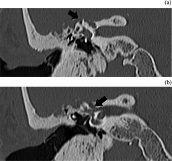
Computed tomography (CT) and magnetic resonance imaging revealed a right supralabyrinthine petrous bone cholesteatoma, with erosion of the superior semicircular canal and the roof of the internal auditory canal (Figure 1).
Coronal computed tomography scans showing (a) the supralabyrinthine cholesteatoma eroding the superior semicircular canal (arrow), and (b) the cholesteatoma eroding the internal auditory canal (arrow).

Despite 30 years of hearing deprivation, good responses were recorded during a left promontory stimulation test. Our suggestion to the patient was to eradicate the petrous bone cholesteatoma and to rehabilitate the contralateral ear with a cochlear implant (CI). Due to the high risk of post-operative right-sided deafness, and the patient's short experience of bilateral deafness following his sudden right-sided hearing loss, we decided to perform a left cochlear implantation first, and then to proceed with a combined approach (transmastoid and middle cranial fossa) for treatment of the right petrous bone cholesteatoma.
In June 2009, the patient underwent left cochlear implantation (using a Digisonic SP device; Neurelec-MXM, Vallauris, France), with full insertion of the electrodes through the round window. The previous radical cavity was cleaned of all epithelial debris and obliterated with abdominal fat, after blind sac closure of the external auditory canal. The post-operative period was uneventful.
One month later, the CI was activated, with all electrodes functional and impedances in the correct range.
Five months later, the patient gained a 40 per cent score for open set two-syllable word recognition, and an 85 per cent score for sentence recognition. Considering these good performances only a few months after implantation, the surgical plan for the right ear was changed: the decision was taken to eradicate the cholesteatoma via a translabyrinthine approach, together with insertion of a second CI.
In December 2009, the patient was operated upon by the senior author (MF). The right petrous bone cholesteatoma was removed via a translabyrinthine approach combined with a subtotal petrosectomy. During the same surgical session, right cochlear implantation was performed (using another Digisonic SP device; Neurelec-MXM), achieving full insertion of electrodes with no complications.
The right superior semicircular canal and the roof of the internal auditory canal were found to be eroded by the cholesteatoma, which was also in contact with a large area of the middle fossa dura and with the labyrinthine portion of the facial nerve. After removal of the cholesteatoma, the dura in contact with the matrix was coagulated using bipolar diathermy, in order to devitalise any residual disease.
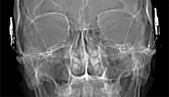
Post-operatively, the patient experienced mild unsteadiness which subsided within a few days. He had no tinnitus, and normal facial nerve function. Post-operative X-ray imaging showed correct positioning of both CIs (Figure 2).
Post-operative X-ray confirming correct positioning of both cochlear implants.

The right CI was activated one month later, using the main peak interleaved sampling stimulation strategy, as for the other implant. All electrodes were functional and impedances were in the correct range.
With time, the patient's auditory performance progressively improved. In December 2010, he gained an 85 per cent score for open set two-syllable word recognition, and a 95 per cent score for sentence recognition, using both implants, despite the fact that the performance of his second CI was still improving and had not yet reached that of the contralateral CI.
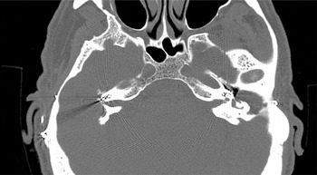
A further CT scan showed no evidence of residual cholesteatoma, and confirmed correct positioning of both electrode arrays (Figure 3).
Axial, control computed tomograph scan showing correct positioning of the electrode arrays in both cochleae.

Discussion
Radical surgical removal is the mainstay of treatment for petrous bone cholesteatoma. In the majority of cases, residual hearing (if any) should be sacrificed in order to achieve total removal of disease. However, patients in whom cholesteatoma affects the only hearing ear are very challenging to manage. To our best knowledge, cochlear implantation in a patient with petrous bone cholesteatoma has not previously been described.
Cochlear implantation is an effective method of hearing rehabilitation in patients with profound hearing loss. After their initial introduction by House in 1972,Reference House2 the performance of CIs has shown progressive improvement. As a consequence, indications for their use have broadened.Reference Arístegui and Denia3
The presence of chronic otitis media in patients with profound hearing loss was initially considered an absolute contraindication to cochlear implantation.Reference Belal4 However, in 1993 Parnes et al. reported the first case of cochlear implantation in a patient with chronic otitis media,Reference Parnes, Gagne and Hassan5 and since then there have been several reports of cochlear implantation in patients with chronically diseased ears.Reference Donnelly, Pyman and Clark6, Reference Gray, Ray and McFerran7
In the presence of chronic ear disease, the question of whether to postpone cochlear implantation to a later surgical stage is controversial. Some authors have proceeded with cochlear implantation and subtotal petrosectomy in the same surgical setting.Reference Bendet, Cerenko, Linder and Fisch8 However, others have preferred to eradicate the disease first, and to postpone cochlear implantation to a second stage.Reference Donnelly, Pyman and Clark6, Reference Gray, Ray and McFerran7 The latter authors argue that staging the procedure avoids the risk of meningitis, and also enables a ‘second look’ at the results of the earlier procedure, during cochlear implantation.
• Petrous bone cholesteatoma is a rare lesion, and may occur in the patient's only hearing ear
• In such cases, cochlear implantation may enable hearing rehabilitation, after total eradication of disease, if the cochlea is spared
• Cholesteatoma removal and cochlear implantation can be achieved as a single-stage procedure, if no active disease is encountered
In the absence of purulent discharge, we believe that a subtotal petrosectomy with abdominal fat obliteration of the surgical cavity ensures a sterile setting for the CI, and may also prevent unnecessary staging. We applied this principle to our patient affected by petrous bone cholesteatoma.
Our initial plan had been to eradicate the cholesteatoma via a combined transmastoid and middle cranial fossa approach. This was changed to a translabyrinthine approach combined with a subtotal petrosectomy, for several reasons: (1) the patient's good auditory performance after contralateral implantation; (2) to improve surgical control by using a larger approach, thus reducing the risk of residual disease; (3) to avoid temporal lobe retraction and the consequent risk of epilepsy; (4) to decrease the risk of cerebrospinal fluid leakage in the event of a dural tear; and (5) to avoid a third additional operation for cochlear implantation, in the case of no hearing preservation after a combined transmastoid and middle cranial fossa approach.
At the time of writing, the patient was trouble-free, with no evidence of residual cholesteatoma on high resolution CT scan. He was a good user of both CIs. As expected, due to the six month delay between the two cochlear implantations, the most recent auditory test showed better performance of the left CI, even though it was inserted in the side affected by a long period of hearing deprivation. The performance of the right CI was still progressively improving, reducing the functional gap between the two implants.
Conclusion
Recent improvements in the performance of CIs have increased their indications. Cochlear implantation can be an effective method of hearing rehabilitation in patients with petrous bone cholesteatoma, following total eradication of the disease, if the cochlea remains intact. Cholesteatoma surgery and cochlear implantation can be undertaken as a one-stage or a two-stage procedure, depending upon the presence of active infection.