Introduction
Haemangiomas are benign, vascular neoplasms which often involve the head and neck region.Reference McGill, Forsen, Mulliken and Cummings1 They typically appear by the age of one year in children and disappear by five or six years of age.Reference Rice and Betsakis2 They are classified into two categories: capillary or cavernous. Capillary haemangiomas consist of tightly arranged blood vessels and usually affect the skin, subcutaneous tissue, mucosal surfaces and lips, but can also be found in organs such as the liver, spleen and kidneys.Reference Robbins and Vasi Sanguigni3 Cavernous haemangiomas are less common. They are composed of large, vascular spaces which are usually bigger, less defined and (usually) deeper than those of capillary haemangiomas.Reference Robbins and Vasi Sanguigni3
Cavernous haemangioma of the external ear canal and tympanic membrane is a rare, benign tumour. Only ten cases have been reported in the literature (Medline search from 1972 to 2007).
We describe a case of cavernous haemangioma which affected only the external auditory canal. The lesion was located in the posterior superior zone and caused almost complete blockage of the external auditory canal.
Case report
A 45-year-old woman presented to our department with a five-month history of left-sided hearing loss and aural fullness. External auditory canal examination revealed a purple mass emerging from the supero-anterior portion of the canal, which occluded almost the entire external auditory canal. The mass appeared soft, non-pulsatile and did not blanch on pneumatic otoscopy. The otoscopic appearance of the right ear was normal. Weber testing at 512 Hz was consistent with a left-sided, conductive hearing loss. There was no lymphadenopathy, and the remainder of the head and neck examination was not significant.
Audiometric testing revealed mild to moderate, left-sided, conductive hearing loss.
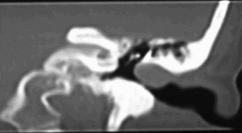
High resolution computed tomography (CT) scanning without contrast showed a soft tissue mass of approximately 1.5 cm diameter lateral to the tympanic membrane, which filled the left external auditory canal. There was no middle-ear involvement or bone erosion (Figure 1).
Axial temporal bone computed tomography scan without contrast, showing a 1.5 cm soft tissue mass in the left external auditory canal.

Puncture of the lesion with a needle resulted in venous bleeding. Under general anaesthesia, an endaural approach was used to access the lesion. The vascular tumour was based antero-superiorly in the lateral external auditory canal and was not in contact with the tympanic membrane. After creating skin flaps, the tumour was excised en bloc by elevating the lesion from the bony canal, without violating the tympanic membrane. The ear canal skin flaps were replaced, the meatal incision was sutured and the ear plugged with Merocel (Medtronic Xomed, Jacksonville, FL 32216 USA).
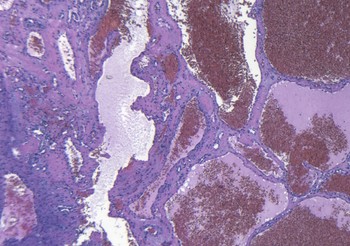
The post-operative period was uneventful. Histological examination revealed the lesion to be composed of large, tortuous, vascular spaces and cuboidal endothelial cells, indicating a cavernous haemangioma (Figure 2).
Photomicrograph showing the typical appearance of a cavernous haemangioma (H&E; ×original magnification 200).

There was no recurrence three months after the operation.
Discussion
The literature shows that cavernous haemangioma rarely affects only the external auditory canal, as in our case; of the 10 previously reported cases of external auditory canal cavernous haemangioma, only three involved no other site (Table I).
Reported Cases of External Ear Canal Haemangioma

Pt = patient; yrs = years; M = male; F = female; EAC = external auditory canal; TM = tympanic membrane
In 1972, Freedman et al. reported the first two cases of cavernous haemangioma of the external auditory canal.Reference Freedman, Barton and Goodhill4 Both patients were men, one 50 years old without symptoms and the other 57 years old with otalgia. Neither patient had significant conductive hearing loss and both had normal representation (surgical appearance or anatomy) of the middle-ear spaces.
In 1983, Andrade et al. published the first case of cavernous haemangioma limited to the tympanic membrane, which was found during routine examination of a 59-year-old man.Reference Andrade, Gehris and Breitenecker5 The benign, vascular neoplasm was excised via an endaural approach.
Also in 1983, Kemink et al. reported a case of cavernous haemangioma in a 52-year-old patient with normal hearing.Reference Kemink, Graham and McClatchey6 The sessile mass extended from the posterior external auditory canal onto the tympanic membrane.
In 1987, Hawke and van Nostrand were the first to report a cavernous haemangioma limited to the external auditory canal and arising from the anteroinferior canal wall.Reference Hawke and van Nostrand7 The lesion was easily excised; subsequent bleeding was controlled by cauterisation.
In 1990, Jackson et al. reported a 60-year-old woman with a ‘mixed’ capillary and cavernous haemangioma in the inferior canal wall.Reference Jackson, Levine and McKennan8 Her only symptom was a 40 dB conductive hearing loss. The lesion was partly removed for biopsy but recurred after two months and filled the external auditory canal. The tumour was then completely removed. The final diagnosis was ‘mixed’ haemangioma.
In 2002, Limb et al. described the case of a 67-year-old Caucasian woman with a one-month history of tinnitus, hearing loss and aural fullness.Reference Limb, Mabrie, Carey and Minor9 A CT scan showed a mass 1.5 cm lateral to the tympanic membrane. Angiography was used in an attempt to identify the blood supply to the mass, but no identifiable blood vessels were found. The tumour was removed via an endaural approach.
In the same year, Reeck et al. documented the case of a 53-year-old man with a three-year history of left-sided hearing loss.Reference Reeck, Yen, Szmit and Cheung10 A violet mass was found, occupying 20 per cent of the osseous external auditory canal in the medial posterior-inferior portion, which did not affect the tympanic membrane. These authors also used the endaural approach to gain access to the mass.
In 2006, Yang et al. reported a 72-year-old woman with a 5 mm diameter lesion of the left external auditory canal, discovered incidentally.Reference Yang, Chiang, Chao and Lee11 A transcanal approach was used to remove the lesion.
Finally, in 2007 Magliulo et al. reported the first case of a cavernous haemangioma invading the external auditory canal, tympanic membrane and middle-ear space via a small perforation in the tympanic membrane.Reference Magliulo, Parrotto, Sardella, Della and Re12 The lesion was 3 mm in diameter, purple, rounded, well defined and localised in the posterosuperior region of the medial external auditory canal. In this case, an endaural approach was used.
Generally, this pathological condition is found accidentally or is revealed following a history of hearing loss. The differential diagnosis includes high jugular bulb, jugular glomus, attic cholesteatoma, aural polyp, arterovenous malformation and carcinoma of the external auditory canal.Reference Wackym, Friedman, Jackler and Driscoll13 Computed tomography is the best imaging technique with which to evaluate the local features of the mass. Angiography is generally not required, especially when the mass derives from the upper part of the external auditory canal. Histological examination should always be performed, above all to exclude malignancy. Complete excision appears to be curative for benign lesions such as cavernous haemangiomas of the external auditory canal, with only one documented recurrence.Reference Jackson, Levine and McKennan8