INTRODUCTION
Partial breast external radiation is a method of treatment that reduces the volume surrounding the tumour to avoid the exposure of most of the residual breast tissue to radiation. The motivation for partial breast external radiation is that it can be used to treat only the breast area that is at the highest risk for recurrence.Reference Njeh, Saunders and Langton 1 Researchers are studying partial breast external radiation after lumpectomy to see how the benefits compare with the current standards of radiation to the whole breast. This technique is designed to increase the dose on the area to be treated and to prevent or reduce radiation to the tissue in the vicinity. The need for exact and rapid practical three-dimensional (3D) dosimetry has become an interesting subject in the field of radiation delivery and treatment system research. Recently, PRESAGE® (Heuris Pharma LLC, Skillman, NJ, USA), a radiochromic 3D dosimeter composed of polyurethane, radiochromic components and halogen-containing free-radical initiators that have an optical attenuation coefficient and change linearly with the absorbed dose, has been used by researchers. The combination of PRESAGE® and an optical computed tomography (CT) scanner has addressed the need to measure the dose in three dimensions.Reference Oldham, Sakhalkar, Guo and Adamovics 2 – Reference Iqbal, Gifford, Ibbott, Grant and Buzdar 15 PRESAGE® has been investigated previously for dosimetric characteristics and also for the PRESAGE®/optical-CT system for 3D dosimetry. These investigations subsequently involved carrying out treatment planning verification, such as ECLIPSE® dose distribution, as a gold standard. PRESAGE® can be carried out in various shapes but most of the previous work has been performed using cylindrical shapes. The present study uses a breast-shaped anthropomorphic PRESAGE® dosimeter for verification of three-field dose distribution.Reference Iqbal, Gifford, Ibbott, Grant and Buzdar 15 The present study also uses qPRESAGE® dosimeters to build on earlier work by applying the PRESAGE®/optical-CT system for the verification of three-field delivery, in which the accuracy of commercial treatment planning systems is less well known. Comparison was performed using the Pinnacle3 v 9.0® (Philips Laboratories, Milpitas, CA, USA) treatment planning system and GAFCHROMIC® EBT2 film to verify the PRESAGE®/optical-CT system for external beam partial breast 3D dosimetry.
MATERIALS AND METHODS
Optical-CT scanning and PRESAGE® dosimeter
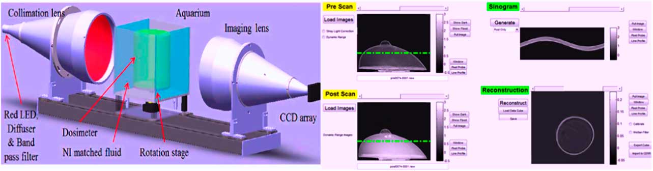
The PRESAGE® dosimeter was moulded from a pre-mould mixture comprising a solvent, leuco dye and free-radical initiator. The formulation of PRESAGE® used in this study had an effective atomic number (Z eff) of 7.6 provided by Heuris Pharma and a physical density of 1.07 g/cm3 measured by the Pinnacle3 treatment planning system. A medium broad-beam optical CT scanner (DMOS-RPC), developed for joint research between the Department of Radiation Oncology, Duke University Medical Center, Durham, NC, USA and Radiological Physics Center, Houston, TX, USA was used for scanning the PRESAGE® dosimeter. Projections of ~80µ from each dosimeter were collected at intervals of 1° over a full 360°, both before and after irradiation. We calculate the 3D reconstruction of the attenuation coefficients across the dosimeter. Scans normalised the calculated dose distribution and 3D comparison was made with a treatment planning system.Reference Newton, Thomas, Ibbott and Oldham 16 The DMOS-RPC scanner shown in Figure 1 (left) consists of a matched telemetry source and image lenses provide visual field.Reference Iqbal, Gifford, Ibbott, Grant and Buzdar 15 In graphical user interface, the top-left quadrants of projection screen images are associated with painting on preliminary irradiation, and the bottom-left projection screens are associated with the image scan after irradiation. Correction for stray light is applied to each pre-scanning irradiation and post-irradiation scanning projection before the renovation. Transverse images were reconstructed with filtered-back projection of voxel distance of 1 mm as shown in Figure 1 (right).
DMOS scanner (left); reconstruction of graphical user interface of DMOS (right).

Treatment planning and delivery
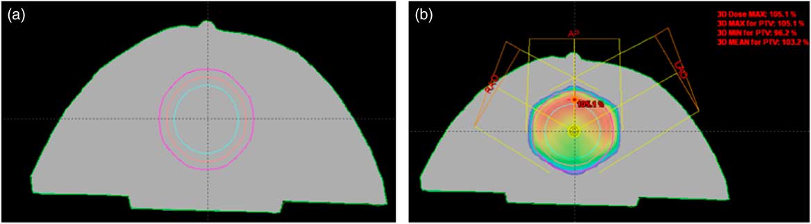
CT slices with a thickness of 3 mm were acquired using the breast PRESAGE® dosimeter on a GE CT scanner (GE Healthcare Technologies, Waukesha, WI, USA). A single CT scan was performed before irradiation to assess any changes in the optical density (OD). CT data were exported to treatment planning Pinnacle3 workplace, in which the treatment plan was created using the collapsed Cone convolution algorithm with a resolution of 3 mm. Formation was carried out in such a way that the contour of the breast was described 1 mm below the surface and the gross tumour volume (GTV) was contoured in a central slice of the volume of the breast phantom of 62 cm3. Clinical tumour volume (CTV) volume was prepared using a 1 cm margin all around the GTV and planning tumour volume (PTV) was constructed using a 1 cm margin CTV, as shown in Figure 2a.
(a) CT slice indicating the regions of interest for the partial breast case.  , gross tumour volume;
 , clinical tumour volume;
 , planning tumour volume. (b) Three-field plan with dose distributions.

A three-field partial treatment plan was designed with a 15° enhanced dynamic wedge on a pair of oblique fields to deliver a dose distribution of 300 cGy to PTV with gantry angles of 0, 60 and 300° using six X-ray, 500 Gy/min dose rate and with a field size of 8×8 cm2, as shown in Figure 2b.
Independent EBT film measurement
Film dosimetry has been working as a powerful tool for checking the radiotherapy treatment and for quality assurance for several years.Reference Devic 17 Independent verification of dose distribution was carried out using GAFCHROMIC® EBT2 film (ISP Corp., Wayne, NJ, USA). Temporal stability, independence, comfort and self-oriented development are the basic reasons for using EBT2.Reference Iqbal, Gifford, Ibbott, Grant and Buzdar 15 , Reference Newton, Thomas, Ibbott and Oldham 16 Once the treatment plan was implemented, the PRESAGE® dosimeter was cut into two levels, which correspond approximately parallel to the axial planes, and pieces of EBT2 film were placed between the sections of PRESAGE®. Dosimeter films inserted were irradiated with the same three-field plan. Films were digitised using a 48-bit transmission–reflection flatbed photo scanner, Epson–10000XL (Epson America, Inc., Long Beach, CA, USA). Each film was recorded in transmission mode, and the red channel was used for the examination: for example, red light and a maximum response at 633 nm.Reference Brown, Bailey, Willowson and Baldock 12 , Reference Newton, Thomas, Ibbott and Oldham 16 , Reference Devic, Seuntjens and Sham 18
Data registration and analysis
Transverse image by distributing doses of RPC-DMOS and treatment plan Pinnacle3 have been exported to the CERR, Computational Environment for Radiotherapy Research, programme (Memorial Sloan Kettering Cancer Center, New York, NY). The calculated dose distribution of Pinnacle3 was compared with the measured value from PRESAGE® and EBT2 film. EBT2 scans were analysed using Image J software (National Institutes of Health, USA). All quantitative analysis of the datasets was limited to a slice-by-slice analysis through the line profiles, gamma two-dimensional (2D) map and DVHs.Reference Samuel, William, Corey, Sua and Oldham 4 The percentage of uncertainty of PRESAGE® and EBT2 films was calculated using this formula:
 $$\eqalignno{&{\rm Percentage}\,{\rm uncertainty}{\equals}\cr\qquad{{{\rm Standard}\,{\rm deviation}\,{\rm of}\,{\rm optical}\,{\rm density}} \over {{\rm Mean}\,{\rm optical}\,{\rm density}}}{\times}100$$
The distance to the agreement acceptance criterion of 3 mm was used to match dose grid resolution calculated in Pinnacle3. Comparisons with EBT2 GAFCHROMIC® film were used to verify the accuracy of PRESAGE® and 3D comparisons with PRESAGE® and Pinnacle3 were also performed.
RESULTS
Film and PRESAGE® calibration
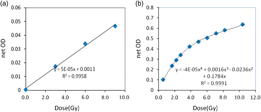
Figure 3a is the OD to dose curve for the PRESAGE® dosimeter and Figure 3b is the calibration curve that was applied to the EBT2 film to convert OD to dose. The radiochromic response was linear with a sensitivity of 0.0057 OD change for a 1 mm path length.
(a) The PRESAGE® linearity dose curve. (b) Optical density (OD) to dose curve used for EBT2 films.

The uncertainty in the net optical density was derived from three sources of error associated with the mean pixel value for each region of interest (ROI): (1) changes in the scanner lamp output after warm-up (2) variation in pixel value across the ROI due to statistics and beam non-uniformities and (3) differences in the background optical density for each 5×5 cm2 piece of film. Changes in the scanner lamp output were investigated by performing consecutive scans of a single piece of film.
DVH comparison
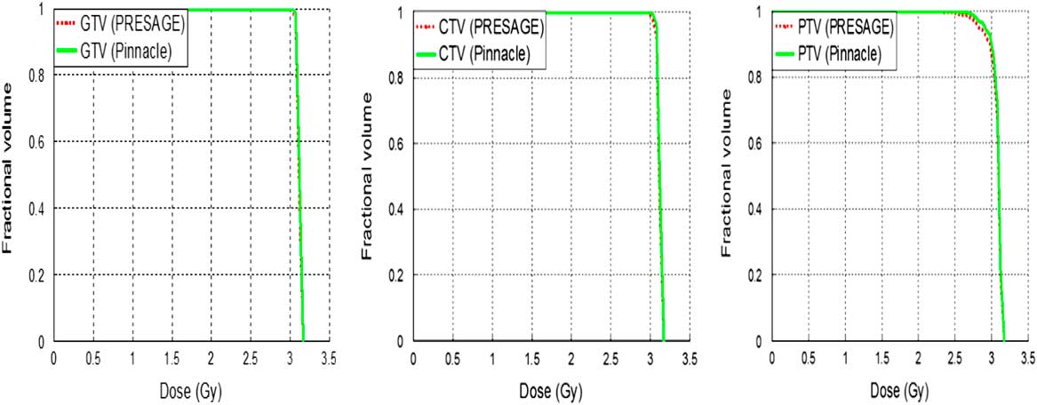
It was determined that the X-ray CT dose did not produce any change in OD of the breast PRESAGE® dosimeter. Figure 4 illustrates the target volume DVH comparison of the three-field breast plan between PRESAGE® and Pinnacle3. The advantage of PRESAGE®/optical-CT was to produce 3D dosimetry. DVHs were plotted between the PRESAGE® and dose distribution of Pinnacle3 treatment planning. In addition, an interpretation of these differences can be elucidated using multiple deliveries. The PTV DVH of PRESAGE® and Pinnacle3 showed 2.2% dose difference whereas CTV and GTV show 1.5 and 0.8%, respectively, as shown in Figure 4.
Gross tumour volume (GTV), clinical tumour volume (CTV) and planning tumour volume (PTV) dose–volume histograms (DVH) comparison between the PRESAGE® and Pinnacle3 planning dose.

Isodose line profiles
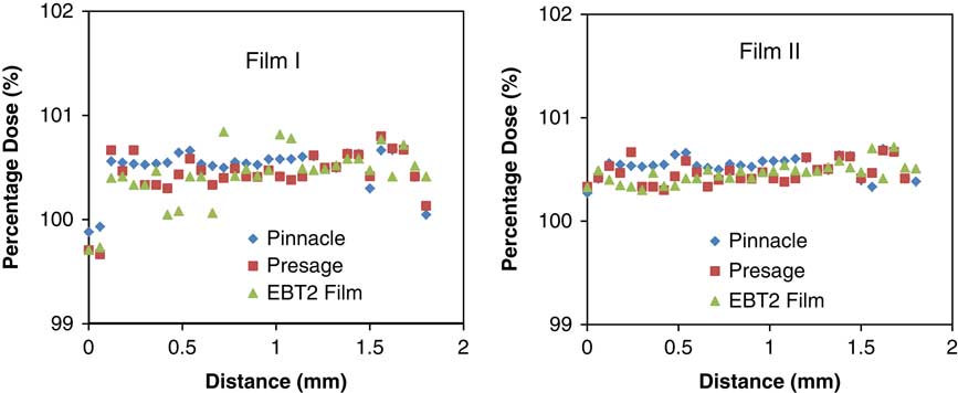
Figure 5 is the 2D dose distribution in the selected two levels. Independent 2D dose measurements in two selected planes made using EBT2 film facilitates the resolution of any differences between PRESAGE®/optical-CT and Pinnacle3 distributions. A total of two sets of isodose line graphs show agreement between all three distributions with a maximum difference of 1.5%. The average spread in all three systems is 1.2% with standard deviation of 0.56%.
Line profiles of the Pinnacle3, PRESAGE® and EBT2 film dose distributions of axial slice.

Gamma map comparison
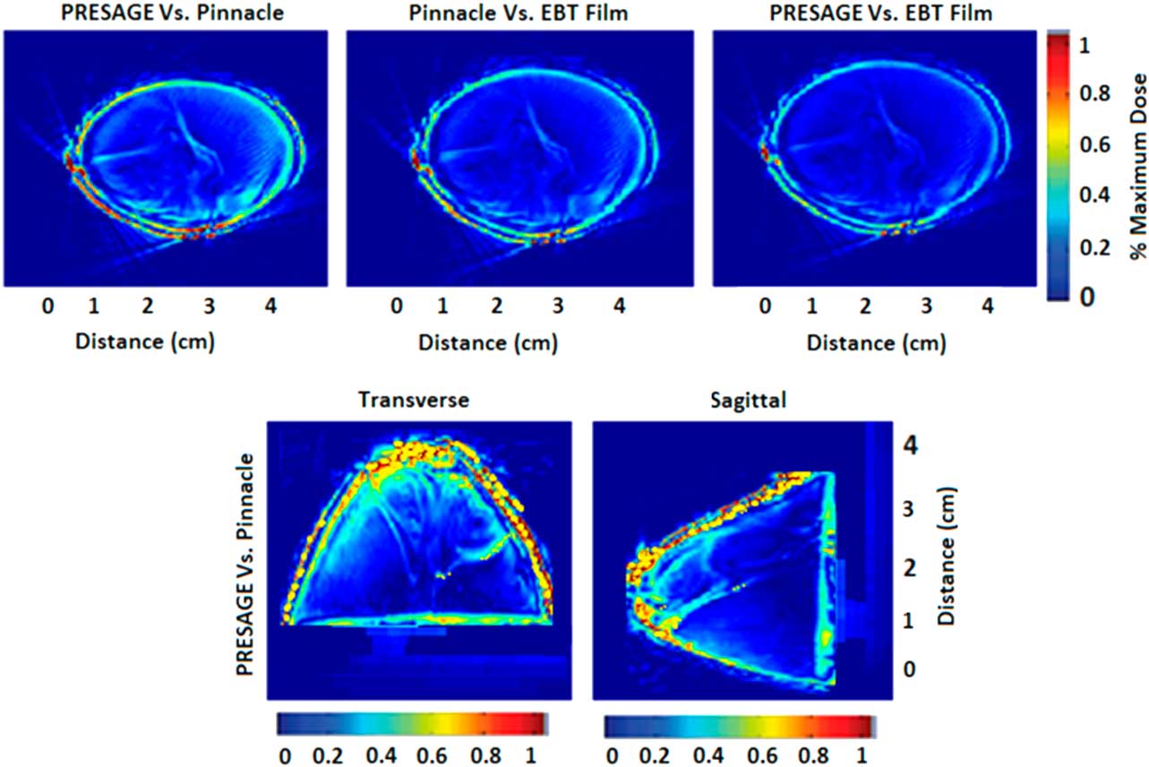
Figure 6 is the gamma comparison of PRESAGE®/optical-CT, EBT2 film measurement and dose calculation from the Pinnacle3 treatment system at ±3%/±3 mm criterion. The values for the axial 2D gamma comparisons of EBT2 versus PRESAGE®, PRESAGE® versus Pinnacle3 and EBT2 versus Pinnacle3 were 97.6% and 97.4% and 95.3%, respectively.
Gamma maps in the axial plane. Gamma distributions for all three systems. Greater than 95% of comparison points passed ±3% of ±3 mm criterion among the three distributions. PRESAGE® and Pinnacle3 gamma distributions in three orthogonal planes.

DISCUSSION
The percentage of uncertainty in GAFCHORMIC® EBT2 films was 1.8% in the reference dose of 3 Gy. Contributions of imperfect uniformity and uncertainty in the repeatability of the dose, as well as the contribution of the uncertainty in the fit of the dose–response curve were expected and could not be avoided.Reference Aland, Kairn and Kenny 19 Pixel mean values for both small and large region of interests described above were found to vary by less than ±0.06% (1σ) after completion of the lamp-heating process. The standard error of the mean value was used to estimate the uncertainty associated with the change in pixel value throughout ROI. This error was very small (less than ±0.01%) because of the large number of pixels associated with each ROI.
The contribution of noise in areas that are used to measure the dose in EBT2 film was also assumed to be negligible, and it was estimated as the average standard deviation in the region of interest across pieces of EBT2 film exposed to 3 Gy.Reference Aland, Kairn and Kenny 19 , Reference MacDougall, Miquel and Keevil 20 , Reference Iqbal, Gillin, Summers, Dhanesar, Gifford and Buzdar 21 As for the percentage of uncertainty in the PRESAGE® dosimeter, it was found to be 0.8% of the reference dose of 3 Gy. We therefore conclude that PRESAGE® can be used as a relative dosimeter with normalisation to the point that corresponds to the 100% isodose distribution area of the planned dose. The power of the linearity response of PRESAGE® did not impose any limitation on the analysis of data and there was no evidence of a fault-volume effect.Reference MacDougall, Miquel and Keevil 20
The PRESAGE® PTV DVH indicates that the delivered dose was somewhat less homogeneous than that calculated using the Pinnacle3 treatment planning system, with a maximum of 2.2% of small regions of the relationship above and below the dose occurring near the edge of the breast phantom. The average difference in the target volume was 1.5% with a standard deviation of 0.49%, and a part of this difference is real, whereas some of it was due to artefacts in the PRESAGE® distribution.Reference Iqbal, Gifford, Ibbott, Grant and Buzdar 15
The Pinnacle3 distribution was more uniform, with less noise than any of the measured distributions. The gamma map of comparisons between PRESAGE® and Pinnacle3 dose distribution was 97.4%. The gamma map of the axial 2D comparisons of EBT and PRESAGE® and of EBT against Pinnacle3 were 97.6 and 95.3%, respectively.
The majority of the errors in all three comparisons occur near the edge of the dosimeter in the outer 3 mm. These results confirm previously published data from Oldham M and Iqbal k.Reference Iqbal, Gifford, Ibbott, Grant and Buzdar 15 , Reference Oldham, Sakhalkar, Guo and Adamovics 22 , Reference Iqbal 23 As shown in this challenging region, PRESAGE® and EBT doses were incorrect because of edge artefacts, and the treatment planning system dose was also likely to be inaccurate because of the difficulty to model the structure region. If the outer edge of 3 mm is ignored, the value rises to 96% for the comparison of 3D PRESAGE® with treatment planning system, which corresponds to a close agreement for such a complex plan.Reference Oldham, Sakhalkar, Guo and Adamovics 22 , Reference Iqbal 23
CONCULSION
Anthropomorphic PRESAGE® was created in the form of breasts and measurements were obtained and compared with the treatment planning system Pinnacle3 and GAFCHROMIC® EBT2 film in external beam therapy. Gamma map comparisons showed that all three distributions agreed for more than 95% of the comparative points at ±3%/±3 mm gamma criteria. Line profiles of the EBT2 film, Pinnacle3 and PRESAGE® were found to be within a 1.5% difference of the dosimeter. The PTV DVH of PRESAGE® and Pinnacle3 showed 2.2% dose difference, whereas CTV and GTV showed 1.5 and 0.8%, respectively. To our knowledge, the presented work establishes the possibility of fashioning PRESAGE® into an anthropomorphic shape for verification of 3D partial breast dosimetry, and it provides groundwork for future investigations into more complex anthropomorphic PRESAGE® phantoms; moreover, artefacts that were produced at the edge of dosimeter because of attenuation of laser light were unaffected by the dose distribution inside the breast anthropomorphic phantom.
Acknowledgements
This work was supported by the NIH Grant No. 5R01 CA 10083. The authors are very thankful to Higher Education Commission of Pakistan, who provided the scholarship for the completion of Ph.D. research.