


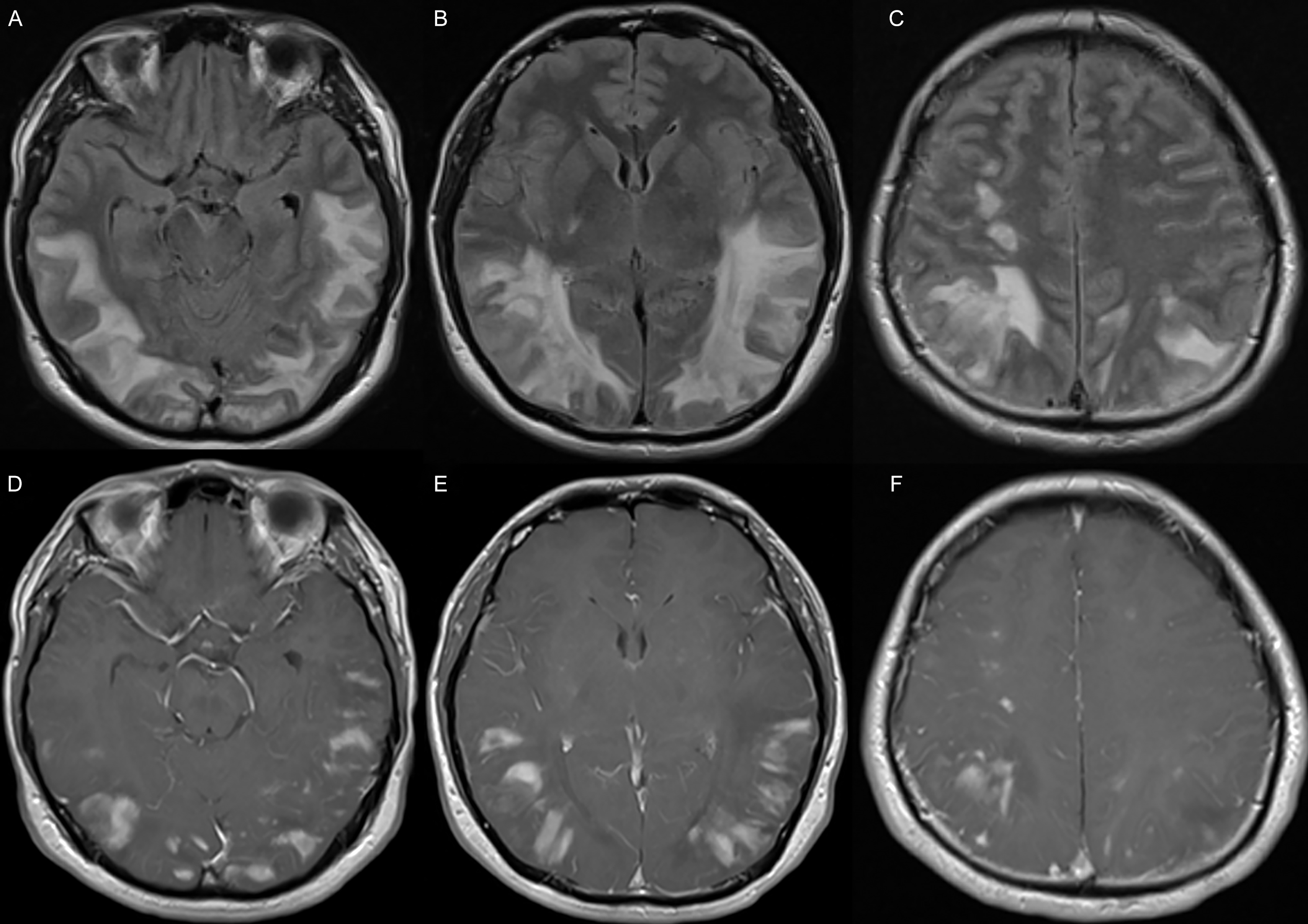
A healthy 36-year-old male presented to the emergency department with a 2-week history of bifrontal headaches, partial cortical blindness, visual agnosia, right–left disorientation, dyscalculia and a generalized tonic-clonic seizure from sleep. He was admitted to the neurologic intensive care unit for a reduced level of consciousness. A CT scan demonstrated symmetric, posterior predominant, bilateral cortical and subcortical hypoattenuation. MRI with gadolinium demonstrated extensive, non-diffusion-restricting T2/fluid-attenuated inversion recovery (FLAIR) hyperintensity extending throughout the parietal and occipital lobes and into the posterior frontal and temporal lobes (Figure 1A–C) with patchy enhancement in the affected areas (Figure 1D–F). Electroencephalogram demonstrated diffuse slowing without epileptiform discharges. Comprehensive inflammatory/autoimmune, infectious, toxic and metabolic workup was unrevealing and included numerous specialized tests including antibody, complement testing and an atypical antineutrophil cytoplastic antibody panel. His initial CSF was acellular with elevated protein (0.65 g/L). Follow-up CSF was acellular with normal protein; however, CSF flow cytometry showed a subset of T-cells (20% of all lymphocytes), which were characterized by the presence of CD5/CD2 and loss of CD7/CD3, with no expression of CD4/CD8. They were negative for CD56 and PD-1. These results were suggestive of a T/NK-cell lymphoproliferative disorder. Further imaging included CT chest/abdomen/pelvis, conventional intracranial angiogram (negative) and positron emission tomography (PET). PET brain showed abnormal hypermetabolism throughout the right posterior frontal, bilateral parietal, bilateral temporal and bilateral occipital cortices. PET body showed mild fluorodeoxyglucose (FDG) activity diffusely involving the thoracic aorta, bilateral subclavian arteries, bilateral superficial and deep femoral arteries and two moderately FDG-avid pulmonary nodules in the left lung; the largest (10 mm) demonstrated central cavitation. There was mild FDG activity involving non-enlarged bilateral inguinal and axillary lymph nodes. CT chest/abdomen/pelvis did not demonstrate additional abnormalities. Ophthalmologic assessment found reduced visual acuity (20/400 oculus uterque) and delayed choroidal filling oculus dexter, with lobular areas of choroidal hypoperfusion and non-perfusion.
Pre-treatment axial brain MRI with FLAIR sequence (A–C) and T1 post-gadolinium enhancement (D–F) at the level of the temporal lobes (A and D), basal ganglia (B and E) and above the lateral ventricles (C and F) demonstrating extensive edema extending throughout the parietal and occipital lobes, with additional extension into the posterior frontal and temporal lobes and irregular patchy enhancement in the affected areas.
Given significant central nervous system (CNS) involvement, a tissue diagnosis was sought, and a right parietal brain biopsy was performed. This revealed a T-cell monoclonal population with cytotoxic features, in keeping with secondary involvement of the brain by an indolent circulating lymphoproliferative disorder of large granular lymphocytes. It demonstrated a mixed lymphohistiocytic population with inflammatory features diffusely distributed in white matter with angiocentric foci and endothelial swelling. This infiltrate featured nodular aggregates of histiocytes partially resembling granulomas with focal histiocytic palisading. Immunohistochemical phenotypic assessment confirmed a T-cell-rich background, with CD4+ and predominantly CD8+ cells showing cytotoxic features and phenotypic aberrancies. These T-cells were accompanied by diffusely distributed CD68+ histiocytes. T-cell receptor (TCR) gene rearrangement studies confirmed a clonal T-cell population. For further diagnostic support, the patient also had a biopsy of a hyperpigmented macule on the lower limb, an erythematous plaque on the left forearm, bronchoalveolar lavage, left lung wedge resection and bone marrow. The neoplastic cells were also seen in the lung, skin, peribronchial lymph node, peripheral blood and bone marrow with a predominant intrasinusoidal/intravascular distribution. TCR gene rearrangement studies confirmed a clonal T-cell population (beta and gamma) in the lung, peribronchial lymph node, peripheral blood and bone marrow but not in the cutaneous sample. The patient was diagnosed with T-cell large granular lymphocyte (T-LGL) leukemia.
The patient was initially treated with multiple courses of high-dose intravenous (IV) steroids followed by an oral steroid taper. He continued to be symptomatic with higher-order visual processing deficits and memory difficulties and persistent enhancing lesions on MRI. Pending pathology results and following consultation with the many specialties, he was started on cyclophosphamide 800 mg/m2 IV monthly for six cycles. He continued to be symptomatic, and so after pathological diagnosis was made, weekly intrathecal methotrexate 12 mg with hydrocortisone 40 mg for 14 cycles was added. One month following the initiation of this treatment, he developed worsening deficits. He was admitted to the hospital for myeloablative cyclophosphamide (600 mg/m2/dose IV days 1, 2, 4, 6 and 8 with IV methylprednisolone 1000 mg). Following this and up to the current day, he has had clinical improvement with stability and is maintained on dexamethasone 1 mg twice daily. An allogenic stem cell transplant was considered but not offered. His symptoms as of last follow-up are reduced processing speed, difficulties with word retrieval, associative prosopagnosia, imbalance and headache. He has been in follow-up for 33 months, and the latest MRI shows new enhancing cerebellar lesions with persistent multifocal nodular enhancement (Figure 2). The time interval between his first MRI and the latest MRI is 32 months, with his latest MRI being 22 months after myeloablative cyclophosphamide.
Post-treatment axial brain MRI with FLAIR sequence (A–D) and T1 post-gadolinium enhancement (E–H) at the level of the cerebellum (A and E), temporal lobes (B and F), basal ganglia (C and G) and above the lateral ventricles (D and H) demonstrating enhancing lesions in the cerebellar hemispheres, with persistent multifocal nodular enhancement bordering right and left parietal-occipital encephalomalacia with ex vacuo dilatation of the occipital horns of the lateral ventricles.

T-LGL leukemia is a hematologic malignancy that typically presents as recurrent infections with cytopenia, though up to 1/3 of patients may be asymptomatic. Constitutional symptoms with fatigue are seen in 20–30% of cases. Reference Lamy, Moignet and Loughran1,Reference Loughran2 Conditions associated with LGL leukemias include autoimmune cytopenias, B-cell lymphoid neoplasms, myelodysplastic syndrome and autoimmune diseases/connective tissue disorders. Reference Loughran2 The tissues typically involved include blood, bone marrow, liver and spleen, with skin and lymph node involvement being less common. Reference Loughran2,Reference Lamy and Loughran3 Nervous system involvement is rare and mainly relates to peripheral neuropathy. Reference Zhang, Shah and Loughran4 CNS involvement is even rarer and has been limited to case reports, varied in presentation and limited to patients with pre-existing disease. One patient with known disease developed intermittent dizziness with evidence of disease in CSF but no radiographic finding, Reference Liu, Fan, Zhao, Xu and Li5 whereas another patient with known disease presented with headaches, gait difficulty and visual disturbance with bilateral subcortical hyperintensities and evidence of disease in CSF and vitreous biopsy. Reference Cheung, Ip, Iu, Lee and Kwong6 Likewise, there have also been reports of seizure and encephalitis as a manifestation, again with known disease. Reference Ichikawa, Noguchi and Masuda7 Additionally, choroidal infiltration has been described as an initial ocular manifestation of T-LGL leukemia in a patient presenting with photophobia and panuveitis. Reference Sarny, Beham-Schmid and El-Shabrawi8
In conclusion, we describe a patient without known T-LGL leukemia whose initial clinical manifestation of T-LGL leukemia was significant CNS involvement. Based on our review of the literature, this is the first such case described. He was found to have additional clinically silent disease with pathological evidence in blood, lung tissue, lymph node and bone marrow in the absence of an associated autoimmune condition, hepatomegaly or splenomegaly. He had a partial response to therapy and continues to be followed clinically and radiologically.
Author contributions
JM conceptualized the study and is the main clinician for the case. GC and JM comprehensively reviewed the case. JM, JD and MT provided expert analysis of investigations. GC prepared the first draft. JM, JD and MT critically revised the final manuscript.
Funding statement
This study received no funding.
Competing interests
None of the authors has any conflicts of interest to disclose.


 Open access

