

Introduction
Investigations into the neural basis for spatial navigation in the rat began with the discovery by O’Keefe and Dostrovsky (Reference O’Keefe and Dostrovsky1971) that a place cell in the hippocampus fires when the rat forages in a particular location within an enclosure. O’Keefe (Reference O’Keefe1991, p. 230) interpreted the finding as evidence for a spatial system that represents environments ‘within an allocentric spatial framework, that the ordering principles of the system are the Euclidean distance and direction between places within the framework, and that the system is localized within the nervous system to the hippocampal region’. O’Keefe and Nadel (Reference O’Keefe and Nadel1978) likened this system to the cognitive map described by Tolman (Reference Tolman1948) which provides the rat with an internal representation of the environment, including pathways between landmarks. O’Keefe and Burgess (Reference O’Keefe and Burgess1999) stated that the basic tenet of the cognitive map theory is that, in the rat, the processing and storage of spatial information is the primary role of the hippocampus. This theory was supported by the finding that animals with lesions in the hippocampus were unable to find a hidden platform in a tank filled with water while those with neocortical lesions performed normally on the task (Morris, Garrud, Rawlins, & O’Keefe, Reference Morris, Garrud, Rawlins and O’Keefe1982).
An important addition to the model came with the discovery of head direction cells in the postsubiculum, cells that encode the animal’s directional heading in the horizontal plane (O’Keefe, Reference O’Keefe1991; Ranck, Reference Ranck1984; Taube, Reference Taube2007; Taube, Muller, & Ranck, Reference Taube, Muller and Ranck1990). This was followed by the discovery of grid cells in the dorsal medial entorhinal cortex (dMEC), one synapse upstream from the hippocampus ((Hafting, Fyhn, Molden, Moser, & Moser, Reference Hafting, Fyhn, Molden, Moser and Moser2005)). Hafting et al. (Reference Hafting, Fyhn, Molden, Moser and Moser2005) reported that these cells fired at multiple locations, forming a grid of regularly tessalating triangles that covered the entire surface of the enclosure. Hafting et al. (Reference Hafting, Fyhn, Molden, Moser and Moser2005) considered that their findings pointed to the dMEC as part of a neural map of the spatial environment, and Moser, Kropff, and Moser (Reference Moser, Kropff and Moser2008) suggested that the dMEC contains the elements of a metric system for spatial navigation, with similar properties to the cognitive map of O’Keefe and Nadel (Reference O’Keefe and Nadel1978). They considered that the basic unit for the neural map is the rigid triangular/hexagonal structure of the firing fields of the grid cell, and that this enables the network to provide accurate metric information about the animal’s position relative to points in the environment. Steffenach, Witter, Moser, and Moser (Reference Steffenach, Witter, Moser and Moser2005) found that lesions to the MEC were associated with deficits in spatial navigation; lesions that included the dorsolateral band of the entorhinal cortex completely disrupted recall of a spatial navigation task that had been learned before the lesion. Additionally, Hales et al. (Reference Hales, Schlesiger, Leutgeb, Squire, Leutgeb and Clark2014) found that lesions to the MEC caused a profound impairment in spatial navigation on the Morris water maze.
O’Keefe has consistently argued that the cognitive map theory can be applied to humans as well as to rats (Burgess, Maguire, & O’Keefe, Reference Burgess, Maguire and O’Keefe2002; Burgess & O’Keefe, Reference Burgess and O’Keefe2003; Hartley, Lever, Burgess, & O’Keefe, Reference Hartley, Lever, Burgess and O’Keefe2014; O’Keefe, Reference O’Keefe2014; O’Keefe & Nadel, Reference O’Keefe and Nadel1978), and he and colleagues have undertaken a number of experiments on spatial navigation in humans, interpreting the results in terms of the cognitive map theory. In an experiment by Maguire et al. (Reference Maguire, Burgess, Donnett, Frackowiak, Frith and O’Keefe1998), subjects learned the layout of a shopping centre and they then navigated from one location to another from memory. Regional cerebral blood flow was measured using positron emission tomography. This method showed significant activation of the right hippocampus. It was also found that the accuracy of heading towards the goal was associated with activation of the right hippocampus and the right inferior parietal cortex. Following the cognitive map theory, it was concluded that these results demonstrated that the hippocampus provides a representation of space that allows the animal to compute the direction from any start location to any goal. They argued that the right inferior parietal cortex uses this information to compute the correct body turns to enable movement towards the goal (Maguire et al., Reference Maguire, Burgess, Donnett, Frackowiak, Frith and O’Keefe1998). The authors emphasized that this interpretation is based on the cognitive map theory stating that ‘Our interpretation of the hippocampal role in navigation is concordant with neuronal responses in rats and with models of how the hippocampus guides rats’ navigation, from which our measure was specifically derived, despite the species difference’ (Maguire et al., Reference Maguire, Burgess, Donnett, Frackowiak, Frith and O’Keefe1998, p. 923).
Ekstrom et al. (Reference Ekstrom, Kahana, Caplan, Fields, Isham, Newman and Fried2003) found place cells in the human hippocampus that were activated as the participants explored and navigated a virtual town, a finding that Burgess and O’Keefe (Reference Burgess and O’Keefe2003) regarded as evidence that the cognitive map theory applies to humans as well as rats. More recent studies using functional magnetic resonance imaging have demonstrated correlations between neuronal activity in the human hippocampus and spatial navigation using two dimensional virtual reality tasks (Hassabis et al., Reference Hassabis, Chu, Rees, Weiskopf, Molyneux and Maguire2009), and Howard et al. (Reference Howard, Javadi, Yu, Mill, Morrison, Knight and Spiers2014) found that activity in the human entorhinal cortex was correlated with the Euclidean distance component of a vector to the goal, while cells in the posterior hippocampus were associated with distance along a path. Banta Lavenex, Colombo, Ribordy Lambert, and Lavenex (Reference Banta Lavenex, Colombo, Ribordy Lambert and Lavenex2014) concluded that there is abundant experimental evidence from rats, monkeys and humans supporting the theory that the mammalian hippocampus allows precise metric coding of spatial information, which they described as a hallmark of cognitive maps.
An alternative explanation for the neural basis of spatial navigation in the human begins with the study by Paterson and Zangwill (Reference Paterson and Zangwill1944). They presented two cases of traumatic brain injury involving the right parieto-occipital area. In these cases, there was a piecemeal approach to constructions or drawings. In this disorder, the patients ‘typically drew complex objects or plans detail by detail with little apparent grasp of the whole and poor articulation of the parts’ (Paterson & Zangwill, Reference Paterson and Zangwill1944, p. 356). This led to an extreme disorientation in the plan as a whole. They commented that the patients were unable to represent the room as a unified framework in which individual fittings and articles of furniture were properly located. Individual objects were introduced piecemeal and only those in close proximity were spatially interrelated in the plan. Paterson and Zangwill (Reference Paterson and Zangwill1944) also presented a number of drawings of a bicycle that showed severely disordered spatial relationships. They commented on a failure to articulate the parts accurately, and drew attention to a difficulty in which the frame was drawn in the frontal plane and the wheels in the horizontal. A third case was described a year later when, in addition to the visuo-constructional difficulties, the patient demonstrated a severe impairment of spatial orientation (Paterson & Zangwill, Reference Paterson and Zangwill1945).
Following these initial studies, the piecemeal approach was studied more systematically. McFie, Piercy, and Zangwill (Reference McFie, Piercy and Zangwill1950) presented seven tumour cases, all of which were confined to the right hemisphere apart from one bilateral case of vascular origins. They described the condition as visual–spatial agnosia. They also identified a number of associated disabilities, including a visual–spatial impairment and disorders of topographical orientation and memory. In all cases, there were severe deficits in visuo-constructional abilities that were evident in tasks requiring participants to construct block designs, in cube counting and in drawings. The spontaneous drawings of a bicycle were frequently impaired, characteristically showing faulty orientation and articulation of the parts, confusion of planes and simplification of outline. They commented that the patients could represent the individual parts of a complex object, such as a chair or a bicycle, in a recognizable fashion but appeared unable to relate or assemble these components to form a coherent whole (McFie et al., Reference McFie, Piercy and Zangwill1950).
Hécaen, Penfield, Bertrand, and Malmo (Reference Hécaen, Penfield, Bertrand and Malmo1956) studied 17 cases in which there was excision of cortical tissues for the relief of intractable epilepsy. They identified nine cases of visual–spatial agnosia, which they described as an apractognosia, implying a disorder of movement and action. All nine cases resulted from lesions of the right cerebral hemisphere. Five cases were described in detail; they all showed some degree of visuo-constructive disturbance that could be demonstrated by copying or spontaneous drawing of a house, cube, star or bicycle. As far as the cases in which apractognosia was observed, they noted that ‘there was always a large removal, mainly parietal but also extending posteriorly and inferiorly. In all these cases the parieto-temporo-occipital junction was involved in the excision…we find overlapping in an area involving the gyrus supramarginalis and partially the gyrus angularis and the posterior part of the first temporal convolution’ (Hécaen et al., Reference Hécaen, Penfield, Bertrand and Malmo1956, p. 422). In their final statement on localization with regard to the apractognosia syndrome, they included the whole of the right parieto-temporo-occipital area.
Semmes (Reference Semmes1965) explored the relationship between spatial disorientation on a locomotor test of map reading and on two tests involving the ability to recognize two and three-dimensional shapes by touch. Semmes found that in the brain-injured subjects with left or right parietal damage, the subjects who performed well on the spatial orientation test discriminated shapes almost as accurately as the normal control subjects whereas those who were poor on spatial orientation were also poor at discriminating tactual shapes. On the basis of these findings, Semmes argued that the locomotor test of spatial orientation and the shape discrimination tests had in common the appreciation of spatial relationships regardless of the modality transmitting the information.
Some experimental support for the concept of a spatial coordinate system organized along supramodal lines came from the finding of Corkin (Reference Corkin1965) of a high correlation between a tactual and a visual form of a stylus maze. Milner (Reference Milner1965) administered the visual form to a group of 74 patients who had suffered the removal of cortical tissue for the relief of focal epilepsy and Corkin (Reference Corkin1965) administered a tactually guided maze to 53 of Milner’s group who had undertaken the visual maze. Corkin (Reference Corkin1965) considered that the high correlation supported the idea that spatial abilities are not represented in a modality-specific manner.
Milner (Reference Milner1965) reported the case of J. Du who had suffered a right parietal lobectomy, which included most of the supramarginal gyrus. Milner (Reference Milner1965) reported that J. Du found both the visual and tactual maze tasks extremely difficult and stressful; it took her 52 trials to learn the visual maze and she referred to the maze as a ‘jungle’. This suggests that J. Du’s problems on the maze tests might have been caused by a fragmentation in the sense of space (Hécaen et al., Reference Hécaen, Penfield, Bertrand and Malmo1956; McFie et al., Reference McFie, Piercy and Zangwill1950; Paterson & Zangwill, Reference Paterson and Zangwill1944, Reference Paterson and Zangwill1945).
Milner (Reference Milner1965) described a severe impairment in the ability to learn new spatial information following bilateral medial temporal lobectomy, and she referred particularly to the performance of H.M. who failed to learn the visual maze after more than 215 trials. Milner also reported a significant impairment following removal of the right hippocampus. She described the case of P.A. who, with a radical excision of the right hippocampus, made 528 errors and required 78 trials to learn the maze, whereas the case of L.B. with removal of the lateral surface of the right temporal lobe but with sparing of the right hippocampus obtained a normal score (Milner, Reference Milner1965). Patients with left hippocampal excision performed normally on the maze (Milner, Reference Milner1965). Milner (Reference Milner, Whitty and Zangwill1966) considered that the failure to learn the maze was essentially a defect of consolidation that prevented any new learning from taking place.
Stuart (Reference Stuart1995) proposed that the maze tests of Milner and Corkin could be viewed as measures of spatial navigation and that the principal factors in these tests include the ability to integrate spatial information provided by the right parietal area, and the ability to learn and retain new spatial information provided by the right hippocampus. In view of the high correlation between the tactile and the visual forms of the maze (Corkin, Reference Corkin1965), it was proposed that the two mazes had in common the ability to learn a complex sequence of arm movements in two or three-dimensional space. This led to the development of the Tactual Learning Test in which the blind or blindfolded subject learns a sequence of arm movements, a spatial pattern, which is then recalled by means of a drawing or tracing with the finger. A set of two-dimensional spatial patterns ranging from simple to complex was devised. The level of difficulty of the spatial patterns was established using sighted children under blindfold, such that most of the children in the group (18/20) aged between 8.3 and 9.3 years (mean 8.6 years) completed this test successfully. Five of twelve younger subjects, aged between 6.6 years and 6.11 years, succeeded with the most complex design (Stuart, Reference Stuart1989). It was hypothesized that congenitally blind subjects with difficulty in spatial navigation would perform poorly on this test either from a difficulty in integrating spatial information or from an impairment in the learning of new spatial information. It was argued that the former is necessary but not sufficient condition for the latter.
This model was used for an experiment on spatial navigation in a group of 25 children who had been blind from birth and six subjects who lost their sight around the age of 18 months as the result of retinoblastomas (Stuart, Reference Stuart1995). For this experiment, the fragmented sense of space was identified using the Tactual 3D Constructions Test, a test adapted from the method devised by Benton and Fogel (Reference Benton and Fogel1962) for the assessment of parietal disorders. Most of these blind children had no difficulty in completing the constructions. There were, however, some children who showed a profound impairment on the test, particularly those suffering from retinopathy of prematurity and others with genetic disorders causing blindness. Some of these children were completely unable to establish any spatial relationship between the elements of the construction. These children also performed poorly on the Tactual Learning Test with difficulty recalling even the simplest design having four changes in direction. It was found that these subjects also suffered from an extremely severe impairment in their topographical orientation. Another group performed normally on the Tactual 3D Constructions Test but poorly on the Tactual Learning Test. It was hypothesized that the former group had damage to the parieto-occipital area while the latter had a memory impairment caused by damage to the right hippocampus.
Aim of the Study
The aim is to test the hypothesis that the processing of spatial information in the human occurs in the right parietal lobe and not in the right hippocampus as suggested by the cognitive map theory. It is proposed that damage to the right parietal area will lead to a fragmented sense of space, and this will be seen as a fragmented copy of the Rey figure; this fragmentation will affect performance on tests of spatial memory in both the visual and the tactual modalities. It is also proposed that both memory tests, the Tactual Learning Test and the Visual Reproduction Test, will be sensitive to injury occurring as the result of hippocampal damage of various aetiologies, but there will be no fragmentation seen in this group’s copy of the Rey figure (Rey, Reference Rey1959). It is hypothesized that these two clinical groups, right parietal and right hippocampus, will be impaired in relation to controls, and in relation to participants with injury to the left hemisphere. There is another clinical group who have damage to the right hemisphere outside the right parietal area and who make a normal copy of the Rey figure; it is anticipated that this group will perform better on both memory tests compared with those with right posterior parietal damage.
Method
Participants and Procedure
This study was undertaken at the Royal Talbot Rehabilitation Centre and was approved by the Austin Health Human Research Ethics Committee. Sixty-three subjects participated in this study, and all provided informed consent. For the subjects with focal brain damage, the lesions were verified on CT scan; the lesions were drawn onto a set of templates created from the atlas of Matsui and Hirano (Reference Matsui and Hirano1978) using projections 15° from the orbitomeatal line. There were 12 subjects who had suffered a left hemisphere stroke (LHS). There were 21 subjects with right hemisphere pathology. Thirteen of these subjects had suffered strokes in the right hemisphere outside the right parietal area (RHS); eight of the 21 subjects had lesions involving the right parieto-occipital area as shown on CT scans (RP). Five of these eight subjects had suffered a stroke in the right parieto-occipital area, and three were seen following tumour removal (one involving the right temporoparietal area, one following a right superior parietal meningioma and one having a right occipital lobectomy that included the whole of the right occipital lobe and extended into the posterior parietal region). There were 15 subjects with memory disorders (MD) of mixed aetiology, including bacterial meningitis, cerebral hypoxia, herpes simplex encephalitis, posterior cerebral artery stroke, closed head injury and multiple sclerosis. A group of 15 control subjects (C) also participated in this study. They were matched to the right hemisphere group for age and intelligence as indicated by scores on the Vocabulary and Similarities subtests of the (Wechsler, Reference Wechsler1997). Further details about age, gender, intelligence (as measured by scaled scores for the Similarities and Vocabulary subtests on the Wechsler Adult Intelligence Scale-Third Edition) and time since injury are provided in Table 1.
Details of the Participant Groups

LHS, left hemisphere stroke; RP, right parieto-occipital area; RHS, right hemisphere stroke; MD, memory disorders.
Rationale and Description of Tests
Rey figure
In this study, the piecemeal approach (Paterson & Zangwill, Reference Paterson and Zangwill1944, Reference Paterson and Zangwill1945) was established using the Rey figure (Rey, Reference Rey1959). All subjects made a copy of the original figure (Fig. 1). Copies were assessed for the qualitative appearance of fragmentation and a Fragmentation score was assigned. The Fragmentation score is based on the internal rectangle with its diagonal, horizontal and vertical lines. The number of intersecting lines at the four corners and four midpoints were summed, giving a possible total score of 24 (this does not include the central point where eight lines cross). In the event of unilateral spatial neglect, a Fragmentation score can be assigned to the section to the right of the midline with a maximum score of 13, indicating that there is no fragmentation in the right hemifield.
Rey figure.

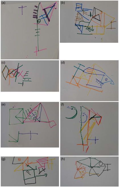
All subjects had Fragmentation scores between 22 and 24, apart from eight subjects whose scores ranged from 0 to 7, forming a cluster of very low scores on this measure. These eight copies are shown in Fig. 2. Each of these eight subjects had injury that included the right parieto-occipital area.
Copies of the Rey figure showing a FRAGMENTED appearance.

Similarities and Vocabulary
These are subtests of the Wechsler Adult Intelligence Scale-Third Edition (Wechsler, Reference Wechsler1997) using scaled scores.
Visual Reproduction Test
This test comprised the Visual Reproduction Test from the Wechsler Memory Scale-Revised (Wechsler, Reference Wechsler1987). There are four diagrams. Each was presented for 10 s. In view of the possibility of unilateral visual neglect, the participants were asked to make sure that they looked to the left. A drawing of the diagram was then made from memory. Scoring was performed according to the manual with a maximum score of 41.
Tactual Learning Test
The test items for the Tactual Learning Test were made from 1.2 mm aluminium welding rods bent into the shapes shown in Fig. 3 and mounted on a plain acrylic sheet measuring 26 by 30 cm. Figure 3 shows the three levels of difficulty as well as the measurements for each design.
Tactual learning test (presented from the point of view of the subject).

The blindfolded subject was told ‘I am going to move your finger around a piece of wire that has been bent into a shape. I will do this three times and then ask you to draw the shape from memory’. If the drawing was not correct, the procedure was repeated and a further drawing was requested. A third trial was given if the shape was still not correct. This gave a possible total of nine exposures to the shape. The preferred hand was used. Sensation (light touch, vibration and joint position) was tested prior to administration of the Tactual Learning Test to ensure that there was no sensory impairment.
Scoring was based on the final drawing at each level of difficulty and reflected the number of changes in direction and orientation of the elements of the figure. Each drawing comprised a number of open rectangles and single lines; each open rectangle was scored 3 if all three sides were drawn correctly and a further point was added for correct orientation. Single lines were scored one if correctly positioned. The final diagram was given a further point if correctly oriented. Maximum scores were as follows: Level 1–7, Level 2–10, Level 3–15, resulting in a total score of 32.
In this study, the verbal tests were presented first, followed by the Rey figure and the Visual Reproduction Test. This was followed by the Tactual Learning Test.
Results
Characterization of Group Differences
This study considered the performance of different groups on two tasks designed to measure different aspects of spatial memory. The tasks were the Visual Reproduction Test and the Tactual Learning Test. A number of covariates were measured: age, Similarities score, Vocabulary score and years of education.
The primary aim was to compare the performance of a number of different groups on the two tasks, the Visual Reproduction Test and the Tactual Learning Test. There were five groups considered for analysis – one control group and four clinical groups with varying forms of brain injury. The groups are described in Table 1, along with the abbreviations used in the remainder of this paper.
It was expected that performance on both the Visual Reproduction Test and the Tactual Learning Test would be worst for the MD and RP groups. The controls, LHS and RHS groups, are expected to perform at similar levels and better than the MD and RP groups.
In the analyses, the groups are ordered in presentation to correspond to the expectations of performance on the Visual Reproduction Test and Tactual Learning Test (from best to worst): C, LHS, RHS, MD, RP.
Statistical Analyses
Table 2 shows the means and standard deviations of the scores on the Visual Reproduction Test and Tactual Learning Test by group. Mean scores on both tests correspond to the ordering expected with the control group doing the best and the RP group doing the worst. However, the variability of performance differed greatly between the groups. On the Tactual Learning Test, for example, the standard deviation of the MD group is almost 10 times that of the Control group. Table 2 also shows the proportions of participants in each group achieving the maximum score. Clearly, there is a ‘ceiling effect’ – almost half of the control group scored the maximum for the Visual Reproduction Test, and over half of all the participants scored the maximum for the Tactual Learning Test.
Visual Reproductions and Tactual Learning Test by group

For the measures shown in Table 2, average performance and spread are related but there is less variation as performance nears the upper limit for a test. In situations such as this, appropriate method of analysis is logistic regression which models the odds of successes on a test. For the Visual Reproduction Test, each participant’s score was interpreted as being the number of successes achieved in a set of 41 (the maximum possible score) increments. For Tactual Learning Test, each participant’s score was interpreted as being the number of successes achieved in a set of 32 (the maximum) increments.
Two logistic regression models are reported: one for Visual Reproduction Test score and one for Tactual Learning Test score, each as a proportion of the total possible score. The primary explanatory variable is group; in addition, four explanatory covariates (age, Similarities score, Vocabulary score, years of education) were controlled for.
Logistic regression provides a test of the null hypothesis of no effect of each explanatory variable on the outcome. It also gives estimates of the relative odds of success for any group relative to a particular group of interest. The odds of success is the ratio of the probability of success to the probability of failure. Groups are compared in terms of their relative odds of success. The comparisons of interest are between the MD group and each of the C, LHS and RHS, and between the RP group and each of the C, LHS and RHS. The covariates are included in each model, but details of the results for the covariates are not reported here; they are available upon request.
Logistic Regression Models
The overall test of group differences was statistically significant in both models (Visual Reproduction Test: F (4,47) = 20.04, P < 0.001; Tactual Learning Test: F (4,47) = 19.32, P < 0.001). Tables 3 and 4 summarize the logistic regressions for the Visual Reproduction Test and Tactual Learning Test, respectively. They provide the odds ratios for comparing groups, and assist in characterising the effect of group on each test performance.
Logistic regression of Visual Reproductions Test scores

Logistic regression of Tactual Learning Test scores

The odds ratios for the MD and RP groups relative to the Controls, LHS and RHS groups are all less than one for the Visual Reproduction Test (Table 3). This indicates that the MD and RP groups had lower odds of success on this test than the other groups. The odds ratios correspond to the expected ordering. For the Visual Reproduction Test, the odds for the RP group are very low compared with Controls: 2% of the Control group odds. For the Tactual Learning Test, the odds relative to the MD and RP groups are generally smaller than for the Visual Reproduction Test (Table 4). For example, the odds for the RP group are less than 1% of the Control group odds.
Note that logistic regression relies on large sample normal approximations, so confidence intervals or P values cannot be presented when the estimated Odds ratio is zero or infinity. This is the case for the LHS group relative to the other groups on the test of Tactual Learning Test; some LHS cases have missing values for the covariates and all included cases have perfect scores.
The logistic regression results described above together allow a characterization of performance on the Visual Reproduction Test and Tactual Learning Test tasks. There is a reasonably consistent pattern of results corresponding to expectations. The MD and RP groups perform poorly in comparison to Controls. For the RP group, the odds of success are at best 1/50th of the Controls. For the MD group, the odds of success are also dramatically lower than Controls (at best around 1/16th). Patients with LHS and RHS perform better than the RP group and the MD on both tests; odds for the clinical groups are less than 30% of that for the LHS and RHS groups.
The Association between the Visual Reproduction Test and the Tactual Learning Test
Next, the relationship between performance on the Visual Reproduction Test and the Tactual Learning Test was examined. Figure 4 shows a scatterplot of the Visual Reproduction Test versus Tactual Learning Test scores for each group. The pattern of association clearly varies substantially by group – this in part arises from the ceiling effects in performance on the tasks. For the Control group, for example, there is little variation on either task.
The primary interest is in the association between the two tests for the MD and RP groups. For both groups, the scatterplot shows reasonable variation in performance on the two tests and a roughly linear relationship. Hence, it is reasonable to summarise the association using a correlation coefficient for the MD and RP groups; a correlation coefficient is less appropriate for the other groups.
Scatterplot showing visual reproductions score versus tactual learning test score.

Table 5 summarises the correlation between the Visual Reproduction Test and Tactual Learning Test scores for the MD and RP groups. There is a moderate positive value for the MD group and a moderate negative value for the RP group. The third column gives a P value for a test of the null hypothesis of no association between the Visual Reproduction Test and the Tactual Learning Test scores. The P values suggest that the correlations observed are consistent with no underlying correlation between performances on the two tests. 95% confidence intervals for the correlation are also presented. Note that these confidence intervals are very wide and indicate that the data are consistent with a wide range of underlying correlations (including positive correlations, a zero correlation and negative correlations). They suggest that it is difficult to accurately estimate the association between the tests within a group with such small samples; hence these P values cannot be interpreted as providing evidence of no association between the tests.
Correlation between Visual Reproductions and Tactual Learning Test scores

Figure 4 shows the scatterplot of the two test results for all groups combined. It shows a strong positive relationship between the Visual Reproduction Test and Tactual Learning Test scores. The correlation between the Visual Reproduction Test score and Tactual Learning Test score for all participants is 0.82 (95% confidence interval for the correlation is 0.73, 0.88). The different groups, however, lie on different parts of the plot; this means that the correlation between the scores arises in part because groups of differing ‘ability’ are included. However, this plot highlights the diminished performance of the RP and MD groups, relative to the other groups, on both tests.
Discussion
In this study, it is proposed that, while the superior parietal lobe is concerned with multimodal integration for the perception of space (Andersen, Reference Andersen1997), in humans, there is a second system for the processing of spatial information, located in the right parieto-occipital area, and functioning at the representational level. The results of this study provide some support for this hypothesis. The main focus of this study was on a group of eight subjects who had sustained damage to the right parietal lobe and whose copy of the Rey figure showed a fragmentation of the spatial relationships in the figure as seen by inspection and by the low Fragmentation score. The results show that damage to the right parietal lobe, together with a fragmentation in the sense of space, is associated with a severe impairment in the learning and recall of spatial information in both the tactile and the visual modalities. This group of participants performed extremely poorly compared with controls on both the Visual Reproduction Test and the Tactual Learning Test. For the right parietal group, performance on the Visual Reproduction Test was only slightly better (2% chance of success) than performance on the Tactual Learning Test (1% chance of success). The small number of participants precluded the calculation of a correlation coefficient for the right parietal group, but the overall group of 63 participants showed a close correlation between the two tests (0.82). The results are affected by the small sample size for the right posterior parietal group, but the tight criterion of a fragmented sense of space for this group strengthens the findings.
The results extend the findings of Hécaen et al. (Reference Hécaen, Penfield, Bertrand and Malmo1956) by demonstrating that the fragmented sense of space seen on the Rey figure affects performance on tests of spatial memory, regardless of the modality in which the information is presented. This supports the hypothesis of Semmes (Semmes Reference Semmes1965; Semmes, Weinstein, Ghent, and Teuber Reference Semmes, Weinstein, Ghent and Teuber1955), who suggested that there is a supramodal centre that is responsible for ideas of directionality in space. However, there is some evidence to suggest that the concept of a supramodal centre in the right parietal area might not fully explain the present results. In this study, the fragmented sense of space was identified using the Rey figure, presented in the visual modality. In a previous study (Stuart, Reference Stuart1995), it was shown that some children with congenital blindness were able to build complex spatial constructions on the Tactual 3D Constructions Test, and they could learn and recall complex spatial patterns on the Tactual Learning Test. This leads to the conclusion that vision is not a necessary condition for the development of the reference frame of conceptual space. It shows that the spatial coordinate system of the right parieto-occipital area can develop in the absence of any visual experience. It suggests that there is no combination of modalities in a supramodal centre concerned with ‘ideas of directionality in space’ (Semmes, Reference Semmes1965). That function is performed by the proprioceptive modality which is primary in spatial processing, as proposed by Hécaen et al. (Reference Hécaen, Penfield, Bertrand and Malmo1956). According to this formulation, visual information may play a part in refining or enhancing the operation of this system on practical tasks, but it is not a necessary condition for its development.
The format of the Tactual Learning Test provides a model for the reference frame of conceptual space. According to this model, an internal map is created by a series of arm movements in two or three-dimensional space; the direction of each movement is defined geometrically in relation to the body’s upright position in relation to gravity (Paillard, Reference Paillard1971). Each movement is geometrically related to the preceding movement, and in its final form all points in the map are related in two and three-dimensional representational space. This representation requires an understanding of the invariant nature of the right angle and rectangular forms, and of the distance between two points. The final representation of the various elements of this internal map includes a grasp of the spatial relationships between all points within these complex patterns.
These results suggest that the proprioceptive modality has developed a web of geometric concepts that constitute the reference frame or spatial coordinate system. The reference frame could be described as a container for this geometric knowledge, but it could also be said that the geometrical knowledge is embodied in the proprioceptive modality. The motor system acts within this system of spatial coordinates to create an internal map that binds together the changes of direction generated by the moving limb, a body part or the whole body. Any change of direction occupies a privileged position within this map as it defines the direction of travel and permits the construction of an internal map that is isomorphic with identified features of the landscape. These locations may contain representations of certain landmarks such as buildings for the sighted traveller or tangible objects for travel in the absence of vision.
According to this formulation, the fragmented appearance of the Rey figure together with the preservation of the ‘small elements’ of the figure such as triangles, squares and circles, as seen in the eight subjects of the right parietal group, can be attributed to a loss of the geometric concepts that bind together the salient points and small elements of the figure. In the Rey figure, the reference frame provides a representation of the spatial relationships that make up the figure. Damage to the right parietal area may cause a loss of the reference frame of conceptual space, leading to the fragmented appearance on the Rey figure.
This reference frame of conceptual space has not developed de novo. Piaget and Inhelder (Reference Piaget and Inhelder1956) described its development in considerable detail, beginning with the representation of a single gesture through to an understanding of the geometric properties of space, including the developmental course for topological, projective and Euclidean concepts.
There is evidence from the Rey figure to suggest that the motor system has the capacity to act within this reference frame to create changes to the motor images, carrying out translations, rotations and distortions through the activities of kinetic and anticipatory images as described by Piaget and Inhelder (Reference Piaget and Inhelder1971). It could also be active during the route planning phase of spatial navigation when alternative routes are considered and then consolidated (Spiers & Maguire, Reference Spiers and Maguire2006). It is proposed that this intimate relationship between the proprioceptive system and the motor system is an extension of the sensorimotor interface for visually guided eye and arm movements seen in Brodmann’s area 5 and 7 of the posterior parietal cortex (Buneo & Andersen, Reference Buneo and Andersen2006; Cui, Reference Cui2014).
It is also proposed that the Tactual Learning Test is a pure test of spatial navigation, as its test items can be walked in a large-scale environment (Stuart, Reference Stuart1995), drawn or traced from a familiar route, or (presumably) drawn in a virtual reality setting, with or without vision, and with or without landmarks. Maps of this sort are used in arid and featureless environments to provide travel directions (Lewis, Reference Lewis1972).
Some residual capacity is seen in the fragmented copies of the Rey figure where the small elements of the figure, such as the circle and cross, are preserved. The participants with fragmented copies also showed some limited preservation of spatial ability on both the memory tests. This residual capacity was observed by Paterson and Zangwill (Reference Paterson and Zangwill1944) and Bohbot et al. (Reference Bohbot, Kalina, Stepankova, Spackova, Petrides and Nadel1998). It is proposed that this residual capacity is provided by the left parietal lobe, where there is a reference frame that provides the spatial basis for writing (Jancke, Kleinschmidt, Mirzazade, Shah, & Freund, Reference Jancke, Kleinschmidt, Mirzazade, Shah and Freund2001; Potgieser, van der Hoorn, & de Jong, Reference Potgieser, van der Hoorn and de Jong2015; Seitz et al., Reference Seitz, Canavan, Yaguez, Herzog, Tellmann, Knorr and Homberg1997) and which is affected in conditions such as ideomotor apraxia (Haaland, Harrington, & Knight, Reference Haaland, Harrington and Knight2000). The geometric contribution in the left parietal lobe is limited to common shapes such as circles, squares and rectangles. This residual capacity is likely to assist spatial navigation for the blind traveller with right hemisphere damage; its absence would, however, cause a more profound topographical disorientation as all spatial processing capacity would be lost.
The results also showed that there was an impairment on both the Visual Reproduction Test and the Tactual Learning Test for the MD group. Unlike the right parietal group, the MD group showed no evidence of any fragmentation of the sense of space on the Rey figure, either by inspection or on the Fragmentation score, but there was an impairment seen on the Visual Reproduction Test and the Tactual Learning Test with a significant association between the two tests. The conclusions are limited in that the subjects in the MD group may have suffered left, right or bilateral damage to the hippocampus. It is obvious that this aspect of the present experiment needs to be repeated using subjects with focal damage limited to the right hippocampus. The results are, however, consistent with the findings of spatial memory impairment on a visual maze following lesions to the right hippocampus as described by Milner (Reference Milner1965), and by its high correlation with the tactual maze test administered by Corkin (Reference Corkin1965); it is also consistent with the report that, following bilateral hippocampectomy, H.M. made an accurate copy of the Rey figure, but had no recall of the figure, even of the ‘small elements’ (Stark, Reference Stark, Andersen, Morris, Amaral, Bliss and O“Keefe2007). The association is further strengthened by the fact that the Tactual Learning Test was derived from the pathway through the maze tests. This makes it a reasonable assumption that a poor performance in the MD group may be attributed to damage to the right hippocampus.
If this theory of the reference frame of conceptual space is correct, it leaves no obvious explanation for the part played by the right hippocampus in the learning of new spatial information in humans. It means that, while damage to the right hippocampus impairs the learning of new spatial information, it does not affect the area responsible for the registration and spatial processing of that information. It follows that, if the place cells in the hippocampus are not responsible for spatial processing, they must perform a different role in spatial learning. It also implies that, while new learning can take place within the hippocampus through the process of replay, the neocortex is unable to initiate this process and must rely on input from the hippocampus.
Wilson and McNaughton (Reference Wilson and McNaughton1994) found that pyramidal neuron pairs that fired together in the CA1 region of the hippocampus during spatial exploration had an enhanced tendency to fire together during subsequent slow wave sleep. This firing during post experience sleep was most pronounced during the high-frequency oscillations in the local field potential known as sharp wave ripples. This finding was extended to the firing of sequences of neurons during sharp wave ripples in the hippocampus (Skaggs & McNaughton, Reference Skaggs and McNaughton1996), and Lee and Wilson (Reference Lee and Wilson2002) found that the replay of sequences of place cells corresponded to temporally compressed versions of movement trajectories. Dupret, O’Neill, Pleydell-Bouverie, and Csicsvari (Reference Dupret, O’Neill, Pleydell-Bouverie and Csicsvari2010) reported that goal related assembly patterns of neuronal firing in the hippocampus were associated with sharp wave ripples, and they concluded that these assemblies represented the formation and expression of new memory traces. It was also found that the suppression of sharp wave ripples in the hippocampus caused an impairment in spatial learning, implying a causal role in spatial memory for sharp wave ripples (Ego-Stengel & Wilson, Reference Ego-Stengel and Wilson2010; Girardeau, Benchenane, Wiener, Buzsaki, & Zugaro, Reference Girardeau, Benchenane, Wiener, Buzsaki and Zugaro2009).
Furthermore, there is evidence to suggest that this spatial learning process involving sharp wave ripples is not confined to the hippocampus. Qin, McNaughton, Skaggs, and Barnes (Reference Qin, McNaughton, Skaggs and Barnes1997) reported that, during periods of slow wave sleep after an episode of extended spatial behaviour, patterns of neuronal correlation that were manifest during the behaviour reemerged in the neocortical and hippocampal circuits as well as in the interactions between these areas. It was later found that, during sleep following a spatial movement task, the firing of neurons in the medial prefrontal cortex occurred simultaneously with hippocampal sharp wave ripples (Peyrache, Khamassi, Benchenane, Wiener, & Battaglia, Reference Peyrache, Khamassi, Benchenane, Wiener and Battaglia2009). Maingret, Girardeau, Todorova, Goutierre, and Zugaro (Reference Maingret, Girardeau, Todorova, Goutierre and Zugaro2016) considered that, during spatial learning, the medial prefrontal cortex and hippocampus were in a fine tuned coordination between sharp wave ripples, delta waves and spindles. Rothschild, Eban, and Frank (Reference Rothschild, Eban and Frank2017) showed that patterned activity in the auditory cortex preceded firing in the CA1 area of the hippocampus during sharp wave ripples, and there was information flow in the reverse direction. Wilber, Skelin, Wu, and McNaughton (Reference Wilber, Skelin, Wu and McNaughton2017) extended these findings to the posterior parietal cortex. They reported that, in the posterior parietal cortex, the learning of a movement sequence and the establishment of a reference frame were accompanied by the coupling of cortical delta waves and hippocampal sharp wave ripples. This led Wilber et al. (Reference Wilber, Skelin, Wu and McNaughton2017) to conclude that the mechanism for consolidation of the memory trace in the neocortex might be a cortical neuronal excitability state shaped by cortical delta oscillations and enhanced hippocampal output during sharp wave ripples.
These findings in the rat provide a possible mechanism for movement learning in the human. In conjunction with the findings from this study, they suggest that the function of the right hippocampus is not to establish a reference frame for spatial navigation, but to provide the right hippocampus with a directional arrow or link to specific areas of spatial movement encoding in the neocortex (the motor image) within the spatial coordinate system, to remember that link through replay within the hippocampus, and to generate the sharp wave ripples required for the process of replay in the neocortex. According to this hypothesis, learning of movement sequences in humans takes place when specific patterns of neurons in the inferior posterior parietal cortex are stimulated by the coupling of cortical oscillations with sharp wave ripples generated by the hippocampus.
In summary, the results of this study show that, in the human, damage to the right posterior parietal cortex may affect the higher levels of the proprioceptive modality, causing a loss of the geometric concepts that bind locations according to the complex concepts of topological, projective and Euclidean space. Damage to this system affects the ability of the motor system to create the internal maps (the movement sequences) that guide travel, with or without visual guidance. In both these forms of travel, changes of direction are usually, but not necessarily, marked by some form of landmark, a building in the former and some tangible object in the latter, whose representation is recruited from other parts of the brain. Some redundancy of function is provided by the left parietal cortex, where the ‘small elements’ of spatial configurations are processed, along with the ability to recall simple spatial configurations involving two or three changes in direction.
O’Keefe and Nadel (Reference O’Keefe and Nadel1978) had a similar model in mind in their initial work on spatial navigation in the rat, but they proposed that spatial processing takes place in the hippocampus. This was later revised to include the contribution of the grid cells in the medial entorhinal cortex (O’Keefe, Reference O’Keefe2014). The results of this study show that this model might not apply to humans. It has been demonstrated that, in humans, the processing of spatial material occurs in the right parieto-occipital area and forms part of the system related to movement and action. From this neuropsychological perspective, the right hippocampus is an engine, tracking specific cortical activity, strengthening synaptic connections, and driving the process of consolidation in the neocortex.
Acknowledgements
The author would like to acknowledge the contribution of Dr Sue Finch of the Statistical Consulting Centre, University of Melbourne, who performed the statistical analysis.
Financial support
None.
Conflict of interest
None.
Ethical standards
The author asserts that all procedures contributing to this work complied with the ethical standards of the relevant national and institutional committees on human experimentation and with the Helsinki Declaration of 1975, as revised in 2008.