Introduction
The lichen genus Maronina (Lecanoraceae) is characterized by a crustose thallus containing a chlorococcoid photobiont, lecanorine apothecia, polysporous asci of a modified Lecanora-type, simple, hyaline, non-halonate ascospores and bacilliform conidia (Hafellner & Rogers Reference Hafellner and Rogers1990; McCarthy Reference McCarthy2004; Kantvilas & Elix Reference Kantvilas and Elix2007). There are three known species, M. australiensis Hafellner & Rogers, M. hesperia Kantvilas & Elix and M. multifera (Nyl.) Hafellner & Rogers, all of which occur in mainland Australia, with the last also known from tropical South America. In their account of the genus, Kantvilas & Elix (Reference Kantvilas and Elix2007) suggested that, on the basis of ascus structure, the genus may be heterogeneous. The discovery of a new species in Thailand, described below, has enabled a reappraisal of the genus using morphological, anatomical and chemical data.
Material and Methods
The study is based chiefly on collections housed in the Natural Medicinal Mushroom Museum Herbarium, Thailand (MSUT), and on selected duplicate and reference specimens in the Tasmanian Herbarium (HO) and the Field Museum (F). Anatomical and morphological observations were undertaken using light microscopy, with thin hand-cut sections mounted in water, 10% KOH (K), lactophenol Cotton Blue and Lugol's iodine with pretreatment with KOH. Ascospore measurements are presented in the format: smallest measurement–mean–largest measurement; single outlying values are given in parentheses. Chemical constituents were identified routinely by thin-layer chromatography (Culberson Reference Culberson1972; Culberson et al. Reference Culberson, Culberson and Johnson1981; Culberson & Johnson Reference Culberson and Johnson1982; Elix & Ernst-Russell Reference Elix and Ernst-Russell1993).
The New Taxa
Maronina orientalis Kantvilas & Papong sp. nov. var. orientalis
Thallus crustosus, corticola, apotheciis lecanorinis et ascis polysporis, ad Maroninam pertinentibus instructus, thallo potius crasso, verruculoso, olivaceo-viridi, acidum alectoronicum continenti, ascosporis 5–8 μm longis, 2–3 μm latis, et paraphysibus 1·5–2·5 μm crassis, ramosae anastomosantibusque recognita.
Typus: Thailand, Wat Tham Chao Pha, Phu Pha Kud, Muk Dahan Province, Nhong Sung District, 16°46′N, 104°43′E, in dry dipterocarp forest, 285 m altitude, 20 June 2009, K. Papong & W. Konhin 6576 (HO—holotypus; F, MSUT—isotypi).
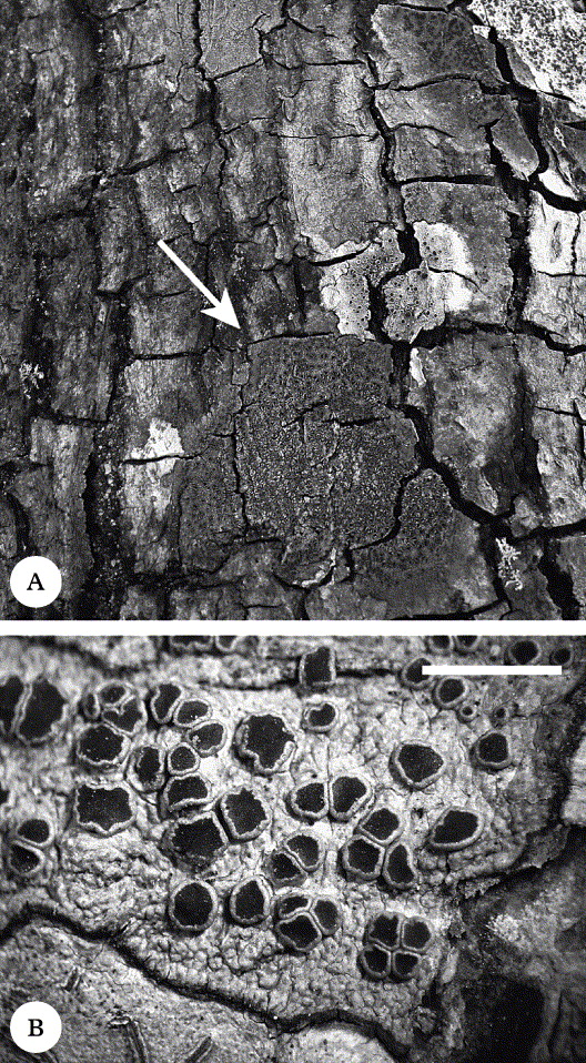
(Fig. 1A & B)
Maronina orientalis var. orientalis (holotype). A, habit on fissured dipterocarp bark; B, detail of thallus and apothecia. Scale = 2 mm.
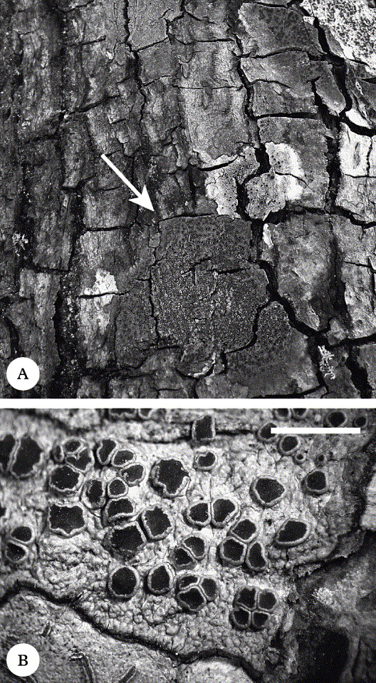
Thallus crustose, continuous, unevenly verruculose, sometimes cracked, dull greyish green to olive-green, more rarely pale greenish grey, lacking soredia and isidia, 60–130 μm thick, ecorticate but with a poorly differentiated outer layer of short-celled, anticlinal hyphae 2–3 μm thick, penetrating the algal layer and overtopped by a brownish epinecral layer c. 5 μm thick; medulla I−, KI−; prothallus dark brown to brown-black, evident as a thin line at the periphery of the thallus; photobiont chlorococcoid, with ±globose cells 5–12 × 4–10 μm.
Apothecia lecanorine, 0·3–1·2(–1·5) mm wide, superficial, basally constricted, very abundant, scattered or densely crowded together, roundish or, more commonly, irregular when adjacent to each other and squashed, often deeply lobed when older; disc matt, brown to dark brown, sometimes a little mottled, epruinose, plane at first, later undulate but never convex; thalline margin entire, persistent, crenulate, a little inrolled when young, in section cupulate, 70–120(–150) μm thick laterally. Hypothecium 30–40(–80) μm thick, colourless, I+ blue. Hymenium 70–80(–100) μm thick, colourless, I+ blue, KI+ blue, highly coherent in water, lax in KOH, overlain by a brownish, highly gelatinized epihymenial layer 2–5 μm thick, inspersed with minute crystals insoluble in KOH. Asci clavate, 45–65(–70) × 17–22 μm, seemingly 32–50-spored, approximating the ‘Maronina-type’: outer wall intensely amyloid; tholus well-developed, with intensely amyloid flanks and a generally rather weakly amyloid, fuzzy, central zone; ocular chamber not developed (see below and Fig. 3). Paraphyses branched and anastomosing, (1·5–)2–2·5 μm wide, wavy, often ± constricted at the septa and hence slightly moniliform; apices not pigmented, not or only occasionally slightly expanded to 3(–4) μm wide, mostly remaining conglutinated in a gel. Ascospores hyaline, thin-walled, non-halonate, narrowly ellipsoid, (4·5–)5–6·4–8 × 2–2·7–3 μm. Pycnidia occasional, immersed, detected by the minute dark ostiole in the surface of the thallus; conidia bacilliform, 6–9 × 1 μm.
Chemistry. Alectoronic acid (major) and α-collatolic acid (minor); medulla K−, KC+ orange-red, C+ orange-yellow, P−, UV+ white.
Etymology. The specific epithet refers to the geographical distribution of the new taxon.
Remarks. This is a very striking lichen, forming conspicuous, dull greenish, extensive thalli to 80 mm or more wide, typically densely beset with lecanorine apothecia. Anatomically it is characterized by the polysporous, Maronina-type asci, a feature that immediately distinguishes it from superficially similar genera such as Lecanora (eight-spored, Lecanora-type asci) and Maronea (polysporous, Fuscidea-type asci). The placement of the new species in Maronina was confirmed only after extensive study of the asci, a task confounded by the fact that despite the abundance of seemingly richly fertile collections, very few apothecia (indeed < c. 2%) contained any well-developed asci with ascospores.
The presence of alectoronic acid and the relatively delicate, branched and anastomosing paraphyses ally M. orientalis with the widespread tropical species, M. multifera. However, that species has different, very delicate asci (see discussion below), and a somewhat different chemistry (no α-collatolic acid, and often with protoconstipatic acid). In addition, the two taxa are superficially distinct, with M. multifera having a paler, smoother thallus and apothecia with a thinner margin. As delimited and discussed by Kantvilas & Elix (Reference Kantvilas and Elix2007), M. multifera is an enigmatic taxon with some chemical variation. With time and further collections, the different populations may require taxonomic reassessment but, on current data, the extensive Thai material studied deserves recognition at specific rank.
Intermixed with some specimens of M. orientalis there is a superficially similar, isidiate taxon. This is described below as a separate variety.
Ecology. This species is common in dry dipterocarp forest at low altitudes (250–350 m) in north-eastern Thailand. It typically grows in well-lit conditions on deeply fissured bark on trunks of various trees of the family Dipterocarpaceae. It associates with a range of crustose lichens, including Lecanora phaeocardia Vain., L. tropica Zahlbr. and members of the Graphidaceae.
Selected specimens examined (total = 42). Thailand: Muk Dahan Province: Nhong Sung District, Wat Tham Chao Pha, Phu Pha Kud, 16°46′N, 104°43′E, 285 m alt., 2009, K. Papong 6514 (HO, MSUT); Phu Pha Kham, 16°46′N, 104°43′E, 310 m alt., 2009, K. Papong & W. Konhin 6601 (HO, MSUT). Sakon Nakhon Province: Phu Pan National Park, next to the repeated Television signal 5 Center, 17°03′44″N, 103°58′20″E, 327 m alt., 2009, K. Papong 6482, 6484, 6485, 6486, 6487, 6491 (MSUT); Phu Pan National Park, Kham Hom Waterfall, 17°03′49″N, 103°58′23″E, 332 m alt., 2009, K. Papong 6488, 6489 (MSUT).
Maronina orientalis var. corallifera Kantvilas & Papong var. nov
Varietas morphologia anatomiaque idem ac varietatis principalis, sed thallo isidiis copiosis, coralloideis, teretiformibus, usque 0·6 mm altis, 0·1 mm latis differt.
Typus: Thailand, Phu Pha Kham, Muk Dahan Province, Nhong Sung District, 16°46′N, 104°43′E, in dry dipterocarp forest, 310 m altitude, 21 June 2009, K. Papong & W. Konhin 6603 p.p. (HO—holotypus; F, MSUT—isotypi).
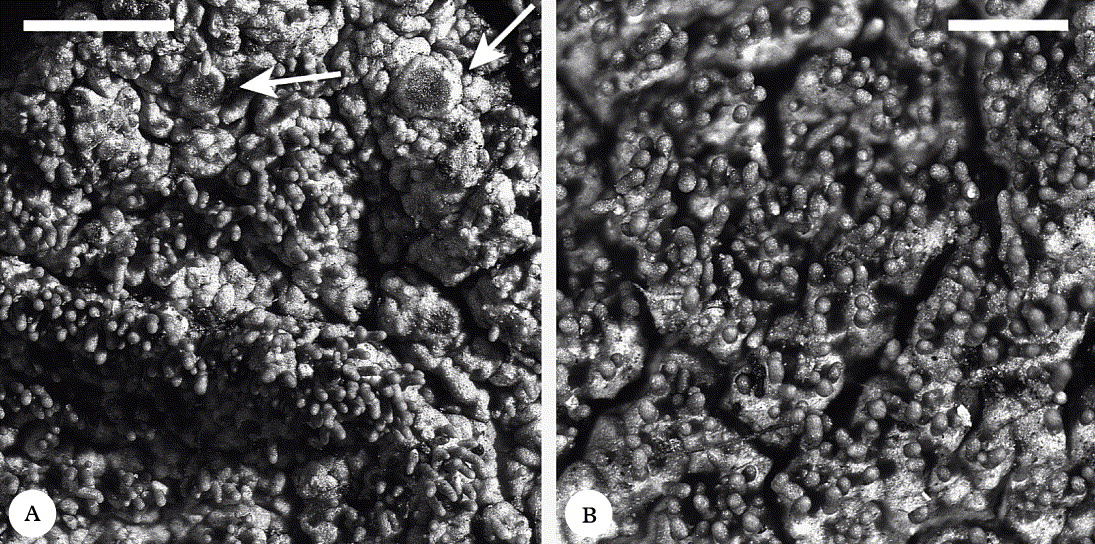
(Fig. 2A & B)
Maronina orientalis var. corallifera (holotype). A, isidiate thallus with apothecia (arrow); B, detail of isidia. Scales: A = 1 mm; B = 0·5 mm.
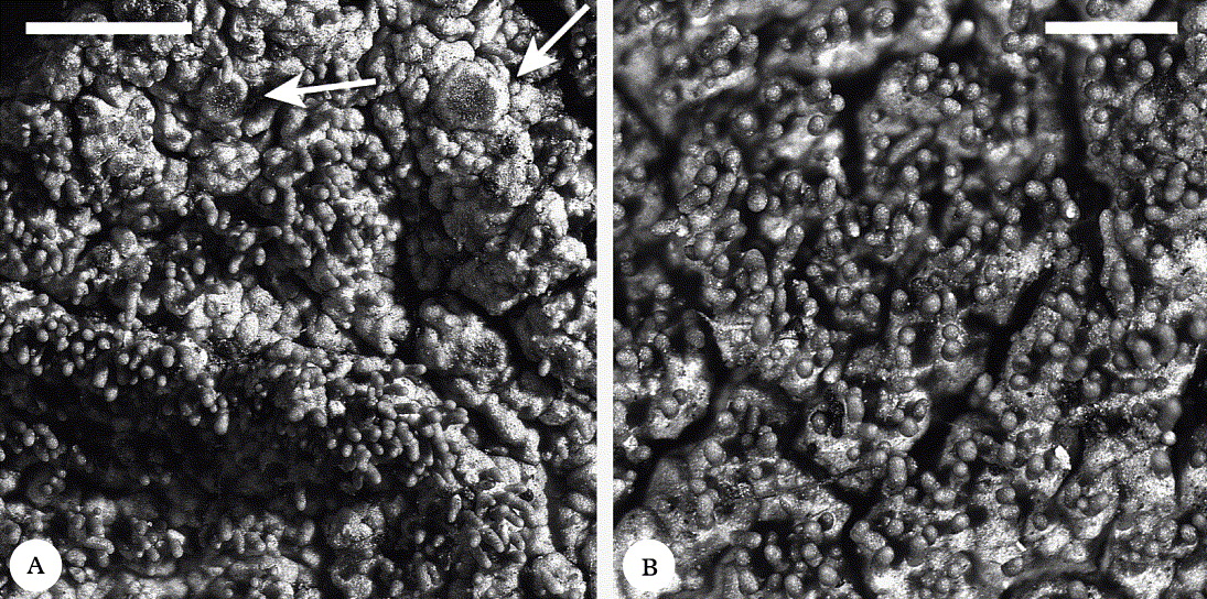
Thallus, apothecia and chemical composition as for the typical variety, but with the thallus isidiate; isidia abundant, scattered or in clumps, terete, 0·25–0·4(–0·6) mm tall, 0·08–0·1 mm wide, simple or coralloid-branched, occurring mainly on the thallus surface but sometimes also on the apothecial margins. Ascospores 7–7·9–9 × 2–2·9–3·5 μm.
Pycnidia not found.
Remarks. This variety grows together with the typical variety on the same pieces of bark, but is easily distinguished by the scattered to clumped coralloid isidia. The seemingly larger ascospores may be an artefact of a smaller sample size or a later stage of development. As with the variety orientalis, the occurrence of fertile asci is extremely rare.
Selected specimens examined (total = 7). Thailand: Muk Dahan Province: Nhong Sung District, Phu Pha Kham, 16°46′N, 104°43′E, 310 m alt., 2009, K. Papong & W. Konhin 6603 (HO, MSUT).
Systematic placement and ascus structure in Maronina
The asci of Maronina were first described by Hafellner & Rogers (Reference Hafellner and Rogers1990) as being a ‘variation of the Lecanora-type’; these authors noted their broad but rather indistinct masse axiale, which they compared to that seen in Protoparmelia badia (Hoffm.) Hafellner. The asci were subsequently investigated by Kantvilas & Elix (Reference Kantvilas and Elix2007) who found that whereas the type of the genus, M. australiensis, and their newly described taxon, M. hesperia, had asci of the type described and illustrated by Hafellner & Rogers (Reference Hafellner and Rogers1990), perhaps appropriately termed ‘Maronina-type’, the asci of the third known species, M. multifera, were different. These they referred to as “approximating the Acarospora-type”. Furthermore, whereas M. australiensis and M. hesperia had simple, robust paraphyses, M. multifera had slender, branched and anastomosing paraphyses.
The discovery of a further species of Maronina, M. orientalis, provided the opportunity to re-examine the asci of all the species. In this reappraisal, the asci of M. multifera were confirmed as being different to those of the ‘core’ of Maronina (i.e. M. australiensis and M. hesperia), but to refer to them as Acarospora-type is not correct. The tholus in M. multifera is well developed but the flanks are intensely amyloid and very highly divergent; they are probably better regarded as a modified Maronina-type or better still, ‘multifera-type’. The curious observation that the asci are very fragile and that the wall of the ascus easily separates from the spore sac, described by Kantvilas & Elix (Reference Kantvilas and Elix2007), was confirmed in this new study.
The asci of M. orientalis display a range of forms (Fig. 3), from the typical ‘Maronina-type’ (well-developed amyloid tholus with ±cylindrical, fuzzy masse axiale; Fig 3A), to asci with the amyloid zone reduced (Fig. 3B), to asci with the amyloid flanks reduced and highly divergent (Fig. 3C), through to asci with the amyloid zone entirely restricted to the flanks of the tholus and so approximating the ‘multifera-type’ (Fig. 3D). The asci of the new species are quite robust and remain intact in the course of the customary staining and squashing technique employed by lichenologists. In that regard they resemble the asci of M. australiensis and M. hesperia. However, the paraphyses of the new species are very similar to those of M. multifera, being slender, branched and anastomosing. Thus M. orientalis can be regarded as bridging the anatomical differences between M. australiensis and M. hesperia on the one hand, and M. multifera on the other, and adds support to the decision to retain all taxa within one, albeit rather variable genus.
Schematic representation of the ascus apex in species of Maronina after treatment with dilute KOH and Lugol's iodine (amyloid parts stippled). A, typical ‘Maronina-type’, as found in M. australiensis and M. hesperia; B, one of the forms found in M. orientalis; C & D: ‘multifera-type’ as found in M. orientalis and M. multifera, with the amyloid zone limited to the flanks of the tholus.

We attempted several times with several specimens to obtain DNA sequence data, but could not generate any reliable sequences. Hence the issue of the systematic placement of the genus will require further study. However, based on anatomy, morphology and chemistry, a close relationship with Protoparmelia is a hypothesis worthy of closer consideration.
This study was financially supported by the Thailand Research Fund and the Research Division at Mahasarakham University and a NSF grant (DEB–0516116) to The Field Museum (PI: HTL). We thank Jean Jarman for providing Figure 2 and preparing the other illustrations for publication, and Imke Schmitt for confirming our chemical results by hplc.