Haematopoietic stem cell transplantation is an important and effective treatment strategy for many malignancies, marrow failure syndromes, and immunodeficiencies in children, adolescents, and young adults. Reference Barriga, Ramírez, Wietstruck and Rojas1 Transplant strategies and supportive care have evolved over the past few decades resulting in improved overall survival. Reference Remberger, Ackefors and Berglund2 Despite advances in supportive care, patients undergoing transplantation are at increased risk to develop cardiovascular co-morbidities in the first weeks of therapy, Reference Dandoy, Jodele and Paff3,Reference Rotz, Ryan and Jodele4 and transplant survivors have a threefold increase in cardiovascular disease over age-adjusted controls. Reference Wingard, Vogelsang and Deeg5 The chemotherapy and radiation utilised in stem cell transplantation can cause cardiac toxicity, which may be early or late after transplantation. Early complications, i.e., in the first 100 days post-transplant, include pericardial effusion, pulmonary hypertension, and left ventricular systolic dysfunction. Reference Rotz, Ryan and Jodele4,Reference Rotz, Powell and Myers6–Reference Friedman, Szmuszkovicz, Rabai, Detterich, Menteer and Wood10 Late cardiac complications include valvular disease, and systolic and diastolic ventricular dysfunction including dysfunction secondary to iron overload. Reference Badawy, Liem, Rigsby, Labotka, DeFreitas and Thompson11 Cardiovascular disease is a leading cause of non-relapse mortality in stem cell transplant long-term survivors. Reference Dandoy, Jodele and Paff3,Reference Majhail, Bajorunaite and Lazarus12–Reference Rotz, Dandoy and Taylor17
Routine scheduled echocardiography is used to monitor for these sequelae throughout the stem cell treatment course. Unfortunately, poor echocardiographic windows due to treatment-related factors, including body habitus, central line placement, bandages, and effusions, often hamper echocardiograms after transplant. Consequently, the uncertainty in measures of function can make longitudinal assessment difficult. Relative to echocardiography, cardiac MRI allows for accurate anatomic visualisation and ventricular function measurement, and is relatively independent of patient factors. Reference Rehr, Malloy, Filipchuk and Peshock18–Reference Natori, Lai and Finn21 One drawback is that complete studies may take upwards of an hour to finish. The study objective was to determine whether a time-abbreviated “rapid cardiac MRI” protocol can be used in this patient population to obtain a reliable assessment of ventricular size and function, pericardial effusion, and estimated right ventricular pressure without increased patient discomfort.
Methods
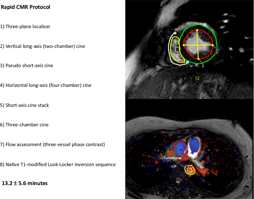
This study was performed as a feasibility study at a single site, Cincinnati Children’s Hospital Medical Center, and was approved by the local Institutional Review Board. Patients who were undergoing initial work up prior to stem cell transplantation or who had recently undergone transplantation were enrolled on a voluntary basis. Within 1 month of a clinical echocardiogram, enrolled patients underwent a research MRI. All MRIs were performed with a 1.5 T Philips scanner (Ingenia; Philips Healthcare, Best, the Netherlands) using a standard-free breathing protocol. Three signal averages were used for respiratory motion compensation; the rapid protocol is illustrated in Fig 1. Volumetric and functional measurements were made using standard cine steady-state-free precession short-axis stack from the mitral valve annulus to the apex (vertical and horizontal long-axis acquisitions were used for reference). Typical parameters for standard cine steady-state-free precession images were a field of view 300 × 112 mm, matrix size 168 × 158 mm, slice thickness of 6–7 mm, pixel resolution 1.8 × 1.8 mm, TR/TE 2.3/1.17 ms, 30 phases per R-R interval, parallel imaging factor 2. Volumetric, functional, and flow data were clinically measured using QMASS MR (Medis® Medical Imaging Systems, Leiden, the Netherlands). Volume and mass were indexed to the body surface area, calculated by the Mosteller algorithm. Ventricular stroke volume was compared to phase contrast derived flow for internal validity. Left ventricular eccentricity indices were measured on short-axis images at the mid-ventricular level at end-systole as a surrogate measure of right ventricular hypertension. Reference Averin, Michelfelder, Sticka, Cash and Hirsch22 Time to complete either the scan only for the echocardiogram or MRI was recorded from the timer on the machine or as documented by the technician. Realising there may be differences in preparation time, this method of determining timing was felt to be more reliable and reproducible. After completion of both the echocardiogram and MRI, patients were given a brief survey to document their perception of the experience of both tests.
Cardiac magnetic resonance protocol.

Statistics are descriptive in nature and include time to complete individual studies and adequacy of imaging. Measures of central tendency and variation including means and standard deviations, medians and interquartile ranges, and numbers and proportions were calculated for all outcomes as appropriate. Intraclass correlation coefficients and Bland–Altman plots were performed comparing left ventricular ejection fraction between MRI and echocardiography. Standard errors and 95% confidence intervals were calculated to obtain tentative population inference for the time required to complete the MRI image analysis.
Results
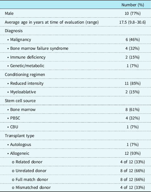
A total of 13 patients were enrolled in the study. The mean age was 17.5 ± 7.7 years (range 9.8–30.6), with 10/13 (77%) male and 10/13 (77%) White patients. Six patients (46%) underwent transplant for malignancy and 4 (32%) for a bone marrow failure syndrome (Table 1). One patient underwent a transplant for acute lymphoblastic leukemia and received total body irradiation. The majority of patients underwent allogeneic transplant (12 of the 13 or 93%). Three patients underwent MRI prior to transplant (range 10–44 days prior to transplant), and the remaining 10 were after transplant at long-term follow-up clinic, at a median of 4.2 years post-transplant (IQR 3.1–5.4 years).
Demographics and disease characteristics in paediatric haematopoietic stem cell transplant patients undergoing rapid cardiac MRI (n = 13).

CBU = cord blood unit; PBSC = peripheral blood stem cell
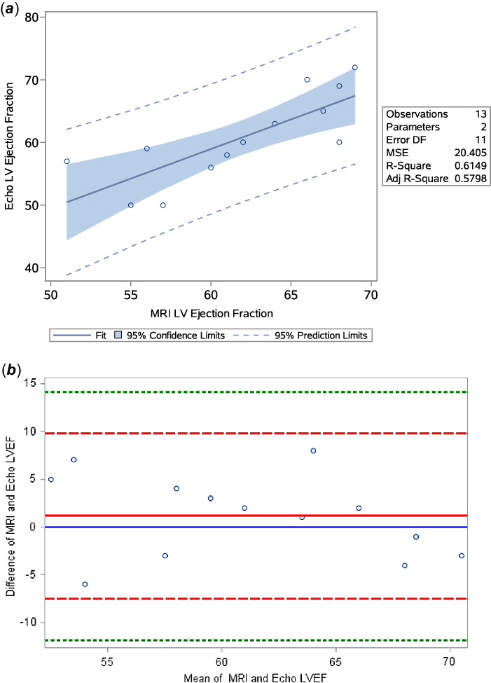
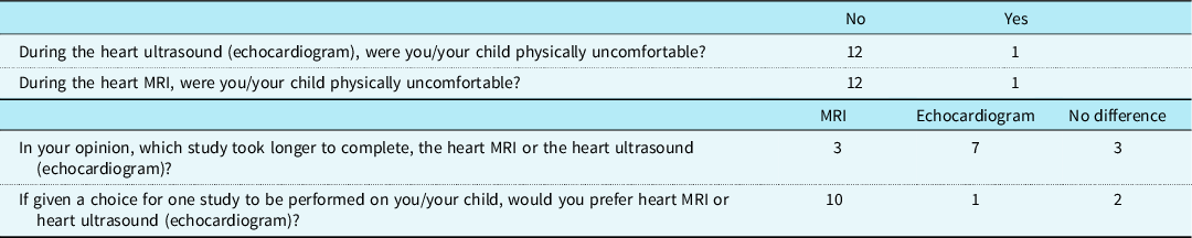
The echocardiogram and MRI were completed in all patients without complications. The mean study time was 13.2 ± 5.6 minutes for MRI and 18.8 ± 5.7 minutes for echocardiogram (p = 0.064). Correlation between ejection fraction by MRI and echocardiogram was good (Fig 2a, Pearson r = 0.78, p = 0.0015). A Bland–Altman plot further describes the agreement between the two modalities (Fig 2b, ICC 0.76; 95% CI 0.47, 0.92). Of note, 4 of the 13 (31%) patients had left ventricular ejection fraction that was different between MRI and echocardiogram by >10%, with 7/13 studies differing by >5% (Fig 2). MRI and echocardiography measures of eccentricity index could be performed in all patients. None of the patients had documented right ventricular hypertension; however, there was not a significant correlation between the two methods regarding systolic eccentricity index (Pearson r = 0.44, p = 0.13). Patients were also given a survey regarding their experiences undergoing both MRI and echocardiogram, with the majority both perceiving that the echocardiogram took longer (7/13) and indicating they would prefer the MRI if given a choice (10/13) (Table 2).
(a) Correlation between left ventricular ejection fraction by echocardiogram (y-axis) and cardiac MRI (x-axis); Pearson correlation r = 0.78. (b) Bland–Altman plot showing difference between left ventricular ejection fraction by echocardiogram and cardiac MRI. The red dashed lines are two standard deviations from the mean.

Patient/parent survey. After completing both echocardiogram and cardiac magnetic resonance testing, patients completed a survey. Data are a simple count.

Discussion
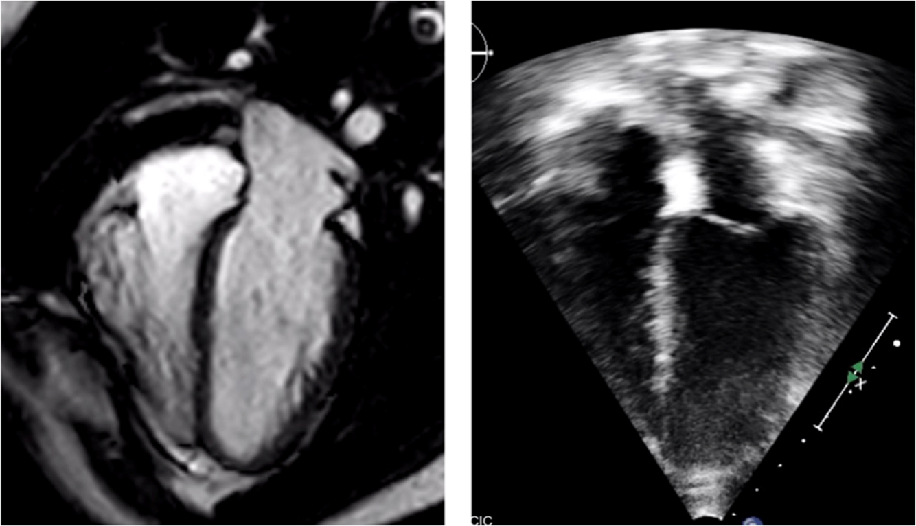
Patients who undergo treatment with haematopoietic stem cell transplantation are at increased risk for a number of cardiac co-morbidities, both during treatment and as survivors. The most common during therapy are the development of pericardial effusion, right ventricular hypertension, and left ventricular systolic dysfunction. Reference Dandoy, Jodele and Paff3,Reference Rotz, Ryan and Jodele4 In our clinical experience, once therapy is started, patients frequently have limited echocardiographic windows due to central line placement, bandages, and effusions. Patient discomfort can also result in imperfect echocardiographic imaging. Limited echocardiography can negatively impact assessment of both anatomy and function. Importantly, this can lead to incorrect estimation of cardiac function hindering medical management (Fig 3).
Representation of four-chamber cardiac MRI (left) and echocardiographic (right) images of the same patient. Note that the well-delineated myocardial blood pool border in the cardiac MRI image is compared with the echocardiographic image. Much of the uncertainty in echocardiographic ejection fraction measurements are secondary to the inability to define this border that is made more challenging by patient factors often present in patients undergoing haematopoietic stem cell transplant.

We sought to establish a time-abbreviated MRI protocol that would obtain the same information collected by echocardiogram albeit with the increased accuracy of precision of MRI without the limitations of imaging windows. This would be of particular use in the early post-transplant period when echocardiographic windows are more likely to be obstructed, the patient may be limited for a time due to illness or other testing and therapies, and the need for repeat studies makes a prolonged session less desirable. We were able to demonstrate that the rapid MRI protocol provided equivalent clinical data regarding left ventricular ejection fraction at a similar time as echocardiogram. We report a statistically strong correlation between echocardiogram- and MRI-measured ejection fraction, in contrast to that shown for survivors of cancer in other studies. Reference Armstrong, Plana and Zhang23 The reason for this discrepancy may be related to sample size. Interestingly, our results showed that one-third (4/13) of patients had greater than 10% and more than half (7/13) had greater than 5% variation of left ventricular ejection fraction between the modalities, which is of potential clinical significance. Again, this is similar to other at-risk children with poor acoustic windows. Reference Soslow, Xu and Slaughter24 Because MRI is considered the “gold standard” in determining left ventricular ejection fraction, this method may actually improve upon the data typically generated by the more commonly used echocardiogram for this patient group. Likewise, MRI is superior to echocardiogram in identifying structural abnormalities and the presence of effusion. One area in which MRI does not currently perform as well as echocardiogram is in the determination of diastolic dysfunction. While it can be a long-term consequence of stem cell transplantation, diastolic dysfunction is generally not an issue in the short term when a rapid protocol would be most useful.
Monitoring for the development of pulmonary hypertension is important in the transplant patient population. We have reported that ∼15% of stem cell transplant patients requiring admission to the ICU after transplant developed clinically significant pulmonary hypertension that is hypothesised to be due to microangiopathic injury. Reference Dandoy, Jodele and Paff3 The standard echocardiographic screening includes measuring the tricuspid regurgitation peak velocity and the left ventricular eccentricity index as indirect measures of the right ventricular pressure. Only a subset of patients has an adequate tricuspid regurgitation velocity envelope for analysis, so in its absence, the eccentricity index is used. Given the technical limitations of measuring peak tricuspid regurgitation velocity by MRI, right ventricular hypertension can be estimated by measuring the left ventricular eccentricity index. There is a good correlation between invasively measured right ventricular pressure and left ventricular eccentricity index as reported in the literature. Reference Haddad, Guihaire and Skhiri25 In clinical practice, an abnormal eccentricity index would generate a clinical decision about whether a cardiac catheterisation was warranted. None of the patients in this study had evidence for right ventricular hypertension, limiting our ability to directly compare MRI and echocardiography in this regard. This may explain why eccentricity index calculations were poorly associated with our study. In thinking about the utility of left ventricular eccentricity index in a patient undergoing therapy who has inadequate tricuspid regurgitation for assessment, limited acoustic windows and central line placement can make it a challenge to obtain a true ventricular short-axis image by echocardiography, a problem not encountered by MRI.
In addition to the standard function metrics and right ventricular pressure estimate, MRI provides accurate measurement of the cardiac index and myocardial tissue characterisation. It has been shown that cardiac index is a very robust measure of the functional capacity of the cardiovascular system and is predictive of outcomes in a variety of scenarios, particularly in the intensive care setting. Reference Khirfan, Ahmed and Almaaitah26–Reference Saugel and Vincent28 Myocardial characterisation by non-contrast can be performed using T1 mapping. Although the analysis was not included in this study due to small patient numbers, myocardial T1 maps can provide estimates of fibrosis, and have shown to be elevated after cardiotoxic therapies. Reference Galan-Arriola, Lobo and Vilchez-Tschischke29 It is possible that this could be a sensitive marker of myocardial injury during the course of stem cell transplantation. It is worth considering that as a patient is further out from transplant, more data on myocardial tissue characterisation, iron deposition, etc., may be desired, making the rapid study without contrast less useful. Another potential clinical limitation is that most data regarding ventricular function after treatment for cancer is derived from echocardiograms, initially reported as shortening fraction and more recently as ejection fraction. While the field is always evolving MRI is not yet regularly used in this setting, thus before substituting MRI for echocardiogram a discussion should occur between providers to ensure that the data generated can be interpreted and used to make clinical decisions for a given patient.
Patient perception is extremely important in this patient population. Patients undergoing haematopoietic stem cell transplantation experience prolonged hospitalisations, multiple painful procedures, and a multitude of diagnostic tests. Although not the primary aim, the majority perception was that MRI was faster and subjectively preferable relative to echocardiography. It is possible that advancements in MRI imaging speed, specifically with compressed sensing, could further shorten MRI scan times. Reference Tolouee, Alirezaie and Babyn30 While the protocol was carried out in patients prior to starting therapy to minimise clinical impact, it is well accepted that the factors that would limit echocardiographic windows in this population would not interfere with data acquisition by MRI. One potential difference between echocardiography and MRI that did not factor in the current study is the need for sedation, as it was required by none of the patients. In general, echocardiography can be done in most patients without sedation, while in young and developmentally delayed patients, sedation may be required in MRI for the patient to lie still and cooperate with breath holding. For the rapid protocol developed by our group, breath holding is not needed, so all that would be required is for the patient to lie still for approximately 13 minutes.
Cost is often raised as a significant difference between echocardiography and MRI, with the latter perceived to be more costly. This is a particularly difficult comparison to make in a way that is meaningful to various practices for a variety of reasons. However, when comparing data using 2020 Medicare fee schedules, there is not a large difference between the studies. According to cms.gov, both the Facility Price and the Non-facility Price for a “transthoracic echocardiogram with Doppler, complete” (Healthcare Common Procedure Coding System 93306) is $211.49, while for “cardiac MRI for morphology” (Healthcare Common Procedure Coding System 75557), it is $325.17. The ability to make a more accurate comparison of costs is difficult as these figures are not likely representative of what will be charged to the patient and the difference between study types is likely variable between centres. Further, patient status (stable outpatient versus critically ill inpatient) could greatly affect the labour involved in transport and monitoring for a study. Finally, if an MRI were used to substitute for an echocardiogram in a patient in need of multiple follow-up studies, even a small difference in cost could be compounded.
The use of rapid MRI extends well beyond the stem cell transplant population. For instance, in Duchenne muscular dystrophy, MRI has become a common modality for assessment of ventricular function. As patients age, the development of scoliosis and body habitus often significantly limit echocardiographic windows. However, due to discomfort being in a supine position for extended periods, standard MRI sequences may not be tolerated and measures to shorten the time of the exam are appropriate. A rapid cardiac MRI could be a reasonable solution for the collection of high-quality data in this population. Reference Lang, Alsaied, Moore, Rattan, Ryan and Taylor31
This study is not without limitations. This was a single-centre study on a specialised patient population. By nature of being a feasibility study, overall numbers were low, and we had relatively low statistical power to determine a significant difference with regard to imaging time and patient experience. This sampling is a fair representation of the diagnoses, preparative regimens, and graft sources of the patients undergoing transplant at our centre. Additionally, it represents the early adolescent and young adult population; however, our study does not include infants and young children. We plan on assessing young children in a future study. Additionally, the patients included in our study were clinically stable and in the outpatient setting at the time of the MRI. We do believe that the rapid cardiac MRI protocol could be used in hospitalised stem cell transplant patients that are able to be transported to the MRI suite, similar to any number of imaging studies regularly performed on this population.
Summary
A rapid cardiac MRI was shown feasible to substitute for an echocardiogram in assessing ventricular function, pericardial effusion, and potentially right ventricular hypertension in patients treated with haematopoietic stem cell transplantation. Cardiac MRI is useful in this patient population where small changes in contractility may be important and echocardiography can be problematic due to limited echocardiographic windows. Rapid MRI protocols can be used for other populations where accurate quantitative cardiac function evaluation is important for clinical management.
Acknowledgements
Special thanks to Suzanne Berger and Alyssa Rohde for their assistance in patient recruitment and data collection for the study.
Financial support
This research received no specific grant from any funding agency, commercial, or not-for-profit sectors.
Conflicts of interest
None.
Ethical standards
This study was approved by the Institutional Review Board at the Cincinnati Children’s Hospital Medical Center.