Introduction
Nowadays, nanoparticle-based drug delivery systems (NDDSs) have been extensively developed for large-scale applications including detection, diagnosis, and treatment of diseases such as cancer, infectious diseases, diabetes, etc. [Reference Luo, Li and Chen1, Reference Torchilin2, Reference Leucuta3] NDDS-based drugs, also known as nanomedicine, have directly improved human health [Reference Ragelle, Danhier, Préat, Langer and Anderson4]. More and more researchers have focused on the design and exploitation of the novel NDDS with enhanced bioavailability, high drug-loading capacity, efficient delivery, and precise release [Reference Alvarez-Lorenzo, Blanco-Fernandez, Puga and Concheiro5, Reference Jia, Kui, Liu, Li, Xu and Sun6]. Over past decades, it has been reported that many nanoparticles are applied as drug carriers or gatekeepers to the construct NDDS, such as inorganic nanoparticles, organic nanoparticles, polymers, carbon nanostructures, host-guest hybrid nanoparticles and biomacromolecules, etc. [Reference Tatsuya and Kunihiro7, Reference Biju8, Reference Singh and Lillard9, Reference Chen, Roy, Yang and Prasad10] Specially, layered double hydroxides (LDHs) and mesoporous (MS) materials are widely considered as ideal materials for drug delivery because they possess many traits within biological systems, such as good biocompatibility, low toxicity, high specific surface, controlled particle size and morphology, tunable composition and functional surface, optimal storage and release rate of drug, etc. [Reference Bi, Zhang and Dou11, Reference Rives, Arco and Martín12, Reference Ma, Feng, Liang, Liu, Zeng and Wang13, Reference Li, Barnes, Bosoy, Stoddart and Zink14] Thus, these advantages have triggered our interest in the architecture of the NDDS by different functional and composed methods based on these two materials.
LDHs are a class of anionic clay materials with two-dimensional layered and hydrotalcite-like compounds. Generally, the chemical formula of LDH is written as  ${\left[ {{\rm{M}}_{1 - X}^{2 + }{\rm{M}}_X^{3 + }{{\left( {{\rm{OH}}} \right)}_2}} \right]^{X + }}{\left( {{A^{m - }}} \right)_{{x / m}}} \cdot n{{\rm{H}}_2}{\rm{O}}$, where M2+ represents a bivalent metal cation, M3+ represents a trivalent metal cation, A m− is an anion (organic or inorganic) with charge m −, and x represents the exchange ratio of the trivalent cation [Reference Tronto, Cardoso, Valim, Marchetti, Vitória and Bentley15]. They possess an anion exchange property in the interlayer to neutralize the positive charge because their divalent ions are partially replaced by trivalent ones. Among various LDH materials, Mg/Al-LDH materials have been found to have extensive application in the NDDS as the host in recent years because of their advantages including good biocompatibility, low cytotoxicity, high loading of drugs, and controllable release of guest molecules [Reference Kriven, Kwak, Wallig and Choy16, Reference Kwak, Kriven, Wallig and Choy17]. However, some drawbacks of Mg/Al-LDH materials, such as easy aggregation in cell culture solution or phosphate-buffered saline solution, and difficult surface functionalization, largely restrict their practical applications [Reference Bao, Yang, Huang, Xu, Hao, Wu, (Max) Lu and Zhao18]. By contrast, MS materials can remedy these defects because of their attractive characteristics including facilitative dual-functional surface (external and internal), stable porous structure, high surface areas, and large pore diameters/volumes [Reference Vyskočilová, Luštická, Paterová, Machová and Červený19, Reference Mathew, Parambadath, Park and Ha20]. More and more MS materials have been used in NDDS construction since MCM-41-type MS was first reported as the NDDS in 2001 [Reference Vallet-Regi, Ramila, del Real and Perez-Pariente21]. Moreover, various gatekeepers have been developed by functionalizing MS materials to achieve precise drug delivery and release under a specific stimulus. Thus, Mg/Al-LDH and MS materials are desirable to exploit the new NDDS by taking full advantages of these materials. They also trigger us to think how to link or assemble these two materials to construct the NDDS.
It is well known that core–shell nanocomposites with an interstitial space between core and shell have attracted much attention because of their structural features and potential applications in the controlled delivery system. In particular, many nanocomposites of mesoporous silica shell surrounding LDH core (LDH@MS) were applied to delivery drugs and shown to have an adjustable slow release effect, good biocompatibility, and good drug loading capacities [Reference Liu, Harrison, Zhou, Li, Yu, (Max) Lu, Qiao and Xu22, Reference Barnabas, Parambadath and Ha23, Reference Aisawa, Sasaki, Takahashi, Hirahara, Nakayama and Narita24]. Thus, it is undoubted that core/shell nanocomposites of LDH and MS are of great interests for practical drug delivery and controlled release applications. Amino acids are biologically important organic compounds and components of natural proteins and enzymes. The intercalation of these biomolecules for LDH has been investigated to prepare novel biomolecule/LDH nanohybrid materials and exploit drug delivery applications [Reference Lampman, Pavia, Kriz and Vyvyan25]. To the best of our knowledge, there has been no report on the synthesis of nanocomposites of mesoporous silica shell surrounding the amino acid-intercalated LDH core (AA-LDH@MS). Therefore, the nanocomposites can be expected for the new carrier materials of the NDDS.
In this article, we tried to concentrate our attentions to construct the NDDS based on the core/shell nanocomposites (AA-LDH@MS) by encapsulating amino acid-intercalated LDH (core) with mesoporous silica (shell) and investigate the delivery process of 5-fluorouracil (5-FU) in vitro. Phenylalanine with a benzyl side chain and histidine with an imidazole side chain were selected as intercalation amino acids for LDH to form the core (AA-LDH) of nanocomposites by the coprecipitation. The ordered mesoporous silica shell was used to encapsulate the AA-LDH core by the surfactant-templating method. The synthesized materials were characterized by X-ray diffraction (XRD), thermogravimetric analyses (TG), Fourier transform infrared (FTIR), and N2 sorption analysis. Drug 5-FU was selected as the targeted molecule to observe the delivery (loading and release) in vitro from the nanocomposites AA-LDH@MS. Moreover, according to these data, the probable mechanism of drug release is proposed by the fitting curve equation method.
Experimental
Reagents and materials
Aluminum chloride hexahydrate (AlCl3·6H2O), magnesium chloride hexahydrate (MgCl2·6H2O), amino acids (phenylalanine and histidine), tetraethyl orthosilicate (TEOS), sodium hydroxide (NaOH), and ammonia solution (NH3·H2O) were purchased from Energy Chemical, Shanghai, China. Hexadecyltrimethylammonium bromide (CTAB) and 5-FU were bought from Sigma-Aldrich, Shanghai, China. All reagents and solvents were of AR grade and used as received without any further purification.
Synthesis of LDH and AA-LDH
The materials LDH and amino acid-intercalated LDH (AA-LDH) were prepared by using a coprecipitation method, as shown in Fig. S1 (seen in Supplementary material). In a typical synthesis of LDH, a mixed metal solution containing 12 mmol of MgCl2·6H2O and 4 mmol of AlCl3·6H2O in 40 mL water was added in a 160 mL of NaOH (0.20 M) aqueous solution under vigorous stirring. The obtained precipitate was gathered by centrifugation and washed twice with large amounts of water. Then, the precipitate was dispersed in the water to form the suspension. Whereafter, they were transferred to a Teflon autoclave and aged for 5 h at 100 °C. After the autoclave was cooled to room temperature, the resulting LDH was collected by centrifuging the suspension in the autoclave and ready for mesoporous silica encapsulation.
The sample AA-LDH materials were synthesized using a coprecipitation technique involving hydrolysis of Mg2+ and Al3+ ions in the presence of amino acids (phenylalanine and histidine). A mixed solution of MgCl2·6H2O (0.03 mol), AlCl3·6H2O (0.01 mol), and amino acid (0.02 mol) in 300 mL water was added into the 500 mL flask. The pH of the reaction mixtures was adjusted and maintained at 10–11 by adding NaOH (8 M) solution. The synthesis system has been carried out under a nitrogen atmosphere to avoid the formation of carbonate anions by atmospheric CO2. The reaction mixtures were stirred for 48 h at 80 °C. The temperature was controlled by a silicon oil bath. The resulting precipitates were collected by centrifugation and washed with the water. The obtained solid products were dried at 60 °C in a vacuum drying oven. The sample was designated as Phe-LDH or His-LDH according to the different amino acid in the reaction mixtures.
Synthesis of nanocomposites (LDH@MS and AA-LDH@MS)
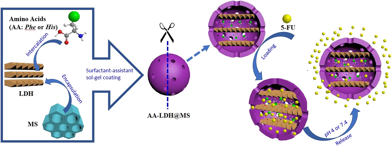
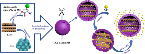
The core–shell LDH@MS and AA-LDH@MS nanocomposites were prepared by the same surfactant-assistant sol–gel coating method in Scheme 1. First, the obtained LDH or AA-LDH material (0.2 g) was added in a solution containing 0.60 g of CTAB, 160 mL of distilled water, 1.80 mL of NH3·H2O, and 120 mL of ethanol to form a uniform dispersion by using ultrasound for 1 h. Next, 0.6 g of TEOS was added dropwise to the above dispersion with vigorous stirring. The reaction mixtures were maintained for 6 h. Then, the product was obtained by centrifugation and washed with distilled water three times. Subsequently, they were dried at 60 °C in a vacuum drying oven for 24 h. To remove the surfactant, the dried samples (1 g) were extracted with the mixed solutions of ethanol (250 mL) and hydrochloric acid (2 M, 5 mL) for 24 h. Finally, the extracted solid products were dried at room temperature and named LDH@MS or Phe-LDH@MS or His-LDH@MS, respectively.
The schematic diagram of preparation of the core/shell AA-LDH@MS (AA denotes phenylalanine or histidine) and loading or release of the drug 5-FU.

Drug (5-FU) loading and release in vitro
5-FU was used as the objective drug molecule to evaluate the loading and release performance of the prepared nanocomposites LDH@MS, Phe-LDH@MS, and His-LDH@MS, as shown in Scheme 1. The 5-FU loading was carried out in a shaking table. 0.1 g nanocomposites LDH@MS, Phe-LDH@MS, and His-LDH@MS were separately mixed with 40 mL of NaOH solution containing 0.4 g 5-FU with a pH of 11.6 in a 100 mL beaker flask. The mixtures were placed in a shaking table concomitant with shaking at 60 °C for 12 h. The solid products were removed by a vacuum pump for filtration. The filtrate was maintained, and 5-FU concentration in the filtrate was measured by a UV-vis spectrometer at a maximum absorption wavelength of 266 nm. To determine the optimum pH value in the loading experiment, the procedure was described as the aforementioned loading process, where the samples were carried out at different pH values. 5-FU concentration in the filtrate was analyzed as the aforementioned method.
The drug release study was performed as following (shown in Scheme 1): 100 mg of nanocomposites (LDH@MS, AA-LDH@MS including Phe and His) was added into 10 mL of hexane containing 30 mg of 5-FU in a conical flask. The suspension was stirred for 24 h in the flask with a bottle plug to prevent the evaporation of hexane. Then, the flask was placed at room temperature for 2 days by removing the plug to evaporate hexane. The dried samples were obtained at the bottom of flask. Next, 0.1 g of the aforementioned samples were placed in a dialysis membrane bag. The dialysis membrane bag with the sample was incubated with stirring in 300 mL of the buffed solution with pH 4 and pH 7.4 as the release medium at 37 ± 0.5 °C. 4 mL of the release medium solution was withdrawn at constant time intervals and analyzed by a UV-vis spectrophotometer at the maximum absorption wavelength of 266 nm. The mechanism (kinetics) of drug release was investigated by fitting the release data using the Korsmeyer–Peppas models.
Results and discussion
Structural analysis of the core–shell nanocomposites
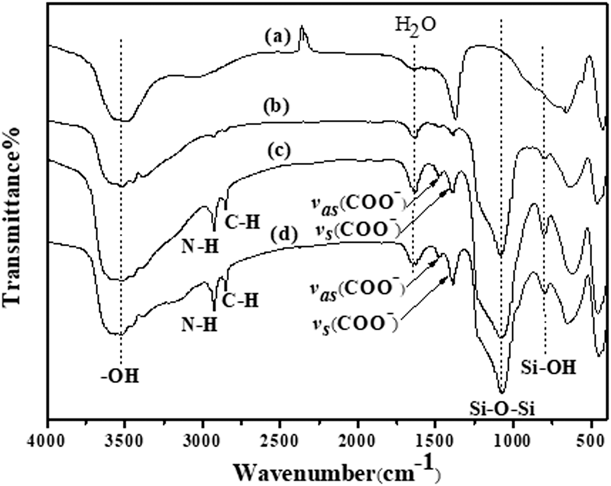
Figure 1 shows FTIR spectra of the materials LDH, LDH@MS, Phe-LDH@MS, and His-LDH@MS. For LDH, a strong and broad peak at approximately 3500 cm−1 is observed in the spectrum. This is assigned to O–H stretching vibrations of the hydroxide groups from the basal plane of LDH and solvated interlayer water. A week peak at 1640 cm−1 is ascribed to the deformation mode of interlayer water molecules. The strong peak at 1370 cm−1 corresponds to the vibration mode of  ${\rm{CO}}_3^{2 - }$ in the gallery of LDH. The peaks at 663 cm−1 and 425 cm−1 in the low-frequency region are attributed to the stretching mode of M–O and the deformation mode of M–O–M vibrations of the LDH lattice (M = Mg and Al). These results are also clearly indicated in the other materials LDH@MS and AA-LDH@MS except the slight shift of wavenumbers. When mesoporous silica was coated on the outer surface of LDH and AA-LDH, two peaks at approximately 1080 and 800 cm−1 are obviously observed in all the core–shell nanocomposites LDH@MS and AA-LDH@MS. They denote the asymmetric stretching vibration of Si–O–Si and the stretching vibration of Si–OH, respectively, which are typical characteristic peaks of ordered mesoporous silica. Besides, two peaks at 2930 and 2850 cm−1, corresponding to N–H and C–H stretching vibrations, are indicated in the spectra of the materials Phe-LDH@MS and His-LDH@MS, which is fully consistent with those observed in FTIR of phenylalanine and histidine. Moreover, the presence of amino acids is further confirmed by two bands at 1480 and 1410 cm−1 related to the asymmetric and symmetric stretching vibrations of the carboxylate (
 ${\rm{RCO}}_2^ -$) group [Reference Veteška, Pospíšil, Melánová, Beneš and Zima26]. These results demonstrate that two amino acids are successfully intercalated into LDH and mesoporous silica is surrounded on their outer surfaces to form core–shell nanocomposites.
FTIR spectra of (a) LDH, (b) LDH@MS, (c) Phe-LDH@MS, and (d) His-LDH@MS.

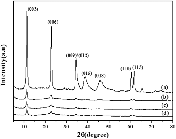
The XRD patterns of pristine LDH and the core–shell nanocomposites LDH@MS, Phe-LDH@MS, and His-LDH@MS are shown in Fig. 2. Several sharp and intense diffraction peaks at 2θ of 11.36°, 22.82°, and 34.60°, corresponding to characteristic basal reflections (003), (006), and (009) of LDH-layered structure, are indicated in Fig. 2. These peaks are also found at the core–shell nanocomposites LDH@MS, Phe-LDH@MS, and His-LDH@MS except the decrease in the intensity and 2θ values. These results illustrate that the structure of LDH is retained in prepared nanocomposites. The intensity of these peaks is decreased, which could be attributed to the interference of the surrounding mesoporous silica shell. When amino acids were introduced into the core–shell nanocomposites, the 2θ of (003) reflection was slightly shifted to the lower value to indicate the expanding layer spacing.
XRD spectra of (a) LDH, (b) LDH@MS, (c) Phe-LDH@MS, and (d) His-LDH@MS.

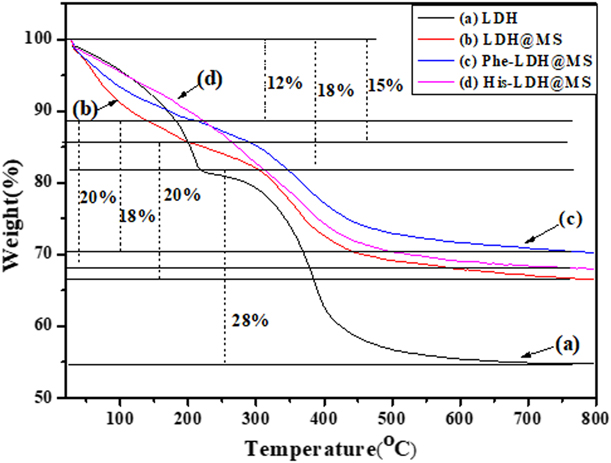
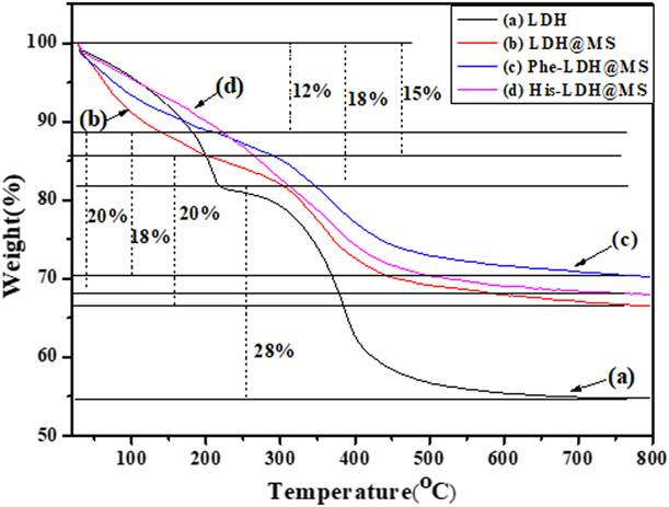
The TG curves of LDH, LDH@MS, and AA-LDH@MS (including Phe and His) are presented in Fig. 3. Two steps of weight loss are showed in the TG curves of LDH and LDH@MS as followings: the first step of weight loss about 18% (pristine LDH) or 15% (LDH@MS) at temperature less than 200 °C is readily assigned to the dehydration of the surface physically adsorbed water and interlayer water. At the higher temperatures between 200 °C and around 600 °C, the second one is a larger weight loss about 28% (pristine LDH) or 20% (LDH@MS), which is attributed to the decomposition of the hydroxide layers through the dihydroxylation. Compared with LDH and LDH@MS, the thermal decompositions of AA-LDH@MS (including Phe and His) indicate three weight loss steps: The first weight loss about 12% from room temperature to 200 °C is as the same as the materials of LDH and LDH@MS; the second one at the temperatures from 250 to 400 °C is a result of the decomposition of amino acid anions and dehydroxylation of the brucite-like layers; the third one at the temperatures from 400 °C to 700 °C is attributed to the combustion of the carbonaceous residue. (Veteška 2008) The weight losses of the last two steps are up to 20% in TG curves of AA-LDH@MS. Obviously, when mesoporous silica was formed in the outer surface of LDH or AA-LDH, all the core–shell nanocomposites showed a very slow and steady weight loss in the TG curves, which is because of the channel metamorphosis exhibited by the material as a function of temperature.
TG curves of (a) LDH, (b) LDH@MS, (c) Phe-LDH@MS, and (d) His-LDH@MS.
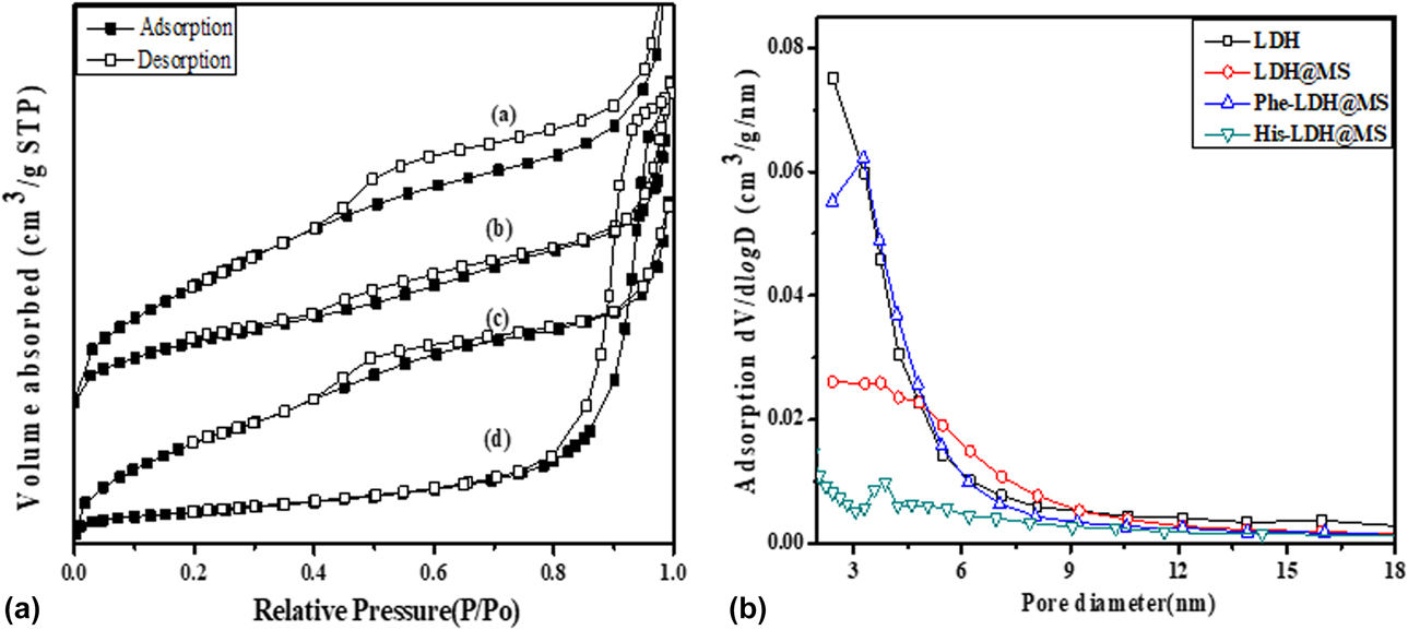
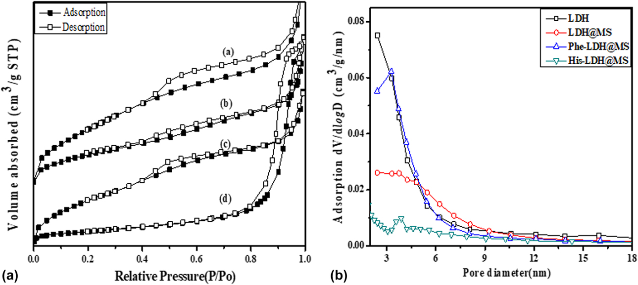
To further know the structure of the core–shell nanocomposites AA-LDH@MS (including phenylalanine and histidine), LDH@MS and pristine LDH, N2 adsorption–desorption analysis was performed to characterize these nanocomposites. Their N2 adsorption–desorption isotherms and pore size distributions are described in Fig. 4. The textural parameters, such as specific surface area (S BET), pore volume, and average pore diameter, are obtained by BET/BJH measurement, which is displayed in Table SI in the Supplementary material. The N2 adsorption–desorption isotherms of all the nanocomposites and pristine LDH exhibited type IV adsorption isotherms with an obvious capillary condensation step according to the IUPAC classification. However, the hysteresis loops reveal the different type in Fig. 4(a) of these samples. The nanocomposites Phe-LDH@MS, LDH@MS, and pristine LDH show H4 type hysteresis loop, whereas the nanocomposites His-LDH@MS indicates H1 type hysteresis loop. These results conclude that the uniform mesopores with narrow pore size distribution are obtained in the nanocomposites Phe-LDH@MS and His-LDH@MS, as clearly shown in Fig. 4(b). The S BET are calculated to be 302 m2/g and 56 m2/g for Phe-LDH@MS and His-LDH@MS, respectively. The pore size distribution exhibits a maximum for the mesopore range centered at 4.7 nm and 5.6 nm for Phe-LDH@MS and His-LDH@MS, respectively, which indicates a uniform mesoporous silica shell.
Nitrogen adsorption–desorption isotherms (A) and pore size distribution (B) of (a) LDH, (b) LDH@MS, (c) Phe-LDH@MS, and (d) His-LDH@MS.
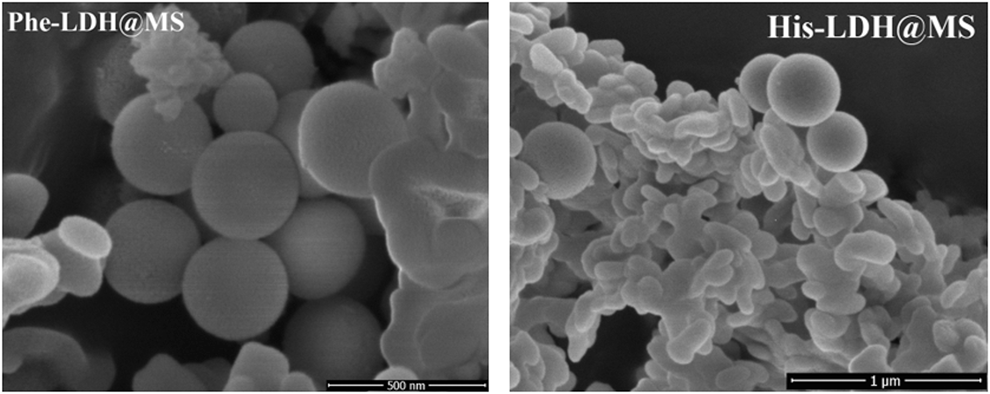
The scanning electron microscopy (SEM) images of the core–shell nanocomposites Phe-LDH@MS and His-LDH@MS are showed in Fig. 5(a) spherical morphology is observed with a mean particle size of 300 nm and 400 nm for Phe-LDH@MS and His-LDH@MS, respectively, which is different from the typical disk-like morphology of pristine LDH. The morphology of the core–shell nanocomposites is related to the amino acids. They display the different particle morphology because of amino acids within the materials. The SEM images indicate that the spherical type is mainly observed in Phe-LDH@MS. However, His-LDH@MS shows the rod particle morphology. Thus, the phenylalanine-intercalated LDH as the core is easily encapsulated by mesoporous silica to form the spherical morphology, compared with histidine-intercalated LDH.
SEM images of Phe-LDH@MS and His-LDH@MS.

Loading of 5-FU in vitro
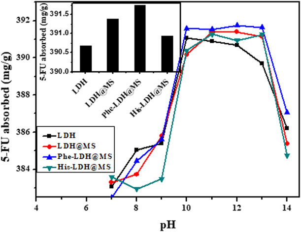
5-FU was selected as a targeted drug molecule for assessing the loading/adsorption and release behaviors of these materials. The loading/adsorbed amounts of 5-FU were determined by UV-vis spectroscopy using the drug solutions before and after loading. Moreover, the loading conditions were optimized by modulating the pH value of drug solutions and the contents of amino acids. Figure 6 shows the absorbed amounts of 5-FU at the prepared materials with the changing of different pH. It is well known that the pH of a drug solution plays a crucial role in loading drug molecules into nanocomposites. In this work, the pH dependence of drug loading was investigated at the range of 7–14 because LDH as the important component of these materials is probably destroyed under low pH values. As shown in Fig. 6, there are three stages of drug loading amounts with the increasing of pH in all the materials: First, the loading amounts of drug are sharply increased at the pH from 7 to 10. The second one at pH of 10–12 presents the steady and slow increase of the absorbed drug. Moreover, the absorbed amounts reached a plateau. When the pH is larger than 12, the absorbed amounts of 5-FU are quickly decreased because of the competition between –OH ions and the anionic drug molecules which are involved in the absorption of 5-FU into the nanocomposites. Furthermore, the mesoporous silica shell can improve the absorbed amounts of 5-FU because the porous channels provide more storage space for the drug. Obviously, the drugs should remain in the pore channels and LDH interlayer to prevent the washout of the drugs on the outer surface. Thus, amino acids were introduced into the interlayer of LDH to enhance the interaction with the drug. The core–shell nanocomposites containing amino acids exhibit high absorbed amounts of drugs at the same conditions of all the materials. Specially, the nanocomposites Phe-LDH@MS display the maximum absorbed amounts of 5-FU. The insert of Fig. 6 indicates that the absorbed 5-FU is up to 392 mg/g at 60 °C for 12 h. The drug loading capacity of Phe-LDH@MS is better than His-LDH@MS because of the following aspects: First, the imidazole groups of histidine may produce electrostatic repulsive force with the pyrimidine group of drug 5-FU. Second, the surface areas of Phe-LDH@MS are larger than those of His-LDH@MS, which provides more interaction spaces with the drug. Thus, the material Phe-LDH@MS exhibits better loading capacity. These results reveal that mesoporous silica and amino acids are the important components in the drug loading of these materials.
Effect of pH on the absorbed amounts of 5-FU at the nanocomposites of LDH, LDH@MS, Phe-LDH@MS, and His-LDH@MS. (the insert: The absorbed amounts of 5-Fu at the optimum conditions pH = 12, T = 60 °C, time = 12 h).

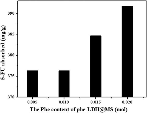
To know the relation between the amounts of amino acid and the absorbed drug, the absorbed amounts of drugs were observed by changing the contents of phenylalanine in the nanocomposite Phe-LDH@MS, as shown in Fig. 7. It is obviously found that the adsorption capacity of the drug also is strengthened with the increasing in the content of phenylalanine. From the result, we can learn that phenylalanine not only expands the interlayer distance to enhance the storage space but also supplies the absorption position to increase the interaction with the drug.
The loading/absorbed amounts of 5-FU at the different contents of phenylalanine in the nanocomposites Phe-LDH@MS.

pH-controlled release of 5-FU in vitro
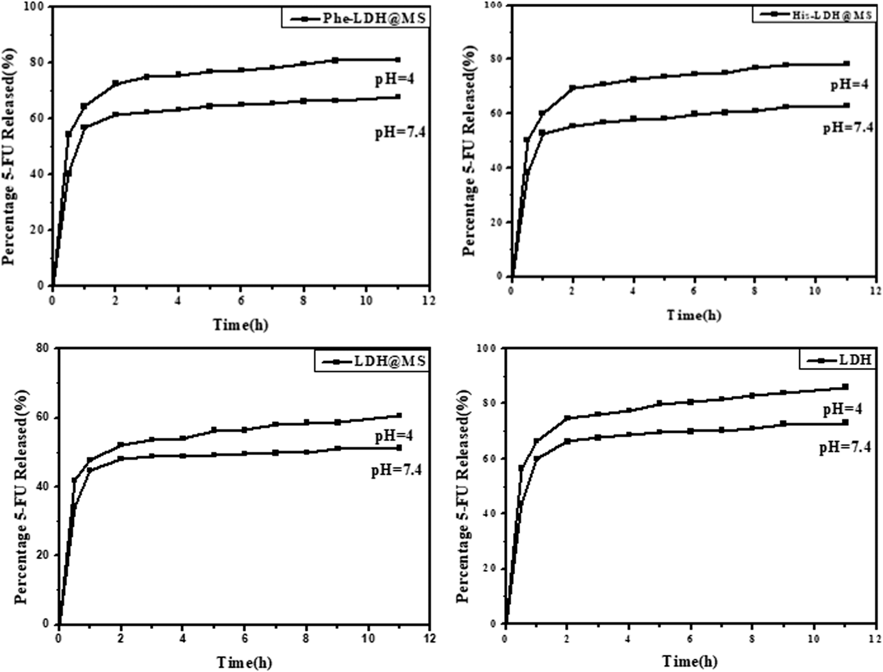
The materials LDH, LDH@MS, and AA-LDH@MS (including phenylalanine and histidine) are dispersed in buffer solution with the different pH values to test their release capacity in vitro. Commonly, the pH value of normal/healthy tissues is about 7.4. However, the pH value in the inflammatory tissues, endosomes, and lysosomes are remarkably acidic (pH < 5), which is lower than the normal tissues. pH values of 4 (simulating endosome/lysosome pH) and 7.4 (simulating normal tissue pH) were respectively used to investigate the pH responsive ability of these materials. The release profiles of 5-FU from these materials at the different pH values are shown in Fig. 8. The dynamic concentrations (C r) of the drug in solution (mg/L) and the corresponding amount (Q r) of the drug remaining in the solid (mg/g) were identified and quantified, as shown in Tables SII–SV in the Supplementary material.
The release curves of 5-FU in vitro from the core–shell nanocomposites: AA-LDH@MS (including phenylalanine and histidine), LDH@MS and pristine LDH at pH 4 and pH 7.4.

Figure 8 shows that the release processes were nonlinear over the time. Moreover, it could be seen that all the materials exhibited discrepant release properties at the different media of pH values. However, the percentage of 5-FU released at pH 4 from these materials was higher than the percentage at pH 7.4. The result reveals that these materials are capable of slower and much controlled release of 5-FU. The similar and sustained release behavior at the different pH values were observed on the release profiles of all the materials. They undergo two stages including the fast release at the short time and the slow release of the drug. For the AA-LDH@MS including phenylalanine and histidine samples, at the initial stage of the release within 2 h, the release profiles exhibit a sharp and sudden removal of 5-FU from the materials, which is because of the departure of the drugs from the pore mouth and pore channels of the shell. When the time is longer than 2 h, a controlled and sustained release is observed because of the slow removal of the drugs mainly from the inner deep core. The release percentage of 5-FU was increased with the decrease in pH. Furthermore, the different amino acids directly affect the 5-FU release percentage. It is clear that the cumulative release percentage of 5-FU arrives to 85% after 11 h at pH 4 and 68% after 11 h at pH 7.4 for the core–shell nanocomposites (Phe-LDH@MS) containing phenylalanine. The lower percentage of 5-FU was released from the core–shell nanocomposites (His-LDH@MS) containing histidine at the same time and pH value of the medium. The aforementioned results indicate that the intercalation of phenylalanine in the LDH core effectively regulates the loading of the drug and controls the release of the drug, which results in the improvement of loading amounts and the release percentage of the drug. In addition, the advantage of amino acids in the release processes is also further confirmed in contrast to the material LDH@MS. On the other hand, compared with the pristine LDH, the cumulative release percentage of 5-FU from the core–shell nanocomposites AA-LDH@MS is reduced after 11 h at the pH 4 and pH 7.4. Nonetheless, the release ratio of 5-FU from the core–shell nanocomposites becomes slow because of the interference of the silanol groups of the outer mesoporous shell. In all, amino acids and mesoporous silicon are the important constituents to develop core–shell nanocomposites as the novel and effective NDDS.
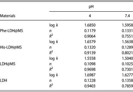
To obtain the exact mechanism of 5-FU release, the kinetic models were used to evaluate the drug release in vitro. Commonly, there are four mathematical models including zero-order, first-order, Higuchi, Korsmeyer–Peppas (K–P) models used to fit the in vitro drug release data [Reference Shoaib, Tazeen, Merchant and Yousuf27]. Specially, the K–P model based on the drug release exponentially to the elapsed time is the main drug release mechanism because it is a simple and semiempiric model. The K–P model is presented by the following equation:  $\log \;{C_{\rm{r}}} = \log \;k + n \times \log \;t$ (k is the kinetic constant, n is the diffusion exponent, and C r is the dynamic concentrations of the drug in solution). The k and n values were calculated from the intercept and slope of the linear plot, as shown in Fig. S2 (seen in the Supplementary material). The relative parameters of the K–P model are given Table I. In the pH 4 medium, the correlation coefficient (R 2) of the K–P model is from 0.9064 to 0.9698 for the 5-FU release from all the samples, which indicates good linearity. However, the data of correlation coefficient R 2 are relatively a small number (from 0.7301 to 0.8021) in the pH 7.4 medium. The results demonstrated the K–P model is fit for the release of 5-FU from the samples. The diffusion exponent n determined from the K–P model is 0.1179–0.1331, 0.1289–0.1320, 0.1025–0.1098, and 0.1228–0.1358 for the 5-FU release from Phe-LDH@MS, His-LDH@MS, LDH@MS, and pristine LDH in the different pH medium, respectively. The exponent n values are less than 0.45, suggesting that 5-FU release from all the samples is Fickian diffusion, which excludes the possibility of erosion or solubilization of the materials. It is well known that the drug 5-FU possesses good solubility in the dilute acids and alkaline aqueous solutions. Thus, the release from the materials toward the solution is because of the concentration gradient between the solid surface and solution and Fickian diffusion. Based on the aforementioned results, the 5-FU release depends on not only the pH value in the medium but also the mesoporous channels and amino acid of the core–shell nanocomposites. The core–shell nanocomposites AA-LDH@MS containing amino acids and mesoporous silica should be as the NDDS for better drug delivery, which is the promising candidate for applications in the pharmaceutical industries.
Kinetic parameters of Korsmeyer–Peppas models for the 5-FU release from the core–shell nanocomposites Phe-LDH@MS, His-LDH@MS, LDH@MS, and pristine LDH.

Conclusions
In summary, a novel NDDS of core–shell nanocomposites was designed and synthesized by using the mesoporous silica (MS) as the shell to encapsulate amino acid–modified LDH (AA-LDH) as the core. Phenylalanine and histidine with aromatic groups were used to intercalate the layer space of LDH, which forms the core of AA-LDH. The anticancer drug 5-FU was selected as a targeted drug molecule to observe the delivery (including loading and release) process in vitro at different pH media. The structure of core–shell nanocomposites AA-LDH@MS was confirmed by the characterization of FTIR, XRD, TG, N2 adsorption–desorption, and SEM. The results indicate the successful coating of mesoporous silica and intercalation of amino acids. The delivery studies in vitro show that the core–shell nanocomposites AA-LDH@MS are suitable for the excellent loading and sustained release of the 5-FU drug. Moreover, the porous channels of mesoporous silica and amino acids make a significant contribution to high loading and slow release of the drug. The kinetic plots of the Korsmeyer–Peppas model further reveal that the 5-FU release from AA-LDH@MS is Fickian diffusion at pH 4 and pH 7.4. As a consequence of the unique core–shell nanocomposites, AA-LDH@MS containing amino acids are used as the promising candidate in the targeted drug release and expected to be as the transporting vehicles for the other biological delivery.
Methodology
FTIR spectra were obtained on a Brucker Vector 22 (Brucker Optics, Faellanden, Switzerland) spectrometer by using a KBr pellet technique over the frequency range from 4000 to 400 cm−1 at 298 K. Powder XRD patterns of samples were recorded in the 2θ range of 5–80° using an RINT-2000 vertical goniometer (Rigaku, Osaka, Japan) diffractometer with Cu Kα radiation (λ = 1.5418 Å) at 40 kV and 20 mA. The TG were collected with an NETZSCH STA449F3 (Netzsch, Hanau, Germany) thermal analysis instruments under airflow with a heating rate of 10 °C/min in the temperature range from 30 °C to 800 °C. Nitrogen adsorption–desorption isotherms were measured on a BELSORP MINI Micrometrics (BEL, Osaka, Japan), and the samples were degassed in a vacuum at 423 K for 6 h before measuring the surface area. The S BET were calculated by the Brunauer–Emmett–Teller (BET) method based on the adsorption isotherm. The pore size distribution was calculated from the adsorption isotherm by the Barrett–Joyner–Hallender (BJH) method. The pore volume was taken at the P/P 0 of 0.973 single point. SEM images were conducted on an FEI Nova NanoSEM 450 (ThermoFisher Scientific, Massachusetts) electron microscope, where the samples were sprayed with a thin gold film before the characterization. The UV-vis absorption spectra of the samples were conducted on a Beijing Purkinje General Instrument (Purkinje, Beijing, China), UV-1810.
Supplementary material
To view the supplementary material for this article, please visit https://doi.org/10.1557/jmr.2019.324.
Acknowledgments
This work was supported by Undergraduate Innovation Program of Jiangsu Province (No. 201810291179X), Nanjing Tech University (No. 2018DC656), and Science Foundation of Qiuzhen School (No. 2018XJKJ59).