Introduction
Chronic osteomyelitis of the frontal bone with subperiosteal abscess, also known as Pott's puffy tumour, is a rare complication of sinusitis, which was first described in the eighteenth century by Percival Pott.Reference Marshall and Jones1,Reference Sharma, Sharma, Gupta, Kocher and Kumar2 The infection spreads directly into the bone or there is retrograde thrombophlebitis of the valveless diploic veins.Reference Akiyama, Karaki and Mori3,Reference Lawson, Reino and Deeb4
Both medical and surgical treatments are employed in treating this condition.Reference Akiyama, Karaki and Mori3,Reference Lawson, Reino and Deeb4 External surgical approaches include frontal sinus trephination, frontal sinus obliteration, cranialisation, craniotomy, and a Riedel operation where the anterior table of the frontal sinus is removed.Reference Lawson, Reino and Deeb4 Recent advances in the endoscopic drainage of the frontal sinus, such as the Draf III procedure, have added an alternative dimension in treating this condition.Reference Agrawal and Garg5,Reference Jung, Lee, Park and Lee6 Post-operatively, eight weeks of antibiotics are usually required.Reference Jung, Lee, Park and Lee6
Treating Pott's puffy tumour can be a major challenge when the osteomyelitis is extensive, involving not only the outer table but also the inner table of the frontal sinus.Reference Akiyama, Karaki and Mori3 In this situation, after debridement, the bony defect will need reconstruction to protect underlying brain tissue, and to prevent cosmetic step deformity or frontal bossing.Reference Akiyama, Karaki and Mori3,Reference Lawson, Reino and Deeb4 Both autologous bone grafts (calvarial, rib) and alloplastic materials can be used. Unfortunately, placing autologous grafts or alloplastic implants into the infected wound is risky, leading to higher rates of infection and graft removal. In the presence of infection, delayed cranioplasty is performed as a secondary procedure after debridement and antibiotic treatment.Reference Agrawal and Garg5
In the case described below, we employed a novel technique of modified distraction osteogenesis to the cranial vault, to induce new bone formation across the skull defect, over a period of three months. One case has been presented in the literature of a small temporal defect repaired using a stock (not customised) internal cranial distractor.Reference Cho-Lee, Naval-Gías, González-García, Martos-Díaz, Muñoz-Guerra and Sastre-Pérez7 To our knowledge, this technique has not been applied to large cranial defects utilising custom external distraction devices.
How we do it
A 55-year-old male was referred to the rhinology clinic with 3-month history of frontal headache, bilateral nasal blockage and intermittent frontal swelling. These symptoms recurred after discontinuing antibiotics. He had been treated by his general practitioner with various antibiotics (one week of clarithromycin, followed by co-amoxiclav and subsequently doxycycline). He then had a further four-week course of clarithromycin. A complete ENT examination that included flexible nasoendoscopy revealed normal findings. There were no clinical signs of frontal inflammation, nor visual or neurological signs.
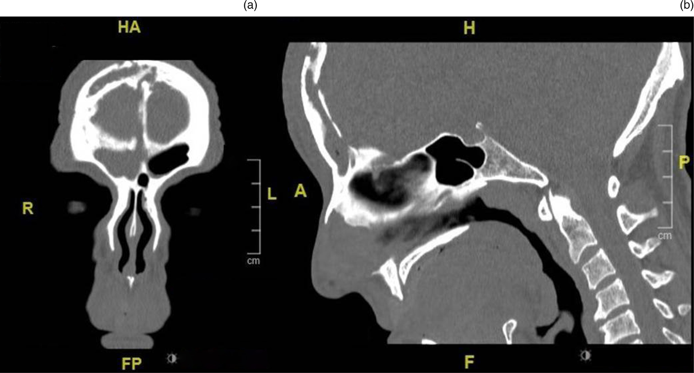
Computed tomography (CT) scanning of the sinus (Figure 1) confirmed extensive frontal osteomyelitis. The patient declined admission for intravenous antibiotics or surgery. Following discussion with microbiologists, he was commenced on empirical doxycycline and budesonide nasal rinses. Subsequent magnetic resonance imaging revealed a 15 ml subgaleal collection, enhancement underlying dura and a right-sided parietal collection.
(a) Coronal and (b) sagittal computed tomography scans of the paranasal sinus, showing extensive chronic infective changes originating in the right frontal sinus, with destruction of the inner and outer tables of the vault, and associated soft tissue swelling in the frontal scalp tissues. Appearances are reminiscent of Pott's puffy tumour, but in a more chronic state. HA = head/anterior; H = head; R = right; L = left; A = anterior; P = posterior; FP = feet/posterior; F = feet

Pus was aspirated and the culture grew Streptococcus milleri, which was sensitive to penicillin and clarithromycin. The patient's white cell count was raised, at 14.30 × 10⁹/l (normal range = 4.00–11.00), with a neutrophil count of 9.0 × 10⁹/l (normal range = 2.0–7.5). C-reactive protein was 12 mg/l (normal range = 0–10). His human immunodeficiency virus blood test results were negative, as were the findings of sputum cultures incubated for 42 days to test for tuberculosis. The patient was admitted for intravenous antibiotics (benzylpenicillin plus metronidazole).
The case was discussed at the skull base multidisciplinary team (MDT) meeting in light of the expected large defect after the debridement of bone sequestrum. Several options were explored, including long-term antibiotics and frontal sinus surgery (endoscopic or open approach, with or without bony debridement, with or without reconstruction of the calvaria). The MDT recommended debridement and reconstruction after medical treatment. Because of infection, we opted for either a delayed cranioplasty or our novel custom technique of distraction osteogenesis. After discussion, the patient chose the latter option.
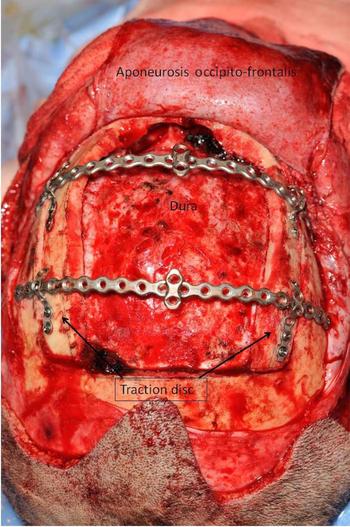
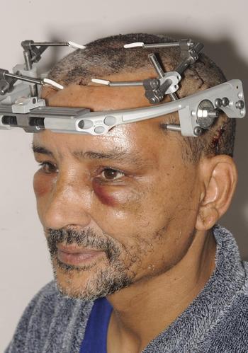
A bicoronal approach and craniectomy of the diseased bone was performed, resulting in a bony defect measuring 94 mm × 44 mm across the frontal and parietal bone (Figure 2). Two non-diseased bony plates, 10 mm in width, were created on each end of the defect, called ‘transport discs’. These were freed from the surrounding skull, but left attached to underlying dura to preserve the blood supply. Two 2.0 mm titanium reconstruction plates (Synthes Matrix Mandible™) were customised to bridge horizontally across the gap in the upper and lower parts of the defect. They were secured to the squamous parietal skull bone outside of the transport segments, to guide the traction osteogenesis in same curvature of the missing skull. Distraction wires (0.5 mm soft, pre-stretched stainless steel) passed from the two transport discs through customised metal holes at two points, inferiorly and superiorly at the midline. The stainless steel anchorage wire then perforated though skin to an external frame (Figure 3). This avoided the wire cutting through the scalp (cheese wire effect) as the transport discs progressed. The external frame had four distraction points, two on each side, which were fully adjustable in multiple planes (Synthes Midface Distraction Kit).
Intra-operative view showing the bone defect after craniotomy, with the generation of traction discs on both sides of the defect.

Cranial distractor with external frames.

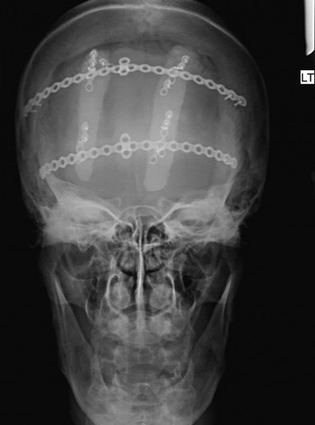
The device was activated 1 week post-operatively by turning the distraction screws three times a day at a rate of 1.5 mm/day during the first 10 weeks post-operatively. The transport disc closure rate was initially monitored through serial dental cone beam CT two weeks apart in the first two months, and subsequently with X-rays of the facial bones (Figure 4) every two to three weeks. The turning rate of the distraction devices was adjusted using the imaging scans as a guide. The external frame was removed at week 15.
X-ray of the facial bones showing a traction disc. LT = left

Histology revealed chronic fibrosis and inflammation, extensive bone remodelling, and new bone deposition both within the medullary cavity and the surface of the cortical bone. Most of the bone showed necrosis, with the absence of osteocytes. There was no bacteria growth on microscopy.
The patient had an uneventful recovery. A post-operative CT scan of the head ruled out any collection and confirmed the position of the traction discs. He was discharged from the hospital after 2 days on 4 weeks of intravenous ceftriaxone (administered 4 g daily for 2 weeks, then 2 g daily for another 2 weeks, through a peripherally inserted central catheter line) and oral metronidazole (400 mg three times daily).
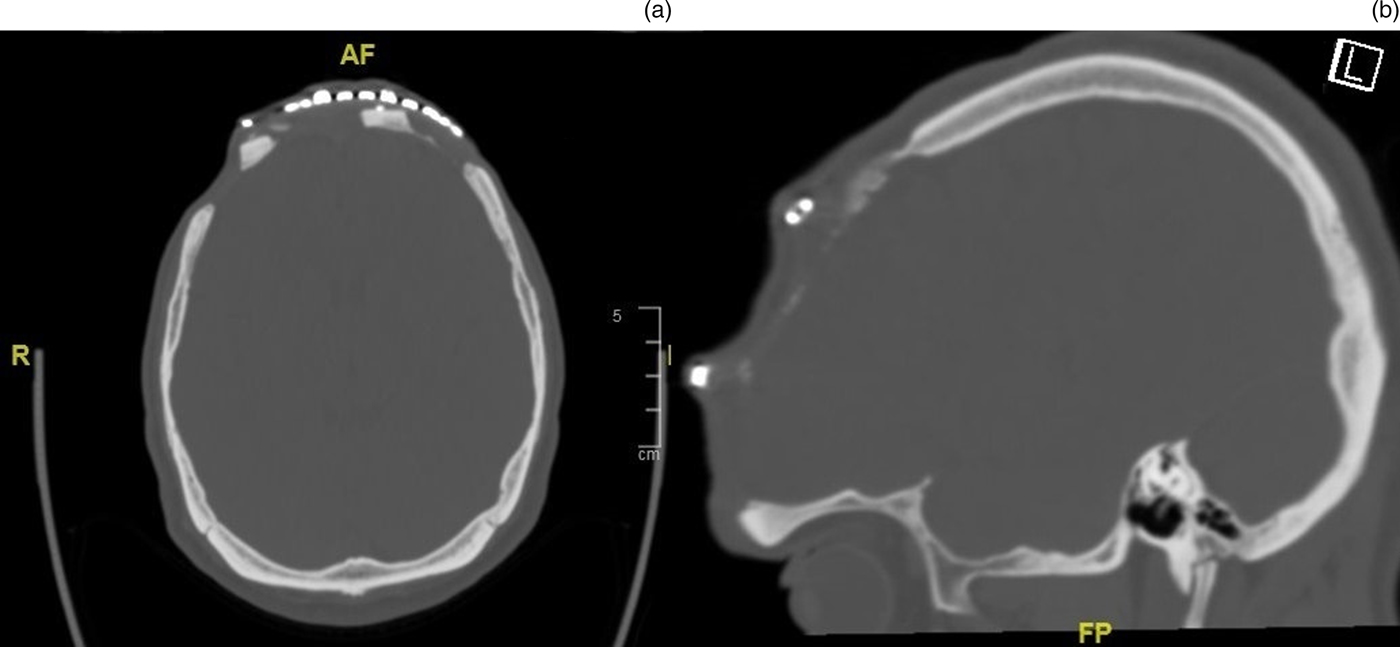
Repeat cone beam CT scanning conducted after one year (Figure 5) showed the new bone formation. It is thinner than natural skull thickness; however, it adds to the protection of the underlying brain.
(a) Axial and (b) sagittal cone beam computed tomography scans after one year, showing new bone formation between the transport disc medially and the cranial calvarial bone laterally. AF = anterior/feet; R = right; FP = feet/posterior

Discussion
Osteomyelitis of the frontal sinus is rare in the post-antibiotic era, with 0.5 per cent incidence reported in 649 retrospective cases.Reference Marshall and Jones1,Reference Sharma, Sharma, Gupta, Kocher and Kumar2 Intranasal recreation drugs such as cocaine or methamphetamine increase the susceptibility to infection.Reference Akiyama, Karaki and Mori3 S milleri was the commonest (51 per cent) cultured organism.Reference Marshall and Jones1 Other frequently reported pathogens are Staphylococcus aureus and Streptococcus intermedius.Reference Akiyama, Karaki and Mori3 Lower oxygen concentration in the frontal sinus results in polymicrobial involvement, including anaerobic bacteria. Therefore, broad spectrum antibiotic cover, such as third-generation cephalosporin and metronidazole, should be used in the peri-operative period and for up to six to eight weeks after the surgery.Reference Akiyama, Karaki and Mori3,Reference Lauria, Fernandes, Brito, Pereira and Chone8
There are cases where the infected bone must be debrided. If the skull base defect is extensive, cranioplasty may be considered using reconstructive techniques ranging from autografts to allografts. Cranioplasty is generally recommended early (within three months) after craniectomy for cases without infection (e.g. road traffic accidents). Satisfactory dissection planes in early surgery have been advocated.Reference Andrabi, Sarmast, Kirmani and Bhat9 Where there is infection, reconstruction is generally delayed for at least six months, to avoid the recurrence of infection, which is monitored clinically and via radionuclide imaging (triphasic bone scanning and/or indium white blood cell scanning). There is still a high failure rate despite these precautions.Reference Lawson, Reino and Deeb4,Reference Verbon, Husni, Gordon, Lavertu and Keys10
Cranial vault distraction osteogenesis is a new technique, employed over the last two decades. It is used mainly in paediatric and adolescent populations because of the premature closure of calvarial sutures, known as craniosynostosis. It was first described when McCarthy et al., in 1992, used it to lengthen the mandible by stimulating neo-vascularised bone formation.Reference McCarthy, Schreiber, Karp, Thorne and Grayson11
Distraction osteogenesis offers significant advantages over a traditional vault remodelling technique. The gradual expansion of the vault occurs over weeks. In addition, the new technique minimises the need for bone grafting with its associated donor site morbidity.Reference Ong, Harshbarger, Kelley and George12 In our case, it was crucial to keep the traction discs attached to underlying dura, in order to maintain the nutrition and vitality of transport segments. The curved nature of the frontal bone was a real challenge when designing equal traction forces for the mobile transport discs without rotation or dislocation from underlying dura.
The rate of distraction varies with age. The younger the age, the quicker the bone heals, hence the quicker the distraction rate. The rate of distraction is 1.2 mm/day and 0.9 mm/day for children up to two years and for those aged two to eight years, respectively, and is 0.6 mm/day for adolescents.Reference Ong, Harshbarger, Kelley and George12 In this case report, the device was turned every two weeks at a rate of 0.6 mm/day. The slow rate of distraction of the two traction discs from the surrounding calvarial bone promoted the osteogenesis in the created lateral gaps, until the traction discs met in the midline.
Potential complications include dural injury, device failure and loosening, infection, bleeding associated with dural sinus injury, and wound dehiscence.Reference White, Evans, Dover, Noons, Solanki and Nishikawa13 In a recent cranioplasty study, 15.3 per cent of patients encountered complications of haematoma, seizures, bone resorption and sunken bony plates.Reference Andrabi, Sarmast, Kirmani and Bhat9 About 50 per cent of the patients required revision surgery. The complication rate was higher in delayed cranioplasty than at six months after the initial craniectomy.Reference Andrabi, Sarmast, Kirmani and Bhat9 There were no reported complications in this case report. The scalp wound healed nicely. The wires and frame did not add any further risk for re-infection in the surgical bed.
The authors recognise that with improved surgical techniques and antibiotics, the need for frontal bone resection is rare. However, there will be a subset of patients who may require extensive resection. Adding this technique to existing cranioplasty armamentarium is therefore a viable novel option.
Conclusion
Pott's puffy tumour is a rare complication of sinusitis. Distraction osteogenesis of the anterior cranial vault represents another option in the armamentarium of cranioplasty, particularly when reconstructing extensive skull vault defects.
Acknowledgements
The authors would like to thank Andrew Richmond and Jason Watson (maxillofacial prosthetists).
Competing interests
None declared