Truncus arteriosus is a rare congenital cardiac defect characterised by a single arterial root that divides into a pulmonary trunk and ascending aorta. The ductus arteriosus is commonly absent or small. A conoventricular septal defect resides typically between the limbs of the septal band. Rarely there may be a complete subarterial conus with discontinuity of the intervalvular fibrosa between the truncal and mitral valves.Reference Anderson 1 Approximately 25% of patients with truncus arteriosus have a right aortic arch. Distinguishing truncus arteriosus from tetralogy of Fallot with pulmonary atresia – a form of pulmonary atresia with ventricular septal defect – can be challenging on fetal echocardiography.Reference Gotsch 2 In this study, we report a newborn with many features of truncus arteriosus but with an atretic main pulmonary artery. To our knowledge, a similar pattern has only been reported once before.Reference Tongsong 3
Case
Fetal echocardiography at 36 weeks of gestation demonstrated a single outflow tract overlying a large ventricular septal defect. Normal-sized branch pulmonary arteries appeared to arise from a short common stump from the posterior aspect of the single arterial root. The female infant was born at 39 weeks of gestation with a birth weight of 3.5 kg. The patient was admitted to the neonatal intensive care unit for further evaluation.
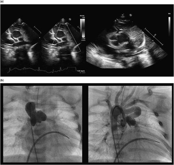
On examination, the infant appeared well without dysmorphic features. There was a grade III/VI systolic murmur at the left sternal border. Echocardiography showed pulmonary atresia with a large conoventricular septal defect (Fig 1a). A single arterial valve appeared to be tricommissural. No main pulmonary artery could be identified, but there were normal-calibre, confluent branch pulmonary arteries. A moderate persistent ductus arteriosus arose from the left subclavian artery and had continuous aortopulmonary flow. There was a right aortic arch with mirror-image branching. The arterial root was rotated with respect to the coronary artery position; the left main coronary artery arose anteriorly, and the right coronary artery had a rightward take-off. There was an aneurysm of the left coronary sinus of Valsalva; furthermore, there was an aneurysm of the mitral–truncal intervalvular fibrosa. On the basis of these findings and decreasing oxygen saturation, prostaglandin infusion was started. At cardiac catheterisation, angiography was consistent with the echocardiographic findings (Fig 1b).
( a ) Echocardiography demonstrates confluent pulmonary arteries with no flow in a normal-sized main pulmonary artery. There is a left sinus of Valsalva aneurysm and rotated aortic root. ( b ) Cardiac catheterisation demonstrates a dilated aortic root with a sinus of Valsalva aneurysm. Right aortic arch with mirror-image branching supplies a patent ductus arteriosus.

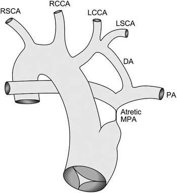
At surgery, an atretic stump of the pulmonary trunk arose from the left/posterior aspect of the single arterial root and was adherent to the left coronary sinus aneurysm; two normal-calibre branch pulmonary arteries originated from the atretic pulmonary trunk (Fig 2). On cardiopulmonary bypass, the ductus arteriosus was ligated and divided. A right ventricle-to-pulmonary artery conduit was constructed from a composite, 5-mm, ringed polytetrafluoroethylene (PTFE) graft proximally and femoral vein homograft confined within a 6-mm external stent distally. The ventricular septal defect was left open because of lack of subaortic tissue and potential for aortic insufficiency. Furthermore, repair of the mitral–truncal aneurysm was deferred to allow patient growth in the absence of valvular dysfunction. The patient came off bypass easily and recovered without complication.
Schematic of patient anatomy demonstrating truncus arteriosus with the atretic main pulmonary artery (MPA), right aortic arch with mirror-image branching, and a persistent ductus arteriosus (DA) from the left subclavian artery (LSCA). The left common carotid artery (LCCA), the right common carotid artery (RCCA), and the right subclavian artery (RSCA) are also shown.

Discussion
We present a rare variant of truncus arteriosus type I in which there was atresia of the main pulmonary artery, with pulmonary blood flow supplied by a persistent ductus arteriosus. The differential diagnosis also included tetralogy of Fallot with pulmonary atresia and confluent branch pulmonary arteries. This case demonstrates several anatomical features that may occur with either diagnosis, including aortic root dilatation,Reference Carlo, McKenzie and Slesnick 4 rotated coronary origins,Reference Anderson 5 and right aortic arch. Properties favouring truncus arteriosus were the single overriding truncal valve and atretic main pulmonary artery originating from the left/posterior aspect of the truncal root. Taken together, these anatomical lesions suggest a variant of truncus arteriosus.
Tongsong et alReference Tongsong 3 reported prenatal diagnosis of a very similar case. Fetopsy following termination of pregnancy demonstrated an atretic main pulmonary artery emanating from the single arterial root and giving rise to continuous branch pulmonary arteries supplied by a persistent ductus. Their case lacked the aortic root pathology and right aortic arch reported in our case. Schofield and AndersonReference Schofield and Anderson 6 reported a case of truncus arteriosus with pulmonary atresia, but the pulmonary blood flow was supplied by aortopulmonary collaterals. We were unable to identify any previous report with the exact anatomy of the case reported here.
Truncus arteriosus and tetralogy of Fallot with pulmonary atresia share many features and may be difficult to distinguish on fetal echocardiography. In the rare case reported in this study, a forme fruste of truncus with atretic main pulmonary artery, this distinction was even more confounded. Successful initial palliation was accomplished with placement of right ventricle-to-pulmonary artery conduit and ligation of the ductus arteriosus.
Acknowledgements
The authors acknowledge Dr Stephen P. Sanders for his review of the manuscript. They also acknowledge Emily Harris for her assistance with figures.
Financial Support
This research was supported in part by funding from Philip Greer and the Greer Family Foundation.
Conflicts of Interest
None.