Introduction
Thus far, 14 morphotypes of Hysterothylacium larvae, numbered I to XIV, and 16 species, including H. aduncum, H. cenaticum, H. cornutum, H. chrysostomi, H. leptaspi, H. macquariae, H. makairi, H. murrayense, H. pelagicum, H. scomberoidei, H. scomberomori, H. sebae, H. tasmaniense, H. tetrapteri, H. thalassini and H. zenis, have been described from fish in Australian waters (Cannon, Reference Cannon1977; Beumer et al., Reference Beumer, Ashburner, Burbury, Jetté and Latham1982; Bruce & Cannon, Reference Bruce and Cannon1989; Bruce, Reference Bruce1990a, Reference Bruceb; Bruce et al., Reference Bruce, Adlard and Cannon1994; Moravec & Justine, Reference Moravec and Justine2005; Shamsi et al., Reference Shamsi, Gasser and Beveridge2013, Reference Shamsi, Poupa and Justine2015). In Australia, unlike adult Hysterothylacium spp. where no molecular data are available, Hysterothylacium larval types have been reported and described in detail. Hysterothylacium spp. are marine ascaridoids, the larvae of which cannot be identified to species based solely on morphological characteristics. Although first and second internal transcribed spacers (ITS-1 and ITS-2, respectively) sequence data are available for most Hysterothylacium larval types reported in Australia, due to the lack of comparable data for adult Hysterothylacium spp. the specific identities of these larval types are still unknown. The aim of this paper was to examine selected species of Hysterothylacium in adult form from selected Australian fish, both morphologically and genetically, in order to provide a basis for specific identification of larval stages.
Materials and methods
Collection and examination of nematodes
Adult Hysterothylacium spp. were collected from three species of fish, including Yellowtail amberjack, Seriola lalandi (n = 3), blue marlin, Makaira mazara (n = 1) and striped marlin, Kajikia audax, previously known as Tetrapterus audax (n = 3) collected from the eastern coast of New South Wales, Australia. Nomenclature for fish in this study is based on www.fishbase.org. Small pieces of the mid-body of individual nematodes were removed with a scalpel for molecular studies and the rest of the nematode was cleared in lactophenol for morphological examination (Shamsi et al., Reference Shamsi, Norman, Gasser and Beveridge2009b). Most specimens were not morphologically in the best of condition. Identification was based on the morphology of the lips and alae, size of spicules, and number and distribution of pre- and post-cloacal papillae (Bruce & Cannon, Reference Bruce and Cannon1989; Bruce, Reference Bruce1990a, Reference Bruceb). Measurements are given in millimetres unless stated otherwise. All specimens have been deposited in the South Australian Museum, Adelaide (designated by the prefix AHC).
Molecular analysis
For molecular analyses, genomic DNA from nematodes was isolated by sodium dodecyl-sulphate/proteinase K treatment, column-purified (Wizard™ DNA Clean-Up, Promega, Madison, Wisconsin, USA) and eluted into 30 μl of water. ITS-1 and ITS-2 were amplified by polymerase chain reactions (PCR) with primer sets SS1-NC13R and SS2-NC2, respectively, under the same conditions as detailed previously (Shamsi et al., Reference Shamsi, Norman, Gasser and Beveridge2009a, Reference Shamsi, Norman, Gasser and Beveridgeb). Samples with host DNA or without DNA were included in the PCR as negative controls; no amplicons were produced in the PCR from these samples. An aliquot (5 μl) of each amplicon was examined on a 1.5% w/v agarose gel, stained with Gelred and photographed.
Representative specimens from each taxon were subjected to the sequencing of the ITS-1 and ITS-2 regions after amplicons were purified over mini-columns (Wizard™ PCR Prep, Promega), eluted in 30 μl of water. The same primers as for PCR were used for sequencing. Sequences were aligned using the computer program ClustalX (Thompson et al., Reference Thompson, Gibson, Plewniac, Jeanmougin and Higgins1997) and then adjusted manually. The pairwise comparison of sequence differences was calculated using the formula D = 1 − (M/L) (Chilton et al., Reference Chilton, Gasser and Beveridge1995). Polymorphic sites were designated using International Union of Pure and Applied Chemistry (IUPAC) codes. Phylogenetic trees were constructed using the neighbour-joining (NJ) method based on the combined ITS-1 and ITS-2 sequence data.
Results
Hysterothylacium australe n. sp.
Taxonomic summary
Type materials
Holotype (AHC 45365), allotype (AHC 47782), paratypes (seven females, AHC 45364 and AHC 45366).
Type host
Yellowtail amberjack, Seriola lalandi Valenciennes, 1833 (Perciformes, Carangidae).
Type locality
Port Augusta, South Australia.
Site in host
Stomach.
Description
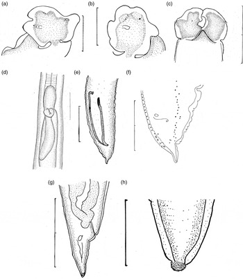
Lips consisting of three labia, one dorsal (fig. 1a) and two ventrolaterals (fig. 1b), with short interlabia between them (fig. 1c). Labia pedunculate, with flanges. Dorsal labium with two double cephalic papillae (fig. 1a). Papillae located at level of flanges. Subventral labia with one cephalic papilla and one amphid each.
Hysterothylacium australe n. sp.: (a) dorsal labium (scale bar = 0.2 mm); (b) subventral labium (scale bar = 0.2 mm); (c) lateral view of lips (scale bar = 0.2 mm); (d) intestinal caecum and ventricular appendix (scale bar = 0.4 mm); (e) spicules (scale bar = 0.4 mm); (f) posterior end in male, lateral view (scale bar = 0.2 mm); (g) and (h) posterior end in female (scale bars = 0.8 and 0.1 mm, respectively).

Male (n = 1). Body length 13.1, width 0.46. Labia 0.12 long, 0.11 wide. Nerve ring 0.52 from anterior end. Excretory pore 0.56 from anterior end. Oesophagus 1.76 long, 13% of body length. Ventriculus 0.16 long, 0.1 wide. Ventricular appendix 0.76 long, 43% of oesophageal length. Intestinal caecum 0.36 long, 20% of oesophageal length, 47% length of the ventricular appendix (fig. 1d). Spicules unequal (fig. 1e), left spicule 0.58 long, 4.4% of body length, right spicule 0.76 long, 5.8% of body length. Ejaculatory duct 1.52 long. Thirty-seven unpaired pre-anal papillae. Medioventral pre-anal papilla single, larger than other papillae. Twelve pairs post-cloacal papillae (fig. 1f). Tail with fine terminal spines, 0.2 long, 1.5% of body length.
Females (n = 8). Body length 42.6 (38.4–51.2), width 1.08 (0.95–1.26). Dorsal and subventral labia 0.23 (0.22–0.24) and 0.250 (0.24–0.26) long, respectively. Interlabia 0.09 (0.08–0.10) long, 0.11 (0.10–0.11) wide. Nerve ring 0.80 (0.72–0.90) from anterior end. Excretory pore 0.98 (0.86–1.39) from anterior end. Oesophagus 3.92 (3.63–4.20) long, 9% (8–10%) of body length. Ventriculus 0.27 (0.20–0.32) long. Ventricular appendix 1.32 (0.96–1.58) long, 33% (24–38%) of oesophageal length. Intestinal caecum 0.72 (0.52–1.04) long, 18% (13–25%) of oesophageal length, 58% (36–94%) of length of ventricular appendix. Tail gradually tapering, bluntly rounded, with terminal nipple (fig. 1g) covered with short spines (fig. 1h), 0.74 (0.57–0.95) long, 2% (1–2%) of body length. Vulva 14.3 (11.8–16.8) from anterior end, 34% (31–39%) of body length. Eggs 0.06–0.07 × 0.05–0.07.
Genetic characterization
Four representative specimens were selected and sequenced for ITS-1 and ITS-2. The length of the ITS-1 region was 436 bp and the G + C content was 51.4%. All ITS-1 sequences belonging to this species (GenBank accession numbers: HE862216–9) were identical. The ITS-2 region was 286 bp long. No sequence polymorphism was detected in ITS-2 and the G + C content was 53.5%. All ITS-2 sequences belonging to this species (GenBank accession numbers: HE862223–5) were identical. No ITS-1 or ITS-2 sequences belonging to adult Hysterothylacium spp. in GenBank were identical to those in the present study.
H. kajikiae n. sp.
Taxonomic summary
Type materials
Holotype (AHC 47777), allotype (AHC 47778), paratypes (two males and five females, AHC 34983 and AHC 34984).
Type host
Striped marlin, Kajikia audax (Philippi, 1887) (Perciformes: Istiophoridae).
Type locality
Nelson Bay, New South Wales, Australia.
Site in host
Stomach.
Description
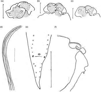
Body annulated. Alae originate at base of interlabial grooves. Lips consisting of three labia, one dorsal (fig. 2a) and two subventrals, with interlabia between them (fig. 2b). Labia pedunculate, with deep post-labial grooves (fig. 2b) and prominent lateral flanges (fig. 2a). Pulp pedunculate (fig. 2a), bilobed anteriorly (fig. 2a, b). Dorsal labium with two lateral cephalic papillae (fig. 2a). Subventral labia with amphid and lateral papilla (fig. 2b, c). Interlabia less than half length of labia (fig. 2b). Excretory pore at level of nerve ring (fig. 2d). Ventriculus present. Intestinal caecum long, up to 1.2 times length of oesophagus.
Hysterothylacium kajikiae n. sp.: (a) dorsal labium (scale bar = 0.1 mm); (b) and (c) subventral labia (scale bars = 0.1 mm); (d) anterior end in an immature male (scale bar = 0.83 mm); (e) posterior end, immature male, ventral view (scale bar = 0.2 mm); (f) spicules in mature male (scale bar = 0.21 mm).

Mature male (n = 1). Body length 50.4, width 0.33. Nerve ring 0.32 from anterior end. Oesophagus 5.57 long, 11% of body length. Ventriculus 0.30 long. Ventricular appendix 1.44 long, 26% of oesophageal length. Intestinal caecum 4.48 long, 80% of oesophageal length, 3.11 times length of the ventricular appendix. Spicules filiform, alate; left spicule 8.84 long, right spicule 9.12 long, 18% of body length. Body annulation prominent anterior to cloaca, with interrupted longitudinal ridges (caudal crests of Bruce & Cannon, Reference Bruce and Cannon1989) between row 7 and 8 to row 15 of pre-anal papillae, separated by a very narrow but distinct field.
Immature males (n = 2). Body length 15.7 (14.3–17.1), width 0.58 (0.32–0.84). Labia 0.07 (0.06–0.08) long. Interlabia 0.03 long. Nerve ring 0.45 (0.44–0.46) and excretory pore 0.50 (0.48–0.52) from anterior end, respectively. Oesophagus 2.50 (2.24–2.76) long, 16% of body length. Ventriculus 0.10 (0.08–0.12) long. Ventricular appendix 1.02 (0.40–1.64) long, 44% (14–73%) of oesophageal length. Intestinal caecum 1.82 (1.60–2.04) long, 73% (71–74%) of oesophageal length, 3.04 (0.98–5.10) times length of ventricular appendix. Tail 0.18 long, 1% of body length. Spicules visible in one specimen; left spicule 1.81 long, right spicule 1.86 long, 11% of body length. Pre-anal papillae 9 and 10 pairs. Pair of double papillae next to cloaca. Four pairs of post-cloacal papillae.
Females not gravid (n = 6). Body length 28.1 (10.2–54.6), width 0.35 (0.26–0.48). Labia 0.10 (0.05–0.13) long and 0.12 (0.10–0.14) wide. Interlabia length and width 0.05 (0.02–0.06) and 0.05 (0.05–0.05), respectively. Nerve ring and excretory pore 0.45 (0.34–0.62) and 0.45 (0.39–0.50) from anterior end, respectively. Oesophagus 3.23 (1.60–4.83) long, 13% (9–17%) of body length. Ventriculus 0.18 (0.08–0.29) long. Ventricular appendix 3.97 (1.56–9.24) long, 1.09 (0.80–1.91) times oesophageal length. Intestinal caecum 2.93 (1.08–5.88) long, 1.02 (0.68–1.22) times oesophageal length, 84% (0.64–1.19) times length of the ventricular appendix. Tail 0.73 (0.30–1.36) long, 3% (1–4%) of body length.
Genetic characterization
Four representative specimens were selected for the sequencing of the ITS-1 and ITS-2 regions, respectively. The ITS-1 region was 429 bp long (GenBank accession numbers: HE862220–1). No sequence polymorphism was detected in the ITS-1, and the G + C content was 50.4%. The length of the ITS-2 region was 271 bp (GenBank accession numbers: HE862226–9). No sequence polymorphism was detected in the ITS-2 and the G + C content was 50.6%. No identical sequences were found in GenBank for these specimens.
H. brucei n. sp.
Taxonomic summary
Type materials
Holotype (AHC 47779), allotype (AHC 34984), paratypes three immature males (accession number AHC 47785) from the stomach of Makaira mazara (Jordan & Snyder, 1901) (Perciformes: Istiophoridae), and one immature male, one mature male and two non-gravid females (AHC 34986) from the stomach of Kajikia audax (Philippi, 1887) (Perciformes: Istiophoridae) from Nelson Bay, New South Wales, 2005.
Type host
Striped marlin, Kajikia audax (Philippi, 1887) (Perciformes: Istiophoridae).
Type locality
Nelson Bay, New South Wales, Australia.
Site in host
Stomach.
Etymology
The species is named after Dr Neil Bruce. He discovered several new species of Hysterothylacium in Australia.
Description
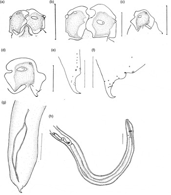
Body annulated. Lips consisting of three labia, one dorsal and two subventrals, with three interlabia (fig. 3a, b). Labia pedunculate with deep post-labial grooves and prominent lateral flanges (fig. 3c, d). Pulp pedunculate, bilobed anteriorly. Dorsal labium with two lateral papillae. Subventral labia each with amphid and one lateral papilla. Interlabia shorter than labia. Excretory pore between tip of caecum and nerve ring. Ventriculus present. Intestinal caecum longer than ventricular appendix, approximately same length as oesophagus.
Hysterothylacium brucei n. sp.: (a) lips in immature male, lateral view (scale bar = 0.1 mm); (b) lips in mature male, lateral view (scale bar = 0.1 mm); (c) and (d) subventral labium (scale bars = 0.1 mm); (e) posterior end of immature male, lateral view (scale bar = 0.2 mm); (f) posterior end of mature male, lateral view (scale bar = 0.2 mm); (g) spicules (scale bar = 0.2 mm); (h) anterior end (scale bar = 0.4 mm).

Mature male. Body length 32.3, width 0.53. Labia 0.12 long. Interlabia 0.09 long. Nerve ring and excretory pore 0.52 and 1.0 from anterior end, respectively. Oesophagus 5.04 long, 16% of body length. Ventriculus 0.56 long. Ventricular appendix 3.68 long, 73% of oesophageal length. Intestinal caecum 4.96 long, 98% of oesophageal length, 1.4 times length of ventricular appendix. Ejaculatory duct 2.16 long. Spicules (fig. 3g) unequal and alate, right spicule 1.76 long, left spicule 2.16 long, 5% and 7% of body length and 81% and 100% of length of the ejaculatory duct, respectively. Seventeen unpaired pre-anal papillae. Cuticle anterior to cloaca with interrupted longitudinal ridges (caudal crests of Bruce & Cannon, Reference Bruce and Cannon1989), separated by a distinct field. Tail short, 0.20 long, 0.6% of body length, narrowing suddenly after first pair of post-cloacal papillae, tip acute and bent ventrally (fig. 3f). Pair of double papillae next to cloaca, followed by 5 pairs of post-cloacal papillae.
Immature males (n = 4). Body length 17.8 (11.6–24.8), width 0.48 (0.42–0.55). Labia 0.08 (0.07–0.08) long. Interlabia 0.04 long. Nerve ring 0.37 (0.28–0.46) and excretory pore 0.39 (0.16–0.58) from anterior end, respectively. Oesophagus 4.23 (3.76–4.96) long, 26% (20–33%) of body length. Ventriculus 0.20 (0.20–0.21) long. Ventricular appendix 1.18 (0.68–1.96) long, 36% (21–51%) of oesophageal length. Intestinal caecum 3.83 (3.36–4.56) long, 90% (88–93%) of oesophageal length, 3 (1.8–4.3) times length of ventricular appendix. Tail 0.21 (0.20–0.22) long, 1% of body length. One spicule visible, 1.0 long, 4% of body length. Seventeen and 21 pairs pre-anal papillae visible in two specimens.
Females (n = 2, poorly preserved). Body length 77.0 (70.6–83.4), width 0.97. Labia 0.16 (0.13–0.19) long. Interlabia 0.07 long. Nerve ring 0.68 (0.64–0.72) from anterior end. Oesophagus 7.0 (6.7–7.3) long, 9% of body length. Ventriculus 0.43 (0.42–0.44) long. Ventricular appendix 3.8 (3.7–4.0) long, 55% of oesophageal length. Intestinal caecum 7.2 (6.8–7.5) long, 1.0 times oesophageal length, 1.87 (1.85–1.88) times length of ventricular appendix. Tail 0.74 (0.68–0.80) long, 1% of body length.
Genetic characterization
The ITS-1 and ITS-2 regions of only one specimen were sequenced. The ITS-1 region was 429 bp long (GenBank accession number HE862222). No sequence polymorphism was detected within the ITS-1 and the G + C content was 48.7%. The ITS-2 region was 264 bp long (GenBank accession number HE862230). No sequence polymorphism was detected in the ITS-2 and the G + C content was 47.4%. No identical sequence for ITS-1 and ITS-2 was found in the GenBank database.
Phylogenetic analyses
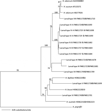
A neighbour joining tree revealed two main clades. All new species found in the present study resolved separately from one another with high bootstrap value. They formed a separate clade from H. aduncum and H. auctum and Hysterothylacium larval types IV and VIII with a high bootstrap value (fig. 4).
Phylogenetic analysis of the combined ITS-1 and ITS-2 sequence data for Hysterothylacium spp., with Anisakis pegreffii as outgroup, using the neighbour-joining method. Bootstrap support values are indicated. Numbers after taxa refer to ITS-1 and ITS-2, respectively. If only one number is presented, it indicates that ITS sequence data were extracted from a bigger region of rDNA. Larval types refer to Hysterothylacium larval types (Shamsi et al., Reference Shamsi, Gasser and Beveridge2013).

Discussion
The genus Hysterothylacium currently consists of more than 70 species and is considered to be one of the largest of the ascaridoid genera parasitizing fish. Li et al. (Reference Li, An and Zhang2007) proposed an identification key to distinguish between 61 species within the genus. Some members of the genus, such as those previously included in the genus Maricostula, including H. cenaticum, H. makairi and H. tetrapteri, were not included in their identification key. Although relatively many Hysterothylacium species have been reported and described in Australia, this is the first genetic characterization of adult Hysterothylacium spp., in which ITS-1 and ITS-2 were sequenced, in the country. As shown in the Results section, specimens examined in the present study can be classified under three distinct species – named as H. australe, H. kajikiae and H. brucei – supported by both morphological and genetic evidence. Phylogenetic analyses of Hysterothylacium species for which comparable sequence data (i.e. ITS-1 and ITS-2) were available in GenBank (fig. 4) supported the distinction of the new species from previously reported taxa.
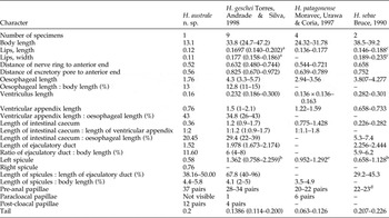
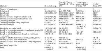
According to the key proposed by Li et al. (Reference Li, An and Zhang2007), adult specimens found in Yellowtail amberjack, S. lalandi, and named as H. australe n. sp., resemble H. geschei Torres, Andrade and Silva, 1998, H. patagonense Moravec, Urawa and Coria, 1997 or H. sebae Bruce, 1990. However, H. australe n. sp. is different from the above-mentioned species based on the differences in the size of the main characteristic features, such as spicules, intestinal caecum, ventricular appendix and labia (for a full list and detailed comparison see tables 1 and 2). In addition, H. australe n. sp. also differs from H. geschei, H. patagonense and H. sebae in the anatomical features of the tail in both males and females, spicules and labia (Bruce, Reference Bruce1990a; Moravec et al., Reference Moravec, Urawa and Coria1997; Torres et al., Reference Torres, Andrade and Silva1998).
Comparison of the morphometric characters of male H. australa n. sp. with males of the most similar species (measurements are given in millimetres unless otherwise stated).

aDorsal labium; bno differentiation between left and right spicules; csubventral labia; dno description of type of papillae; eequal spicules.
Comparison of the morphometric characters of female H. australe n. sp. with females of the most similar species (measurements are given in millimetres unless otherwise stated).

aDorsal labium; bdiameter; cnumber of specimens = 1.
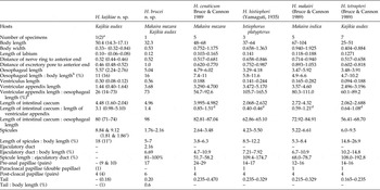
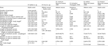
Two more distinct taxa were also found in the present study from a different group of fish, i.e. marlins. Specimens from M. mazara and K. audax in the present study were considered as new species due to differences from known species. To date, H. cenaticum, H. histiophori, H. incurva, H. makairi, H. pelagicum and H. tetrapteri have been found in these fish, and in the closely related species in Australia. Bruce & Cannon (Reference Bruce and Cannon1989) provided a key for differentiation between these species based on the characters of the caudal crests, ratios of ejaculatory duct length to spicule length, and the presence and origin of alae. According to this key, H. kajikiae is most similar to H. tetrapteri and H. brucei n. sp. is morphologically most similar to H. cenaticum, H. makairi, and H. tetrapteri. Both H. kajikiae and H. brucei are different from those species based on a number of main characteristic features, such as anatomy of the tail, ratio of intestinal caecum to ventricular appendix, length of the spicules and their ratio to total body length (tables 3 and 4). In addition to comparing the new species in the present study with those found in the same fish or closely related species of fish, H. kajikiae and H. brucei were also compared with species listed by Li et al. (Reference Li, An and Zhang2007). Based on the identification key by Li et al. (Reference Li, An and Zhang2007) the most similar species to H. kajikiae n. sp. are H. auctum (Rudolphi, 1802), H. ilishae (Yamaguti, 1941), H. sinense Li, An and Zhang, 2007 and H. thalassini Bruce, 1990. However, H. kajikiae n. sp. is different from these species mainly in the length of the spicules and the ratio of spicules to total body length, which is significantly greater in H. kajikiae n. sp. Hysterothylacium brucei n. sp. is most similar to H. cenaticum. Differentiation between H. brucei n. sp. and H. cenaticum has been discussed above.
Comparison of the morphometric characters of male Hysterothylacium spp. found in Australian marlins (measurements are in millimetres; mean followed by the range in brackets unless otherwise stated).

aMature specimen followed by two immature specimens in the brackets; bno mention of sex of the specimens; cone specimen; dcalculated by the author of the present study.
Comparison of the morphometric characters of female Hysterothylacium spp. found in Australian marlins (measurements are in millimetres; mean followed by the range in brackets unless otherwise stated).

As mentioned in the Introduction, the main aim of the present study was to provide comparable ITS sequence data to specifically identify 14 larval types of Hysterothylacium described in Australia and adjacent waters. Alignment of ITS sequences of H. australe found in the present study (GenBank accession numbers HE862216–HE862219 and HE862223–HE862225 for ITS-1 and ITS-2, respectively) with those of Hysterothylacium larval types available in GenBank (Shamsi et al., Reference Shamsi, Gasser and Beveridge2013, Reference Shamsi, Poupa and Justine2015) showed that fourth-stage Hysterothylacium larval type XI from S. lalandi (GenBank accession numbers FN811763 and FN811717 for ITS-1 and ITS-2, respectively) collected from Port Augusta, South Australia, and third-stage Hysterothylacium larval type X from Sphyraena novae-hollandiae (GenBank accession numbers FN811761–FN811762 and FN811714–FN811716 for ITS-1 and ITS-2, respectively) collected from Port Phillip Bay, Victoria have identical ITS sequences, suggesting that these larval types are H. australe. The ITS sequences of H. kajikiae and H. brucei did not match any ITS sequence of other larval types, suggesting that more Hysterothylacium larval types await discovery.
Acknowledgements
The authors would like to thank Dr Julian Peperell for facilitating access to marlins, Dr Kate Hutson, James Cook University for the parasite materials from king fish and Professor Ian Beveridge, The University of Melbourne for his input and guidance.
Financial support
This research received no specific grant from any funding agency, commercial or not-for-profit sectors.
Conflict of interest
None.