Introduction
The fossiliferous potential of teeth relative to other skeletal remains is high due to their composition; consequently, they have become the main focus of paleontological studies, particularly on mammals. Nevertheless, many extinct mammalian groups lack much dental information due mainly to the scarcity of complete and well-preserved dental remains. Historically, the dental characteristics have been used to erect, describe, and contrast mammalian species, as well as to infer different aspects of their way of life, such as their habitat and feeding preferences (e.g., Patterson and Pascual, Reference Patterson and Pascual1968; Fortelius, Reference Fortelius1985; Janis, Reference Janis1988, Reference Janis, Turner and Thulborn1990, Reference Janis1995; Janis and Constable, Reference Janis and Constable1993; Solounias et al., Reference Solounias, Fortelius and Freeman1994; Pérez-Barbería and Gordon, Reference Pérez-Barbería and Gordon1998; Vizcaíno et al., Reference Vizcaíno, Bargo and Cassini2006, Reference Vizcaíno, Cassini, Fernicola and Bargo2011; Townsend and Croft, Reference Townsend and Croft2008; Reguero et al., Reference Reguero, Candela, Cassini, Madden, Carlini, Vucetich and Kay2010; Cassini et al., Reference Cassini, Vizcaíno and Bargo2012a, Reference Cassini, Cerdeño, Villafañe, Muñoz, Vizcaíno, Kay and Bargob, Reference Cassini, Del Pino, Munoz, Acosta, Fernández, Bargo and Vizcaíno2017; Candela et al., Reference Candela, Cassini and Nasif2013; Famoso et al., Reference Famoso, Feranec and Davis2013, Reference Famoso, Davis, Feranec, Hopkins and Price2016). In recent years, these works have been strongly enriched by the publication of other studies focused on deciduous dentition, reconstruction and description of the ontogenetic series, and tooth eruption/replacement patterns of fossil mammals, whose adaptive, taxonomic, and phylogenetic weight is recognized (Smith, Reference Smith, Teaford, Smith and Ferguson2000). Descriptions of the deciduous dentition and eruption patterns refer, for example, to Litopterna (e.g., Cerdeño et al., Reference Cerdeño, Chiesa and Ojeda2008; Bergqvist, Reference Bergqvist2010) and Rodentia (e.g., Vucetich et al., Reference Vucetich, Deschamps, Olivares and Dozo2005, Reference Vucetich, Deschamps, Pérez, Moreira, Barros Ferraz, Herrera and Macdonald2012, Reference Vucetich, Deschamps, Pérez and Montalvo2014a, Reference Vucetich, Deschamps, Vieytes and Montalvob); this information has allowed demonstrating a taxonomic overestimation in some cases (e.g., Hydrochoeriinae rodents) in different localities. Within notoungulates, these studies have markedly increased in the past few years, but the available information concerning ontogenetic series, deciduous teeth, and tooth eruption/replacement patterns is still scarce, considering the remarkable diversity of the group, and shows a great variability (e.g., Scott, Reference Scott1912; Cerdeño and Montalvo, Reference Cerdeño and Montalvo2001; Billet et al., Reference Billet, de Muizon and Mamaní Quispe2008, 2009; Townsend and Croft, Reference Townsend and Croft2010; Billet and Martin, Reference Billet and Martin2011; Cerdeño and Schmidt, Reference Cerdeño and Schmidt2013; Vera and Cerdeño, Reference Vera and Cerdeño2014; Cerdeño et al., Reference Cerdeño, Montalvo and Sostillo2017).
In this context, the members of the subfamily Interatheriinae from the Santacrucian South American Land Mammal Age (SALMA; early–middle Miocene after Cuitiño et al., Reference Cuitiño, Fernicola, Raigemborn and Krapovickas2019, in contrast to previous estimations of early Miocene) of the Santa Cruz Formation (Santa Cruz Province, Argentina) constitute an important study group as they provide a significant number of specimens with a high degree of preservation and involve both juveniles and adults in different ontogenetic stages (with different degrees of dental wear). The information resulting from their study, presented herein, allows completing the data already published for the remaining Interatheriidae (i.e., “Notopithecinae”; see Vera and Cerdeño, Reference Vera and Cerdeño2014) and, therefore, increasing the available information for one of the main notoungulate groups.
The Interatheriinae (Interatheriidae) were small (0.5–10 kg) rodent-like cursorial herbivores (Stirton, Reference Stirton1953; Croft, Reference Croft and Singer1999, Reference Croft2016; Croft and Anderson, Reference Croft and Anderson2008; Cassini et al., Reference Cassini, Cerdeño, Villafañe, Muñoz, Vizcaíno, Kay and Bargo2012b; Elissamburu, Reference Elissamburu2012) morphologically similar to the afrotherian Hyracoidea and comprising protohypsodont and euhypsodont (sensu Mones, Reference Mones1982) forms (Marshall and Cifelli, Reference Marshall and Cifelli1990). This is a broadly represented group in the Cenozoic of South America (Bond, Reference Bond1986; Hitz et al., Reference Hitz, Reguero, Wyss and Flynn2000), known mostly by the representatives of early to middle Miocene (Croft et al., Reference Croft, Anaya, Auerbach, Garzione and MacFadden2009), being recorded from the Tinguirirican SALMA (early Oligocene) to the Huayquerian SALMA (late Miocene). The present study focuses on the comparative study of the deciduous and permanent dentition of the genera Interatherium Ameghino, Reference Ameghino1887 (Ameghino, Reference Ameghino1887a) from the Santacrucian SALMA and Protypotherium Ameghino, Reference Ameghino1885, which has an unusually long stratigraphic range that extends from the Deseadan (late Oligocene) to the Huayquerian SALMA (late Miocene) (Fernández et al., Reference Fernández, Fernicola and Cerdeño2019b) and whose monophyly has been recently questioned (e.g., Vera et al., Reference Vera, Reguero and González-Ruiz2017; Croft and Anaya, Reference Croft and Anaya2020). The obtained results allow the differentiation of several ontogenetic stages for both genera and the establishment of the pattern of eruption and replacement of the deciduous cheek teeth and the eruption sequence of molars. This leads to reinterpreting some taxonomic assumptions and will contribute to further systematic studies on Interatheriinae.
Materials and methods
The studied materials consist of published and, mainly, unpublished juvenile materials assigned to 16 genera within the subfamily Interatheriinae (following the taxonomic proposals by Fernández, Reference Fernández2020): Protypotherium; Interatherium; Patriarchus Ameghino, Reference Ameghino1889; Archaeophylus Ameghino, Reference Ameghino1897; Argyrohyrax Ameghino, Reference Ameghino1897; Choichephilum Ameghino, Reference Ameghino1899; Eopachyrucos Ameghino, Reference Ameghino1901; Cochilius Ameghino, Reference Ameghino1902; Caenophilus Ameghino, Reference Ameghino1904; Progaleopithecus Ameghino, Reference Ameghino1904; Miocochilius Stirton, Reference Stirton1953; Santiagorothia Hitz et al., Reference Hitz, Reguero, Wyss and Flynn2000; Proargyrohyrax Hitz et al., Reference Hitz, Reguero, Wyss and Flynn2000; Federicoanaya Hitz, Billet, and Derryberry, Reference Hitz, Billet and Derryberry2008; Brucemacfaddenia Hitz, Billet, and Derryberry, Reference Hitz, Billet and Derryberry2008; and Juchuysillu Croft and Anaya, Reference Croft and Anaya2020. These specimens are housed at different national and foreign institutions. Most information was gathered from the direct observation of the specimens and original photographs of diverse materials, together with some bibliographic data (e.g., Hitz et al., Reference Hitz, Billet and Derryberry2008 for Federicoanaya and Brucemacfadennia). The most-complete and/or best-preserved juvenile specimens are indicated in Supplementary Data 1, and the majority belong to Protypotherium and Interatherium, which were collected from the exposures of the Santa Cruz Formation, mainly, at the localities of the Río Santa Cruz and at the eastern region of the Santa Cruz Province. The provenance of each studied specimen is also indicated in Supplementary Data 1, although it is necessary to highlight that many of them belong to the “old collections” at certain institutions such as the Museo de La Plata and Museo Argentino de Ciencias Naturales “Bernardino Rivadavia.”
Considering the need of an exhaustive specific revision of the species of Interatheriinae, particularly of the Santacrucian taxa (results in Fernández, Reference Fernández2020 are just partially published), here we present the analysis at generic level, excepting some comments on Protypotherium diversidens (Ameghino, Reference Ameghino1891a) and Eudiastatus lingulatus (Ameghino, Reference Ameghino1891b).
The comparative analysis of the deciduous and permanent teeth is based on the materials assigned to Interatherium and Protypotherium. The deciduous teeth are mainly identified because their permanent counterparts are located below them; this can be observed in the cases where the dental series is incomplete or the bone is lacking around the alveoli. In doubtful specimens, two techniques were used: the cutting of some bone portions and the X-rays, which allow observing the base of the teeth without altering the material. In the particular case of the deciduous incisors and canines, they are identified mainly by the thickness of the enamel and the crown height.
However, the eruption pattern is inferred from the height of the crown and, mainly, the analysis of wear of the deciduous and permanent cheek teeth. Three morphological criteria are taken into consideration: the eruption stage, which ranges from “non erupted” (with the germ in its dental crypt) to “fully erupted”; the absence or presence of roots; and the degree of wear, which is delimited by a combination of characteristics of the occlusal surface, such as the degree of erosion of the main and accessory cusps and the crests/cristids, the formation of the lophs/lophids, the presence/absence and the development of the principal and accessory fossettes/fossettids, the presence/absence and the depth of labial and lingual sulci, and the presence/absence and the development of the cingula/cingulids.
The combination of both the mentioned morphological aspects and the diverse specimens allows reconstructing: (1) the direction of eruption of deciduous and permanent premolars and molars, which can be anterior–posterior or posterior–anterior; (2) the type of replacement of the deciduous and permanent premolars, which can be alternate or sequential; and (3) the time of eruption of M3 relative to the first deciduous premolar being replaced. In addition, the combined data allow differentiating several successive ontogenetic stages for the Santacrucian Interatheriinae Interatherium and Protypotherium, which concern the upper and lower deciduous and permanent cheek teeth. However, we make some comments about the eruption and replacement patterns of the anterior dentition (i.e., dI/i1–dC/c), which are based on a few materials of Protypotherium, due to the lack of preservation of these series in the studied sample.
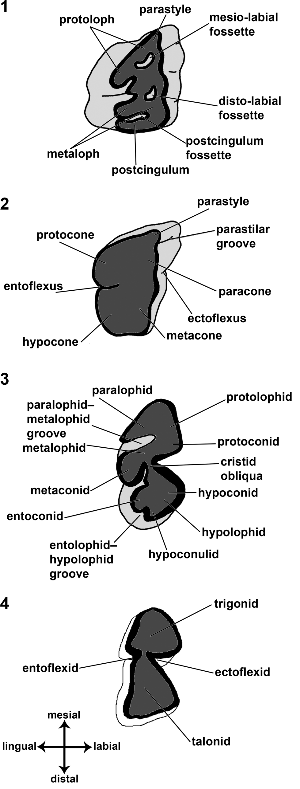
The measurements indicated in this contribution were taken with a Kendo digital calliper (0.02 mm). The photographs were taken with Kodak Digital AZ651, Nikon Micrometrics 318CU 3.2MP CMOS, and Nikon Coolpix L110 cameras. All the nomenclatural acts follow the regulations established by the ICZN (2000). The dental terminology (Fig. 1) follows Hooker (Reference Hooker1986) and Smith and Dodson (Reference Smith and Dodson2003), with some ideas from Reguero et al. (Reference Reguero, Ubilla and Perea2003) for the upper dentition and Vera and Cerdeño (Reference Vera and Cerdeño2014) and Fernández et al. (Reference Fernández, Fernicola and Cerdeño2019b) for the lower dentition.
Schemes of the dental structures mentioned throughout the text. (1) left M1 of Eopachyrucos; (2) left M1 of Protypotherium; (3) right p3 of Eopachyrucos; (4) right m1 of Protypotherium. Modified from Reguero et al. (Reference Reguero, Ubilla and Perea2003); the nomenclature follows Hooker (Reference Hooker1986), Reguero et al. (Reference Reguero, Ubilla and Perea2003), Smith and Dodson (Reference Smith and Dodson2003), Vera and Cerdeño (Reference Vera and Cerdeño2014), and Fernández et al. (Reference Fernández, Fernicola and Cerdeño2019b).

Anatomical abbreviations
C/c, upper/lower canine; dC/c, upper/lower deciduous canine; dI/i, upper/lower deciduous incisor; dP/p, upper/lower deciduous premolar; I/i, upper/lower incisor; LLL, labio-lingual maximum length; M/m, upper/lower molar; MDL, mesio-distal maximum length; P/p, upper/lower permanent premolar.
Repositories and institutional abbreviations
American Museum of Natural History (AMNH), New York, USA; Field Museum of Natural History (FMNH), Chicago, USA; Museo Argentino de Ciencias Naturales “Bernardino Rivadavia” (MACN), Ciudad Autónoma de Buenos Aires, Argentina; Ameghino National Collection at the MACN (MACN-A), Ciudad Autónoma de Buenos Aires, Argentina; Museo de Ciencias Naturales y Antropológicas “Juan Cornelio Moyano” (MCNAM-PV), Mendoza, Argentina; MLP, Museo de La Plata, La Plata, Argentina; Museo Nacional de Historia Natural, Collection of Vertebrate Paleontology (MNHN-BOL), La Paz, Bolivia; Museo Paleontológico “Egidio Feruglio,” Collection of Vertebrate Paleontology (MPEF-PV), Trelew, Argentina; Museo Municipal Padre Molina, Collection of Vertebrate Paleontology (MPM-PV), Río Gallegos, Argentina; Museo de Historia Natural, Collection of Vertebrate Paleontology (SGO-PV), Santiago, Chile; Paleontology Museum of the University of California (UCPM), Berkeley, USA; Florida Museum of Natural History (UF), Collection of Vertebrate Paleontology, Gainesville, USA; and Yale Peabody Museum, Vertebrate Paleontology Princeton University Collection (YPM-VPPU), New Haven, USA.
Results
Description of the deciduous dentition of Protypotherium
A comparative analysis between permanent and deciduous dentition and the main characteristics of the deciduous teeth of Protypotherium is provided below.
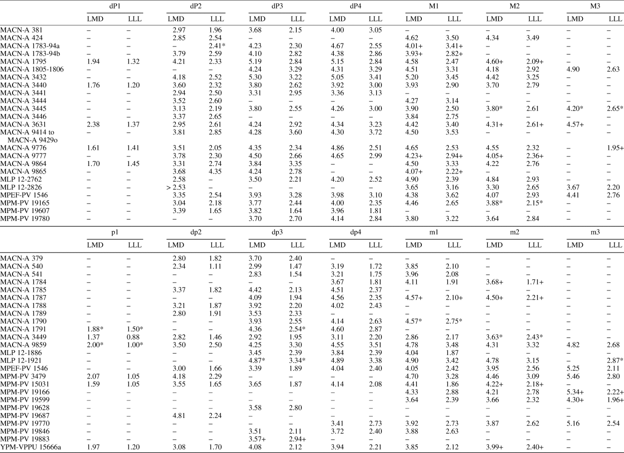
Upper deciduous dentition (Table 1)
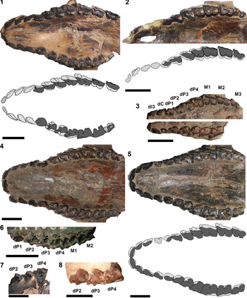
The series dI1–dC is very similar to I1–C, but the deciduous teeth are more fragile, slightly narrower, have a lower crown and a thinner layer of enamel, and are more imbricated (e.g., AMNH 9482, MACN-A 9620, and MPM-PV 3534; Fig. 2.1–2.3) than their permanent counterparts (e.g., YPM-VPPU 15189 and YPM-VPPU 15828; Fig. 2.4, 2.5). Similar to I1–C, the dI1–dC of Protypotherium are uniradiculate and vertically implanted, and their crowns narrow toward the roots; dI1 is mesiodistally expanded (i.e., long), labiolingually compressed (i.e., narrow), subelliptic in contour, and covered with a layer of enamel, which is labially thicker, producing a chisel-like cutting edge with wear; this tooth is vertically implanted; dI2–3 are subequal in size and morphology; dI2–dC exhibit a conspicuous mesiolabial groove, and the distal edge of each tooth always labially overlaps the mesiolabial edge of the posterior tooth; dC is completely incisiviform.
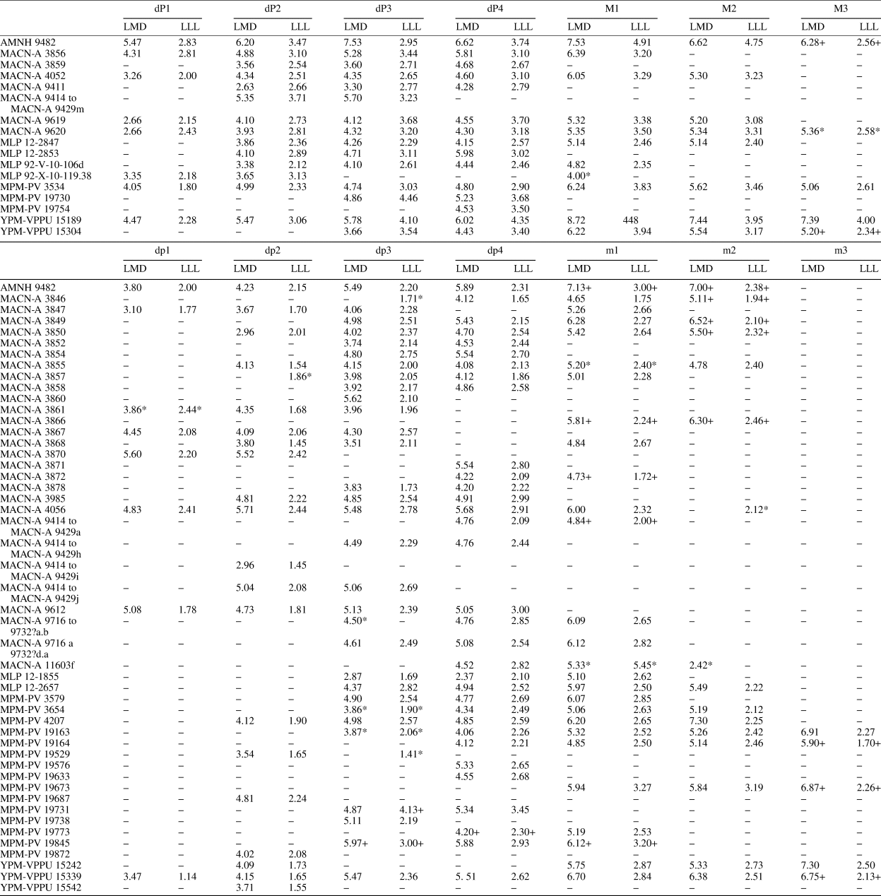
Dental dimensions (mm) of the upper cheek teeth of Protypotherium Ameghino, Reference Ameghino1885. * Approximate measurements due to the bad preservation, articulated material and/or breakage of the specimen; + erupting tooth measurements.

Protypotherium Ameghino, Reference Ameghino1885. (1) AMNH 9482, incomplete skull with deciduous and permanent dentition, occlusal view; (2) left maxillary fragment with alveoli of I1 and I3–C, and I2, dP1–4–M1–3 of MPM-PV 3534, occlusal view; (3) MACN-A 9620, left maxillary fragment with dI3–dP1–4–M1–3 (M3 in eruption) (above), and right maxillary fragment with dI3–dP1–4–M1 (below), occlusal view; (4) YPM-VPPU 15189, almost complete skull with erupting I1–C and dP1–4–M1–3, occlusal view; (5) YPM-VPPU 15828, almost complete skull with full permanent dentition (I1–M3), occlusal view; (6) MACN-A 4052, right maxillary fragment with dP1–4–M1–2, occlusal view; (7) MACN-A 9411, left maxillary fragment with dP2–4, lingual view; (8) MACN-A 3859, left maxillary fragment with dP2–4, occlusal view. (1–6) Scale bars = 10 mm; (7, 8) scale bars = 5 mm. Black with white contour arrows point to the entoflexus; white with black contour arrow points to the roots.

Concerning cheek teeth, dP1 can have a subcilindrical to a subellipsoidal section; it can be mesially and distally imbricated by dC and dP2, repectively, only distally imbricated, or not imbricated at all; when dP1 is barely worn (e.g., AMNH 9482; Fig. 2.1), it has a pronounced labial projection that can be interpreted as a paracone, which becomes indistinguishable as the tooth wears; this tooth is not replaced by any homologue tooth (see Eruption pattern). The dP2–4 of Protypotherium are brachydont and exhibit three roots; the entoflexus and the parastilar groove disappear before the beginning of the roots (e.g., MACN-A 4052 and MACN-A 9411; Fig. 2.6, 2.7). In general terms, dP2–4 are slightly longer and narrower and have a thinner enamel layer than their permanent homologues. The P2–4 (e.g., Fig. 2.4) are not molarized, whereas dP2–4 (e.g., Fig. 2.1–2.4, 2.6, 2.8) are more similar to molars than to permanent premolars. The dP2–4 are very similar to each other, increasing in size in an anterior–posterior direction. The entoflexus of dP2–4 divides the tooth into two lobes, the mesial one that involves the region of the protocone within the protoloph and the distal one, with the hypocone within the metaloph. In P2–4, the protoloph is much smaller than the metaloph, being more mesiodistally and labiolingually compressed, giving a subtriangular contour to these teeth; in dP2–4, this differential development is much less conspicuous, although the protocone column remains smaller than the hypocone column, acquiring a more subrectangular contour. This is more evident in dP4, which is the most molarized tooth of the deciduous premolar series. The entoflexus of P2–4 and dP2–4 is simple, moderately transversally deep, and vertically short, being more persistent as it almost reaches the base of the tooth in P2–4. The entoflexus is mesially placed in P2–4, a feature also observed in dP2–4, but this flexus is more distally displaced. The parastyle and the paracone of P2–4 and dP2–4 are little developed. The parastilar groove is transversally shallow in P2–4 and dP2–4, being much more persistent in P2–4 as it almost reaches the base of the tooth. The metacone is not completely individualized from the paracone, so this region is rather straight.
Lower deciduous dentition (Table 1)
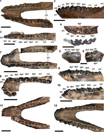
As mentioned for the upper dentition, the di1–dc (e.g., AMNH 9482, YPM-VPPU 15339, MACN-A 637, MACN-A 4056, and MACN-A 9435; Fig. 3) resemble i1–c, except for being more fragile, with thinner enamel, and exhibiting a lower crown than i1–c (e.g., MACN-A 9645 and YPM-VPPU 15828; Fig. 3.11, 3.12). Similar to i1–c, di1–dc exhibit the following features: they are uniradiculated, without spaces separating them; very procumbent (accompanying the morphology of the mandibular symphysis); di1–2 are subcylindrical and contact each other without overlapping. These teeth are divided by a deep transversal sulcus along the crown, giving them a bifid or bicolumnar aspect, with the distal column being more developed than the mesial one. This sulcus gradually disappears to the base of the crown (see Fernández et al., Reference Fernández, Fernicola and Cerdeño2019b for the description of the changes of i1–2 with wear), which becomes subcylindrical in worn incisors. The di3–dc are mesiodistally expanded, labially convex, and lingually excavated by a wide groove, producing a V-shaped tooth in occlusal view. These teeth differ from each other by their size, although the difference is very subtle; di3–dc are imbricated, and the degree of overlapping is variable.
Protypotherium. (1, 2) AMNH 9482, incomplete mandible with deciduous and permanent, occlusal and labial (horizontally rotated) view; (3) left mandibular fragment with di2 (broken)–dp1–4–m1–3 (m3 in eruption) of YPM-VPPU 15339, occlusal view; (4–6) left di1–dp1–3 of MACN-A 637, occlusal, labial, and lingual views; (7) MACN-A 9435, incomplete mandible with deciduous and permanent dentition, occlusal view; (8–10) MACN-A 4056, incomplete mandible of a juvenile with deciduous dentition and molars in eruption, occlusal, lingual, and labial (horizontally rotated) views; (11) MACN-A 9645, incomplete mandible with permanent dentition (lacking left i2–p2), occlusal view; (12) YPM-VPPU 15828, incomplete mandible with permanent dentition, occlusal view. (1–3, 7–12) Scale bars = 10 mm; (4–6) scale bars = 5 mm.

Regarding the deciduous lower cheek teeth, dp1 is caniniform, almost morphologically indistinguishable from dc, and it can be subequal or even longer than its predecessor; dp1 completely lacks a talonid or even vestiges of it; it can also be lingually imbricated with dc. As mentioned for dP1, dp1 do not exhibit a dental replacement (see Eruption pattern). The dp2–4 are morphologically closer to m1–3 (e.g., Figs. 3.1–3.9) than to p2–4 (Fig. 3.10, 3.11). In contrast to p2–4, the dp2–4 are brachydont, with two roots; the ectoflexid and entoflexid end near the base of the crown (e.g., Fig. 3.2, 3.8, 3.9). As in p2–4, the dp2–4 have the mesiodistal length greater than the linguolabial length; they are labially imbricated, so that the paralophid of the posterior tooth overlaps the distal region of the hypolophid of the anterior tooth. The dp2 exhibits (as p2) both trigonid and talonid, but the talonid is insinuated and not as developed as in the remaining deciduous premolars. The dp3–4 are moderately morphologically similar to each other. The subtriangular trigonid and talonid are well defined and separated by the entoflexid and the ectoflexid, the latter flexid being slightly deeper than the former. The trigonid is more developed both mesiodistally and labiolingually than the talonid, particularly in dp3; dp4 is the most molariform deciduous tooth. This differential development between the trigonid and the talonid is much less conspicuous in dp3–4 than in p3–4, although the talonid never acquires the same length or becomes longer and wider than the trigonid (as observed in m1–3). When dp2–4 are barely worn (e.g., MACN-A 4056; Fig. 3.8–3.10), dp2 presents an almost straight and mesiolingually directed protolophid, an undeveloped paralophid lingually directed, and a short transverse metalophid. The paralophid–metalophid groove is transversely deep, but vertically short. The entolophid is already fused with the hypolophid, resulting in a single oblique lophid. The trigonid and the talonid are connected by a straight cristid obliqua that advances mesiolingually until it joins the lingual region of the metalophid. In dp3–4, the protolophid is almost straight and mesiolingually directed. The paralophid is short and distolingually directed. The metalophid is straight and transverse. The protoconid and metaconid are well developed. The paralophid–metalophid groove is transversally deep and vertically short. The cristid obliqua unites to the distolingual face of the metalophid, but this union is more medially displaced than that of dp2. In the talonid, the entoconid is transversely and distally expanded, approaching the hypolophid until it joins it completely. The entolophid and the hypolophid are separated by a conspicuous but vertically short groove.
Description of the deciduous dentition of Interatherium
The main features of the deciduous teeth, as well as a comparative analysis between permanent and deciduous dentition of Interatherium, are given in the following paragraphs.
Upper deciduous dentition (Table 2)
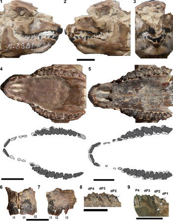
As in Protypotherium, dI1–dC (e.g., MACN-A 3881 and MACN-A 3631; Fig. 4.1–4.4) differ from I1–C in being more fragile, slightly narrower, lower crowned, and with a thinner layer of enamel than their homologous counterparts (e.g., MPM-PV 19500 and YPM-VPPU 15666; Fig. 4.5–4.7). Similar to I1–C, dI1–C are discontinuous due to the presence of diastemata of variable length. The dI1–dC exhibit smooth lingual and labial faces; the enamel distribution is continuous, although it is reduced on the lingual side. These teeth are uniradiculate and decrease in width and length toward the root. The dI1–3 mesiodistally contact each other, without presenting any level of imbrication between them; dI1 is a rodent-like tooth, labiolingually compressed, mesiodistally expanded, and vertically implanted; dI2 presents almost the same compression as dI1, but it is approximately half as long; dI3 is slightly narrower and half as long as dI2; dC is subequal and morphologically similar to dI3.
Dental dimensions (mm) of the lower cheek teeth of Interatherium Ameghino, Reference Ameghino1887 (Ameghino, Reference Ameghino1887a). * Approximate measurements due to the bad preservation, articulation of the material, and/or breakage of the specimen; + erupting tooth measurements.

Interatherium Ameghino, Reference Ameghino1887 (Ameghino, Reference Ameghino1887a). (1–3) MACN-A 3881, almost complete articulated skull and mandible with complete deciduous dentition, left lateral, right lateral, and anterior views; (4) MACN-A 3631, incomplete skull with deciduous and permanent dentitions (both M3 erupting), occlusal view; (5) YPM-VPPU 15666, almost complete skull with permanent dentition, occlusal view; (6, 7) premaxilla with both I1–3 of MPM-PV 19500, anterior and left lateral views; (8) right dP2–4 of MACN-A 3432, lingual view; (9) left dP1–3–P4 of MACN-A 9864, labial view. Scale bars = 10 mm. Black with white contour arrows point to the parastilar groove and/or the entoflexus; white with black contour arrows point to the roots.

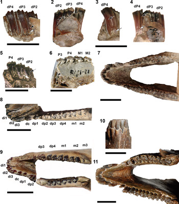
Regarding the deciduous cheek teeth, dP1 does not have the typical molariform morphology of the remaining cheek teeth, but it is not incisiviform either because it is wider than dI1–dC. It is distally imbricated by dP2 and always smaller than dP2, even though there is some variation in the degree of its development. The dP1 does not exhibit any replacement (see Eruption pattern). The dP2–4 of Interatherium are brachydont and exhibit three roots. The entoflexus and the parastilar groove disappear before the base of the crown (Figs. 4.8, 4.9, 5.1–5.5), whereas in P2–4 they occupy the entire crown (Fig. 5.6). The heights of the crowns of dP2–4 markedly decrease with wear (Fig. 5.1–1.5). The dP2–4 are morphologically closer to P2–4, except for being slightly longer and narrower, and the enamel layer is thinner. Similar to P2–4, dP2–4 are subrectangular, longer than wide. These teeth are strongly imbricated; the parastyle of a cheek tooth overlaps with the distolabial region of the metacone column of the mesial tooth. The layer of enamel reduces its thickness in the imbrication contact areas. The dP2 is moderately molarized, differing from dP3–4 in the column of the protocone, mesiodistally shorter than that of the hypocone. The dP3–4 are completely molarized, and dP3 is slightly smaller than the dP4. The ectoloph presents a very pronounced undulation due, in part, to the presence of a deep parastilar groove and a conspicuous separation between the paracone and the metacone. The parastyle and the paracone are well developed; the ectoflexus and entoflexus are medially positioned; the entoflexus is simple, transversally deep, and separates the well-developed protoloph and metaloph, both being subequal, giving a bilobed appearance to the lingual margin.
Interatherium Ameghino, Reference Ameghino1887 (Ameghino, Reference Ameghino1887a). (1) left dP2–4 of MPM-PV 19607, labial view; (2–4) MACN-A 381, left maxillary fragment with dP2–4, lingual, distal, and labial views; (5) MACN-A 3434, right maxillary fragment with dP2–3–P4, occlusal view; (6) MLP 12-1878, left maxillary fragment with P3–M2, lingual view; (7) MPEF-PV 1546, incomplete mandible with complete deciduous and permanent dentition (except for right dp1), occlusal view; (8) YPM-VPPU 15666a, mandible of a juvenile individual with complete deciduous dentition and erupting m1–2 (m3 not yet emerged), occlusal view; (9) MPM-PV 15031, incomplete mandible of a juvenile with deciduous dentition (right m3 in eruption), occlusal view; (10, 11) incomplete mandible with associated permanent dentition of YPM-VPPU 15401, anterior and occlusal views. Scale bars = 10 mm. Black with white contour arrows point to the parastilar groove and/or the entoflexus; white with black contour arrows point to the roots.

Lower deciduous dentition (Table 2)
Similar to the upper dentition, the di1–dc of Interatherium (e.g., MACN-A 3881, MPEF-PV 1546, and YPM PU 15666a; Figs. 4.1–4.3, 5.7–5.9) resemble the i1–c, except for being more fragile, with thinner enamel and a lower crown than their permanent homologous (e.g., YPM-VPPU 15401 and MACN-A 9851; Figs. 5.10, 5.11, 6.1). As in i1–c, di1–3 have a subelliptic crown, with a linguolabial compression near the occlusal surface, and a midlingual groove that does not extend to the base of the crown. These teeth are uniradiculate and are obliquely implanted, directed anteriorly. The di1–3 are subequal and imbricated, without diastema between them. The dc is incisiviform and smaller than the di3, with the crown slightly shorter. It is obliquely anteriorly directed, resting directly on the di3. There is always a diastema of variable length between dc and dp1 as observed between c and dp1.
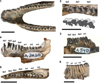
Interatherium. (1) MACN-A 9851, complete mandible with complete permanent dentition, occlusal view; (2, 3) MACN-A 540, right mandibular fragment with dp2–4–m1, occlusal and lingual views, occlusal view; (4, 5) MACN-A 3638, right mandibular fragment with dp1–4–m1–3, lingual and occlusal views; (6) left p3–m3 of MPM-PV 19500, in occlusal view. (1, 3–6) Scale bars = 10 mm; (2) scale bar = 5 mm. Black with white contour arrows point to the entoflexid and/or the ectoflexid; white with black contour arrows point to the roots.

The dp1 is incisiviform without differentiated talonid. It is distinguished from dc mainly for being vertically implanted, and it is not overlapped by any tooth. It is the smallest premolar and does not present replacement, as already mentioned for dP1 (see Eruption pattern). The dp2–4 of Interatherium are brachydont and exhibit two roots; the height of the crown decreases with wear. The entoflexid and the ectoflexid are quite persistent, both disappearing at the same level near the base of the crown (Fig. 6.2–6.5), whereas in p2-4 it reaches the base of the crown (Fig. 6.6). As in dP2–4, the occlusal surface of the dp2–4 does not significantly differ from that in the permanent premolars, except for being slightly longer and narrower and exhibiting a thinner layer of enamel. Similar to p2–4, dp2 has a differentiated trigonid and talonid, although this separation is less pronounced than in dp3–4; trigonid and talonid are connected by the cristid obliqua in the middle region of the metalophid. As in p3–4, the dp3–4 are subequal in size (dp3 hardly smaller than dp4) and completely molarized. The trigonid and the talonid are well defined, forming a bilobed crown. The entoflexid and the ectoflexid are opposite to each other; the ectoflexid is slightly deeper than the entoflexid. The depth of these flexids slightly reduces as tooth wear progresses, and the cristid obliqua joins the middle or slightly lingual region of the metalophid. When dp2–4 are barely worn (e.g., YPM-VPPU 15666a; Fig. 5.8), dp2 exhibits the labial region of the protolophid slightly convex and slightly mesiolingually directed; the paralophid is short, but conspicuous, and lingually directed; there is no evidence of a paraconid in front of the metaconid region. The metalophid is straight and moderately oblique (i.e., distolingually directed); the paralophid–metalophid groove is transversely deep, but vertically short. The entoconid is transversely expanded forming an entolophid, separated from the hypolophid by a shallow, but conspicuous, groove. The hypoconulid is lingually displaced from the entoconid. The dp3–4 are barely worn and exhibit the same general features identified in dp2, except for the presence of a more robust paralophid that is distolingually directed and a more robust metalophid.
Ontogenetic stages of Interatherium and Protypotherium: upper dentition
Five successive ontogenetic stages have been identified among the juveniles of Protypotherium and Interatherium for the upper dentition, which we describe together and summarize in Table 3.
Ontogenetic stages established for upper and lower deciduous postcanine dentitions of Protypotherium and Interatherium. Adapted from Cerdeño et al. (Reference Cerdeño, Montalvo and Sostillo2017).

Stage 1
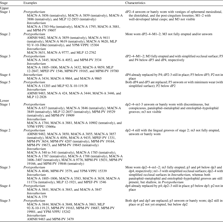
This stage is represented by individuals with immature bone through the entire exposed surface. The dI1–dC–dP1–4 are unworn or barely worn; dP4 is more worn than dP3, and the latter is more worn than the unworn dP2. The molar series in represented by only M1–2, as M3 has not erupted yet. The M1–2 are unworn, barely worn, or with minimum wear (with simplified occlusal surface), with well-developed labial cusps. This stage is observed in MACN-A 3881 (Fig. 4.1–4.3) and MACN-A 1795 of Interatherium, and due to, mainly, the state of wear of the available cheek teeth, the following specimens are included in this stage: MPM-PV 19607 (Figs. 5.1, 7.1) of Interatherium and MACN-A 3856 (Fig. 7.2–7.4), MACN-A 3859 (Fig. 2.8), MACN-A 3886 (Fig. 7.5), and MLP 12-2853 of Protypotherium. MACN-A 1795 is an incomplete palate with deciduous and erupting molars, except for both M3, which have not erupted yet. Likewise, MACN-A 3881 preserves the almost complete skull and mandible in anatomical position, and little can be observed of the occlusal surfaces of the cheek tooth rows; however, it is possible to identify all deciduous and permanent teeth and the absence of both M/m3. MACN-A 3886 presents less worn dP2–4 than MACN-A 3856, MACN-A 3859, and MPM-PV 19607, with unworn dP2 placed deeper inside the alveolus than the already functional dP3 that is, in turn, deeper than dP4. In MPM-PV 19607, MACN-A 3856, and MACN-A 3859, dP2–4 are barely worn, with vestiges of ephemeral mesiolabial, distolabial, and postcingulum fossettes, being more ephemeral in Interatherium (MPM-PV 19607) than in Protypotherium (MACN-A 3856 and MACN-A 3859).
Ontogenetic stages 1–5: upper dentition of Protypotherium (2–5, 7,8) and Interatherium (1, 6, 9–17). Stage 1: (1) MPM-PV 19607, left maxillary fragment with dP2–4, occlusal view; (2–4) MACN-A 3856, maxillary fragment with dP1–4–M1, occlusal, lingual, and labial views; (5) MACN-A 3886, left maxillary fragment with alveoli of C–dP1 and complete dP2 (in eruption)–4–M1–2 (broken), occlusal view. Stage 2: (6) MLP 12-2762, left maxillary fragment with dP1–4–M1–2, occlusal view; (7) YPM-VPPU 15304, fragmented skull with right dP3–4–M1–3 and left dP4–M1–3 (both M3 in eruption), occlusal view. Stage 3: (8) MACN-A 4052, right maxillary fragment with dP1–4–M1–2, occlusal view; (9, 10) left maxillary fragment with dP3–4–M1–3 (broken) (P3 and P4 below dP3 and dP4, respectively) of MPM-PV 19780, mesial and labial views; (11) MACN-A 9859, almost complete skull with deciduous and permanent dentition (alveoli of anterior dentition), occlusal view; (12) right dP2–4 of MACN-A 9859, lingual view; (13, 14) left maxilla with dP3–4–M1–3 of MACN-A 1805 to 1806, occlusal and lingual views. Stage 5: (15) MLP 12-2826, right maxilla with dP1 (alveolus)–dP2–P3–M3, occlusal view; (16) MACN-A 424, right maxillary fragment with dP2–P3–M2 series, occlusal view; (17) right dP2–P3–M1 of MACN-A 3444, occlusal view. (1) Scale bar = 5 mm; (2–17) scale bars = 10 mm. ’ indicates broken tooth; * indicates the dental alveolus; black with white contour arrows point to the parastilar groove and/or the entoflexus; white with black contour arrows point to the roots.

Stage 2
The specimens included in this stage show worn deciduous premolars, with simplified occlusal surfaces and the crown completely exposed. Specimens preserving accompanying molars show that M3 is not fully erupted and/or is unworn. In Interatherium (e.g., MACN-A 3631 and MLP 12-2762; Figs. 4.3, 7.6), the occlusal surface is completely simplified due to the loss of all ephemeral fossettes mentioned in Stage 1; the entoflexus of dP2–4 appears in the whole exposed portion of the crown. In Protypotherium, the mesiolabial fossette of dP2–4 is still present and conspicuous as observed in AMNH 9482 (Fig. 2.1), which is the youngest individual of this stage, but this fossette disappears as wear advances, simplifying completely the occlusal surface of these cheek teeth as observed in MACN-A 3859, MACN-A 9411, MACN-A 9619, MACN-A 9620 (Fig. 2.3), MLP 92-V-10-106d, and YPM-VPPU 15304 (Fig. 7.7). The entoflexus is exposed as in Interatherium.
Stage 3
This stage comprises individuals with worn dP2–4 and M3 fully erupted and worn; all of the occlusal surfaces are simplified. The visible portion of the entoflexus of dP2–4 is shorter in Protypotherium than in Interatherium, not reaching the base of the crown. These conditions are observed in MACN-A 3445, MACN-A 4052 (Figs. 2.5, 7.8), and MPM-PV 3534 (Fig. 2.2) of Protypotherium and MACN-A 3432 (Fig. 4.8), MACN-A 9859 (Fig. 7.11, 7.12), MACN-A 1805–1806 (Fig. 7.13, 7.14), MLP 12-2847, MPEF-PV 1546, MPM-PV 19165, and MPM-PV 19780 (Fig. 7.9, 7.10) of Interatherium. The last specimen mentioned shows P3 and P4 below dP3 and dP4, respectively, and the height of the crown of dP4 is shorter than that of dP3 (Fig. 7.9, 7.10).
Stage 4
This stage gathers a few specimens in which dP4 has already been replaced by P4, whereas dP2–3 are still in place as in the incomplete maxillas MACN-A 3434 (Fig. 5.5), MACN-A 9864 (Fig. 4.9), and MACN-A 9865 of Interatherium. In this stage, P3 is still below dP3, and P2 is not visible below any dP2 of the studied sample.
Stage 5
At this stage, both dP4 and dP3 have been replaced, P3 is unworn or with minimum wear but with simplified occlusal surface, and dP2 is still in place. We include in this stage the specimens MACN-A 11203 and MLP 92-X-10-119.38 of Protypotherium and MLP 12-2826 (Fig. 7.15), MACN-A 424 (Fig. 7.16), MACN-A 3444 (Fig. 7.17), MACN-A 3446, and the skull of AMNH 9483 of Interatherium. In MLP 92-X-10-119.38 and AMNH 9483, P4 has already erupted and presents minimum wear, and P3 is erupting, whereas in MACN-A 424, MACN-A 3444, and MLP 12-2826, P3 presents a complete simplified occlusal surface. In this stage, P2 would be below dP2 as observed in lower dentition (see the following).
Ontogenetic stages of Interatherium and Protypotherium: lower dentition
We have identified five successive ontogenetic stages among the juveniles of Protypotherium and Interatherium for the lower dentition, which are described together and are summarized in Table 3.
Stage 1
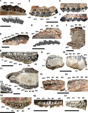
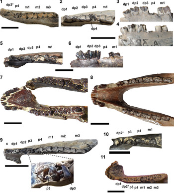
This stage is represented by individuals with deciduous anterior dentition (i.e., di1–dc) and unworn or barely worn dp1–4, dp4 being more worn than dp3, and the latter more worn than the unworn dp2 as in MACN-A 1784 (Fig. 8.1), MACN-A 3881 (Fig. 4.1–4.3), and YPM-VPPU 15666a (Fig. 5.8) of Interatherium. The dp2–4 exhibit conspicuous paralophid–metalophid and entolophid–hypolophid grooves. The molar series is represented by only m1 and m2 because m3 has not erupted yet. In MACN-A 1784 (Fig. 8.1) and YPM PU 15666a (Fig. 5.8), m1 is recently erupted whereas m2 is deeper inside the bone, not fully erupted, with the trigonid more exposed than the talonid. The m1–2 of these specimens exhibit conspicuous paralophid–metalophid and entolophid–hypolophid grooves. Note that the mentioned incomplete mandible and the fragments MACN-A 637 (Fig. 3.4–3.6), MACN-A 3846 (Fig. 8.2), MACN-A 3849 (Fig. 8.3), MLP 12-2657 (Fig. 8.4), MPM-PV 19529, and MPM-PV 19909 of Protypotherium and MACN-A 10902 (Fig. 8.5) of Interatherium are not complete enough to conclusively determine the absence of m3, but the wear stage of the preserved teeth corresponds to this stage.
Ontogenetic stages 1–2: lower dentition of Protypotherium (2–4, 6–11) and Interatherium (1, 5, 12). Stage 1: (1) MACN-A 1784, right mandibular fragment with dp4–m1–2 (m2 in eruption), occlusal view; (2) MACN-A 3846, left mandibular fragment with dp4–m1–2 (m2 in eruption), occlusal view; (3) MACN-A 3849, right mandibular fragment with dp3–4– m1–2 (m2 in eruption), occlusal view; (4) MLP 12-2657, left mandibular fragment with dp3–4–m1–2 (m2 in eruption), occlusal view; (5) MACN-A 10902, right mandibular fragment with dp2–4 barely worn, occlusal view. Stage 2: (6–8) MACN-A 3850, mandibular fragment with dp2–4–m1–3 (m2 and broken m3 in eruption), occlusal, lingual, labial (horizontally rotated) views; (9, 10) MPM-PV 19164, right mandibular fragment with dp4–m1–3 (m3 in eruption), occlusal and lingual views; (11) MPEF-PV 1331, fragmented mandible with badly preserved deciduous dentition with m1–3 (m3 in eruption), occlusal view; (12) MACN-A 1785, left mandibular fragment with dp1 (alveolus)–4, occlusal view. (1, 5, 12) Scale bars = 5 mm; (2–4, 6–10) scale bars = 10 mm; (11) scale bar = 20 mm. White with black contour arrows point to the roots.

Stage 2
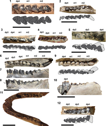
This stage includes the specimens with di1–dp1–4–m1–2 and the m3 not fully erupted, unworn, or with minimum wear: AMNH 9482 (Fig. 3.1, 3.2), MACN-A 3850 (Fig. 8.6–8.8), MACN-A 9435 (Fig. 3.7), MPM-PV 3654, MPM-PV 19164 (Fig. 8.9, 8.10), MPM-PV 19673, and MPEF-PV 1331 (Fig. 8.11) of Protypotherium, and MACN-A 9776, MPM-PV 15031 (Fig. 5.9), and MPM-PV 19166 of Interatherium. The dp2–4 show some wear, although dp2 is the less worn tooth of this series, and the paralophid–metalophid and entolophid–hypolophid grooves are slightly shallower (particularly the latter groove) than those observed in Stage 1. The incompletes mandibles MACN-A 540 (Fig. 6.2, 6.3), MACN-A 541, MACN-A 1785 (Fig. 8.12), MACN-A 1787, MACN-A 1788, MACN-A 3406–3407, and MPM-PV 19846 of Interatherium and MACN-A 3855 (Fig. 9.1), MACN-A 3857 (Fig. 9.2–9.4), MACN-A 4056 (Fig. 3.8–3.10), MPM-PV 4207 (Fig. 9.5, 9.6), and MPM-PV 19845 of Protypotherium are included in this stage despite lacking preserved m3, on the basis of the state of wear of the remaining teeth, particularly the barely worn dp2, and the depth of the lingual grooves.
Ontogenetic stages 2–4, lower dentition of Protypotherium (1–9) and Interatherium (10–12). Stage 2: (1) MACN-A 3855, right mandibular fragment with dp2–4–m1–2, occlusal view; (2–4) MACN-A 3857, right mandibular fragment with dp3–4–m1, occlusal, lingual, and labial (horizontally rotated) views; (5, 6) MPM-PV 4207, right mandibular fragment with dp2–4–m1–2 (m2 in eruption), occlusal and lingual views. Stage 3: (7) MACN-A 4046, left mandibular fragment with p3 (barely worn)–m3, occlusal view; (8, 9) MPM-PV 3579, left mandibular fragment with dp3–4–m1–2 (broken), occlusal and lingual views; (10) MACN-A 11173a, right mandibular fragment with dp3–4–m1, occlusal view. Stage 4: (11, 12) MACN-A 379, right mandibular fragment with dp2–3 (p3 below)–p4, lingual and occlusal views. (1–9) Scale bars = 10 mm; (10–12) scale bars = 5 mm. * indicates the dental alveolus.

Stage 3
The specimens included in this stage exhibit more-worn dp1–4–m1–3, the m3 being fully erupted and with simplified occlusal surface. In Protypotherium, the paralophid–metalophid groove is still present in dp2–4, whereas it is absent in m1–3 (e.g., MACN-A 4046, MPM-PV 3579, YPM-VPPU 15339, Figs. 3.3, 9.7–9.9), and the entolophid–hypolophid groove is absent in dp2–4–m1–3. In Interatherium, both grooves are no longer present (e.g., MACN-A 1805–1806, MACN-A 3583, MACN-A 3638, MLP 12-1921, MPEF-PV 1546; Figs. 5.7, 6.4, 6.5); the entolophid–hypolophid disappears before the paralophid–metalophid groove in dp2, whereas the former disappears shortly after the latter in dp3–4 (e.g., MACN-A 11173a; Fig. 9.10). In addition, and assuming correspondence with the upper dentition (see the preceding), in this stage, p3 and p4 are below dp3 and dp4, respectively.
Stage 4
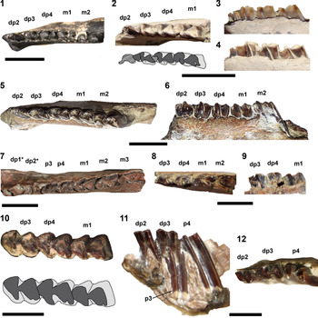
This stage comprises specimens with more worn dp2–3 and m1–3, all with simplified occlusal surfaces, and dp4 is already replaced by p4, which is unworn, barely worn, or with minimum wear: MACN-A 379 (Fig. 9.11, 9.12) of Interatherium and MACN-A 3841 (Fig. 10.1), MACN-A 3843 (Fig. 10.2–10.4), and MACN-A 3847 (Fig. 10.5, 10.6) of Protypotherium. In MACN-A 379, the removal of lingual bone allows registering the presence of the p3 below the functional dp3 and the absence of p2 below dp2 (comparable to the Stage 4 of the upper dentition, see the preceding).
Ontogenetic stages 4–5, lower dentition of Protypotherium (1–6, 8–10) and Interatherium (7, 11). Stage 4: (1) MACN-A 3841, left mandibular fragment with p4 (in eruption)–m3 (broken), occlusal view; (2–4) MACN-A 3843, right mandibular fragment with dp1–3–p4 (in eruption)–m1, occlusal, lingual, and labial (horizontally rotated) views. (5, 6) MACN-A 3847, mandibular fragment with dp1–3–p4 (in eruption)–m1, occlusal and lingual views. Stage 5: (7) MPM-PV 3479, incomplete mandible with left i1–c–dp1 (alveolus)–dp2–p3–m3 and right i1–c and right mandibular fragment with dp1–m3, occlusal view; (8) YPM-VPPU 15242, almost complete mandible with both i1–c series, alveoli of left dp1–2, and right dp1, with complete left p3–m3 series and right dp2–p3–m3, occlusal view; (9) MPM-PV 19163, right mandibular fragment with c–dp1–dp2 (broken)–dp3 (broken and with p3 below)–p4–m3, occlusal view (above), and detail of the remnant of dp3 above p3 (without scale; below); (10) MACN-A 3863, right mandibular fragment with dp2 (alveolus)–p3 (in eruption)–m1, occlusal view; (11) AMNH 9483, left mandibular fragment with p2 (not yet erupted)–m1, occlusal view. Scale bars = 10 mm. * indicates the dental alveolus.

Stage 5
At this stage, both dp4 and dp3 have been replaced; p4 is more worn than in Stage 4; p3 is unworn, barely worn, or with minimum wear; and dp2 is still in place as observed in MPM-PV 3479 (Fig. 10.7) of Interatherium and YPM-VPPU 15242 (Fig. 10.8) and MPM-PV 19163 (Fig. 10.9) of Protypotherium. MPM-PV 19163 presents remnants of the replaced right dp3 stuck to the unworn occlusal surface of p3. We also include in this stage the Protypotherium specimens MACN-A 3844, MACN-A 3848, MACN-A 3863 (Fig. 10.10), MLP 92-X-10-119.23, and MPM-PV 19687, and Interatherium specimens AMNH 9483 and MPM-PV 19901 (Fig. 10.11). MPM-PV 19901 does not preserve dp2, but does the alveolus of its roots, and although p2 is not visible, it is assumed to be already below this alveolus.
Eruption pattern
The dP/p1 are scarcely found, being common that only their alveoli are preserved. Specimens with dP/p2–4 with low wear (e.g., AMNH 9482, MACN-A 4056, MACN-A 637, MPM-PV 15031, and YPM-VPPU 15666a; Figs. 2.1, 3.1, 3.2, 3.4–3.10, 5.9) also bear little-worn first upper/lower premolars, which highly probably erupt after dP/p2; however, in specimens with permanent teeth, the first premolars are usually much more worn, which allows inferring the lack of replacement of this first premolar, here interpreted as dP/dp1 because it erupts with the deciduous dentition.
Regarding the remaining cheek teeth, we refer our results to the three different aspects mentioned in the Materials and methods section.
Direction of molar eruption
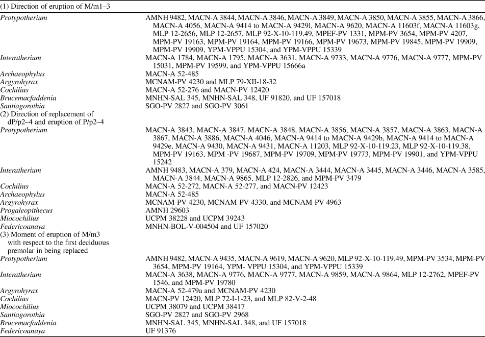
The molars are the first cheek teeth of the permanent series to erupt. The skull and mandible of Protypotherium AMNH 9482 (Figs. 2.1, 3.1, 3.2) and the skull MACN-A 3631 (Fig. 4.4) and mandibular fragment MPM-PV 15031 of Interatherium are appropriate examples: M/m1 are already erupted, in use, and with little wear; M/m2 are recently erupted and barely worn, showing less wear than M/m1; and M/m3 are erupting and unworn. To summarize, these specimens show that the eruption of molars is sequential, erupting in an anterior–posterior direction, from M/m1 to M/m3. This sequence has been observed in other interatheriine genera such as Cochilius, Archaeophylus, Argyrohyrax, Santiagorothia, and Brucemacfaddenia (see Table 4 for the summary of the assigned specimens that allowed inferring this variable).
Specimens assigned to different Interatheriinae genera that allowed inferring completely and/or partially the direction of eruption of molars, direction of replacement and eruption of deciduous and permanent premolars, respectively, and moment of eruption of M/m3 with respect to the first replaced deciduous premolar (or first erupted permanent premolar). Miocochilius from Stirton (Reference Stirton1953), and Federicoanaya and Brucemacfaddenia from Hitz et al. (Reference Hitz, Billet and Derryberry2008).

Eruption of M/m3
The mandibular fragment MACN-A 3638 (Fig. 6.4, 6.5) and the skull MACN-A 9859 (Fig. 7.11, 7.12) of Interatherium show the simultaneous presence of the dP/dp2–4 and M/m1–3 as described in both ontogenetic Stages 3 (see the preceding). The same situation is observed in Protypotherium AMNH 9482 (Figs. 2.1, 3.1, 3.2) and MPM-PV 4207 (Fig. 9.5, 9.6). These and other specimens of both genera provide evidence that M/m3 erupts before the replacement of any deciduous premolar, that is, before the eruption of the first permanent premolar. This pattern has also been identified in other Interatheriinae genera such as Cochilius, Archaeophylus, Argyrohyrax, Progaleopithecus, and Miocochilius (see Table 4 for the summary of the assigned specimens that allowed inferring this variable).
Direction of replacement of dP/p2–4 (and eruption of P/p2–4)
The mandible fragment of Protypotherium MACN-A 3847 (Fig. 10.5, 10.6) shows the p4 barely protruded from the alveolus and fragments of the replaced dp4 stuck to its occlusal surface, whereas dp2–3 are in place, as well as m1 completely erupted and worn. This condition indicates that dp4 is the first deciduous tooth to be replaced. In Interatherium, the mandibular fragment MACN-A 379 (Fig. 9.11, 9.12) shows the p4 fully erupted together with dp2 and dp3, but p3 is observed below dp3, which allows us to infer that dp3 is the second deciduous premolar to be replaced. A similar situation is seen in the upper dentition of Interatherium MLP 12-2826 (Fig. 7.15), in which dP2 is present along with P3–M3. Therefore, both genera show a sequential replacement and eruption pattern, in a posterior–anterior direction, from dP/p4 to dP/p2. This particular sequence has been (completely or partially) observed in other Interatheriinae genera such as Argyrohyrax, Cochilius, Miocochilius, Santiagorothia, Brucemacfaddenia, and Federicoanaya (see Table 4 for the summary of the assigned specimens that allowed inferring this variable).
In specimens where a permanent premolar appears below the corresponding deciduous tooth, we observe that the crown apex of the former inserts inside the open basal end of the deciduous tooth, and just in cases where the former is a little displaced, the occlusal border of the latter is observed.
Comments on the replacement of dI/i1–dC/c—The reconstruction of the eruption and replacement patterns of the anterior dentition can only be inferred from the skull YPM-VPPU 15189 of Protypotherium (Fig. 2.4). This specimen shows the simultaneous presence of dP2–4, M1–3, moderately worn dP1, and erupting I1–C, which provides evidence that deciduous incisors and canines are replaced before the deciduous premolars. Among incisors, YPM-VPPU 15189 shows that I1 is the last incisor to erupt because this tooth is unworn; it is less erupted than I2-C. In addition, AMNH 9482 (Fig. 2.1) shows that dI3 and dC exhibit the same crown exposure and the same wear, whereas dI2 is less exposed and less worn than dI3–dC. This same sequence is also observed in the series I1–C of YPM-VPPU 15189 (Fig. 2.4). After this observation in upper dentition, we assume that the lower would behave in the same way, although it is worth mentioning that MACN-A 637 (Fig. 3.4–3.6) and MACN-A 4056 (Fig. 3.8–3.10) show di3 and dc with the same crown exposure and wear. Therefore, it is inferred that, in Protypotherium, (d)I3/(d)i3 and (d)C/(d)c erupt almost simoultanesly, followed by (d)I2 and, later, (d)I1.
Discussion and conclusions
Deciduous upper and lower dentition in Interatheriinae
The study of the juvenile individuals of Protypotherium and Interatherium makes evident that the deciduous cheek teeth are brachydont and the height of the crown markedly decreases with wear. In general terms, the anterior upper and lower deciduous dentitions (dI/i1–dC/c) of both genera are morphologically close to their permanent homologues, being slightly narrower, longer, and with a thinner enamel layer. This has also been observed in Argyrohyrax, Cochilius, and Federicoanaya (see Hitz et al., Reference Hitz, Billet and Derryberry2008). A difference in Protypotherium, Cochilius, and Argyrohyrax is that dI1–dC are more imbricated than I1–C. Regarding the deciduous cheek teeth, there are differences between Interatherium and Protypotherium. The occlusal surfaces of the dP/p2–4 of Interatherium highly match those of the P/p2–4; this close resemblance has also been observed in Argyrohyrax, Cochilius, Archaeophylus, Progaleopithecus, Miocochilius (personal observation), Federicoanaya, and Brucemacfaddenia (Hitz et al., Reference Hitz, Billet and Derryberry2008). In the case of Protypotherium, the dP/p2–4 are more similar to M/m1–3 than to P/p2–4, a fact that has also been mentioned for Federicoanaya (Hitz et al., Reference Hitz, Billet and Derryberry2008).
However, the permanent premolars of Protypotherium and Interatherium are disposed exactly below the bases of their deciduous counterparts, even appearing continuous, hardly separated by a thin line. Cerdeño et al. (Reference Cerdeño, Montalvo and Sostillo2017) indentified this condition in the hegetotheriid Paedotherium. In this context, Protypotherium and Interatherium add to the examples of groups with balanced wear, which is characterized by a constant position of the occlusal surface (Martin, Reference Martin, Martin and Barnosky1993) in such a way that the intensive wear by an antagonist is in a perfect equilibrium with the tooth eruption (Koenigswald, Reference Koenigswald2011).
Ontogenetic stages of Interatherium and Protypotherium
The ontogenetic series proposed by Cerdeño et al. (Reference Cerdeño, Montalvo and Sostillo2017) for Paedotherium, with three stages for the upper dentition and four stages for the lower, partially fits with what is observed in Interatherium and Protypotherium.
Differences among deciduous cheek-tooth series and the time of replacement and eruption of upper and lower premolars and molars allow us to differentiate five ontogenetic stages among the juvenile individuals of Protypotherium and Interatherium (see the preceding and Table 3). The comparison among these stages shows that there are morphological (and metrical) variations regarding the crown height, the occlusal proportions, the presence of labial and postcingulum fossettes, and the presence of ephemeral grooves. The occlusal proportions change with wear as deciduous teeth become slightly wider toward the base, which is more evident in Interatherium than in Protypotherium. The occlusal surface quickly simplifies due to the loss (e.g., fossettes) and/or reduction (e.g., labial and lingual grooves) of several features; this occlusal simplification is faster in the upper dentition than in the lower of Interatherium and Protypotherium, and faster in the former genus than in latter.
The comparison with other interatheriine taxa allows us to establish comparable ontogenetic stages in different specimens, summarized in Table 3. For example, MACN-A 52-485 of Archaeophylus, AMNH 29603 of Progaleopithecus, and MACN-A 52-272 and MACN-A 52-277 of Cochilius correspond to the Stage 5 of lower dentition, while MLP 72-I-1-23 of Cochilius presents a Stage 2 in upper and lower dentitions. These euhypsodont taxa acquire a simplified occlusal surface more rapidly than the analyzed protohypsodont genera.
Eruption and replacement sequences in the Interatheriinae
Regarding the anterior dentition, and as mentioned in the preceding, only one specimen of Protypotherium reflects that (d)I/i3 and (d)C/c erupt almost simoultanesly, followed by (d)I/i2 and later by (d)I/i1, and all are replaced before the dP/p4. A more complete sample would allow confirming this pattern and testing it in Interatherium.
From the combined mandibular and maxillary data, we can state that dP/dp1 of Interatherium and Protypotherium do not present replacement. This reaffirms what was already suggested (with doubts) for Protypotherium by Sinclair (Reference Sinclair and Scott1909, p. 18), although this author considered this tooth as “P/p1”. In Notoungulata, this tooth is assumed to be rarely replaced (Shockey et al., Reference Shockey, Flynn, Croft, Gans and Wyss2012, p. 20), and it is usually a heavily worn tooth. Within the Interatheriidae, Vera and Cerdeño (Reference Vera and Cerdeño2014) mentioned the presence of dP1 in the notopithecine Antepithecus, but the presence of dp1 and p1 in this genus was doubtful, as well as in Notopithecus; nevertheless, these authors mentioned that the studied specimens seemed to support a replacement of the first tooth. Among Interatheriinae, Simpson (Reference Simpson1932, p. 4) doubted the presence of p1 or dp1 in Cochilius, although according to our personal observations, dp1 seems not to have a replacement; Stirton (Reference Stirton1953, p. 273) mentioned the absence of dp1 and the presence of p1 that erupts with the deciduous dentition in Miocochilius; what Stirton (Reference Stirton1953) considered as p1 is here interpreted as dp1. More recently, Hitz et al. (2000, p. 10) mentioned the lack of replacement of P1 (here interpreted as dP1) on the basis of the extreme state of wear in most specimens of Santiagorothia; later, Hitz et al. (2008, table 2) listed a dp1 and a p1 for Brucemacfaddenia but did not describe or mention this anywhere else in the text, whereas these authors stated the lack of replacement of dP1 in Federicoanaya (Hitz et al., Reference Hitz, Billet and Derryberry2008, p. 455). Larger samples of lower and upper interatherid dentitions in different ontogenetic stages would allow a better understanding of the first premolar behavior and contrasting of the different tendency between notopithecines and interatheriines that seems now to exist.
Concerning the remaining premolar and molar series, we can establish that: (1) the deciduous and permanent premolars erupt sequentionally in the posterior–anterior direction: P/p4→(d)P/p3→(d)P/p2; (2) molars protrude sequentionally in an anterior–posterior direction: M/m1→M/m2→M/m3; and (3) M/m3 erupt before the replacement of dP4, which is the first deciduous tooth to be replaced in Interatheriinae. This timing of eruption of M/m3 is known as “delayed dental eruption” (see Slaughter et al., Reference Slaughter, Pine and Etoh1974; Smith, Reference Smith, Teaford, Smith and Ferguson2000; Billet and Martin, Reference Billet and Martin2011), and it is observed in groups with rapid growth, in the framework of the “Law of Schultz” (Smith, Reference Smith, Teaford, Smith and Ferguson2000), which relates the eruption of M/m3 with respect to the other permanent teeth to the growth rate and lifespan of the organisms. Such are the cases of the hyracoid, tenrecid, and macroscelidean afrotherians, phenacodontid and pleuraspidotheriid “condylarths,” some tillodonts, some primates, some artiodactyls, eulipotyphlans, carnivorans, and scandentians (Slaughter et al., Reference Slaughter, Pine and Etoh1974; Smith, Reference Smith, Teaford, Smith and Ferguson2000; Asher and Lehmann, Reference Asher and Lehmann2008; Holroyd, Reference Holroyd2008, Reference Holroyd2009; Asher and Olbricht, Reference Asher and Olbricht2009; Billet and Martin, Reference Billet and Martin2011; Gomes Rodrigues et al., Reference Gomes Rodrigues, Herrel and Billet2017). Gomes Rodriguez et al. (Reference Gomes Rodrigues, Herrel and Billet2017) stated that the geologically earliest occurrence of this fast eruption of M/m3 seen in Guilielmoscottia (and therefore, in the Interatheriidae) occurred during the late Eocene (approximately 35 Ma). As a result, this modification in the timing of eruption of M/m3, as well as the sequential posterior–anterior eruption pattern of premolars, preceded the crown height increase in the Interatheriidae.
According to our results, there is an evident congruence between the pattern of eruption of deciduous and permanent premolars, and molars with the wear sequence between juvenile and adult specimens. This congruence allows inferring that the masticatory process does not lose the information of the eruptive sequence, and the angle of occlusion between the upper and lower teeth does not affect the wear rate among teeth that differ in positions (see Spinage, Reference Spinage1971).
The knowledge of the replacement and eruption patterns of the Interatheriinae presented here allows making some remarks concerning the species Eopachyrucos ranchoverdensis Reguero, Ubilla, and Perea, Reference Reguero, Ubilla and Perea2003. The holotype MAMC-39 was described as a “left partial dentary with broken c, dp1–p2–3, p4 (talonid), m1–m2, and unerupted m3, and ascending ramus with imperfectly preserved coronoid process” (Reguero et al., Reference Reguero, Ubilla and Perea2003, p. 447). As mentioned by the authors, MAMC-39 shows m2 in full eruption and the lack of m3, not because it is completely absent but because it has not erupted yet. On the basis of the pattern described in this contribution, particularly the time of eruption of molars, it can be interpreted that the premolars present in MAMC-39 are actually deciduous teeth. This juvenile condition would also explain the horizontal ramus of this specimen being lower than the other materials assigned to the species.
Eruption and replacement sequences in other groups of Notoungulata: differences and similarities
As already mentioned, the results presented here increase the knowledge associated to deciduous dentition and the eruption and dental replacement sequence of the cheek teeth in Interatheriinae and, therefore, in Notoungulata.
The direction of the molar eruption of the Interatheriinae matches all the Notoungulata from which the molar eruption sequence is known and, as a matter of fact, most Mammalia. However, as already described, M/m1–3 of the interatheriines erupt completely before the replacement of the deciduous premolars (beginning with dP/p4). This early eruption of M/m3 is also observed in the mesotheriid Trachytherus alloxus Billet, de Muizon, and Mamaní Quispe, Reference Billet, de Muizon and Mamaní Quispe2008 (Billet et al., Reference Billet, de Muizon and Mamaní Quispe2008; Billet and Martin, Reference Billet and Martin2011), the archaeohyracid Archaeohyrax suniensis Billet, Patterson, and de Muizon, Reference Billet, Patterson and De Muizon2009 (Billet et al., Reference Billet, Patterson and De Muizon2009), the hegetotheriid Paedotherium (Cerdeño et al., Reference Cerdeño, Montalvo and Sostillo2017), the notopithecine Guilielmoscottia (Vera and Cerdeño, Reference Vera and Cerdeño2014), and the notohippid Notohippus toxodontoides Ameghino, Reference Ameghino1891 (Ameghino, Reference Ameghino1891c) (M.F., personal observation). By contrast, the Santacrucian toxodontids Adinotherium ovinum (Owen, Reference Owen1853) and Nesodon imbricatus Owen, Reference Owen1847 (Scott, Reference Scott1912), the Miocene mesotheriid “Plesiotypotherium” minus Villarroel, Reference Villarroel1978 (Townsend and Croft, Reference Townsend and Croft2010), and the Eocene notopithecines (except for Transpithecus and Guilielmoscottia) (Vera, Reference Vera2013; Vera and Cerdeño, Reference Vera and Cerdeño2014) present a later eruption of M/m3, being the last permanent cheek tooth to erupt. However, some of the mentioned taxa and other notoungulates show that M/m1–2 erupt before the replacement of the deciduous premolars: Adinotherium ovinum and Nesodon imbricatus (Scott, Reference Scott1912) and the notohippid Notohippus toxodontoides (personal observation; see Hernández del Pino et al., Reference Hernández Del Pino, Fernández, Cerdeño and Fernicola2019) from the Santa Cruz Formation; the Deseadan leontinid Gualta cuyana Cerdeño and Vera, Reference Cerdeño and Vera2015 and the homalodotheriid Asmodeus petrasnerus Seoane and Cerdeño, Reference Seoane and Cerdeño2014 from Mendoza Province; the Eocene Coquenia bondi Deraco, Powell, and López, Reference Deraco, Powell and López2008 from the Lumbrera Formation, Salta Province (Deraco and García López, Reference Deraco and García-López2011); the Eocene Notopithecinae (Vera and Cerdeño, Reference Vera and Cerdeño2014); the Miocene hegetotheriids Paedotherium and Tremacyllus (Cerdeño et al., Reference Cerdeño, Montalvo and Sostillo2017), and Pachyrukhos moyani Ameghino, Reference Ameghino1885 and Hegetotherium mirabile Ameghino, Reference Ameghino1887 (Ameghino, Reference Ameghino1887b) (M.F., personal observation), as well as the Bolivian notohippid Rhynchippus cf. R. brasiliensis Soria and Ferraz de Alverenga, Reference Soria and Ferraz de Alvarenga1989, the mesotheriid Trachytherus alloxus, and the archaeohyracid Archaeohyrax suniensis from Deseadan levels of Salla (Shockey, Reference Shockey1997; Billet et al., Reference Billet, de Muizon and Mamaní Quispe2008, Reference Billet, Patterson and De Muizon2009; Billet and Martin, Reference Billet and Martin2011), and the Miocene “Plesiotypotherium” minor from Cerdas (Townsend and Croft, Reference Townsend and Croft2010).
The peculiar eruption sequence (d)P/p4→(d)P/p3→(d)P/p2) described here for the Interatheriinae was observed in the Eocene henricosborniid Henricosbornia (Vera, Reference Vera2016) and other Typotheria, such as the Eocene Archaeopithecidae Archaeopithecus (Vera, Reference Vera2017), the Notopithecinae (Vera, Reference Vera2013; Vera and Cerdeño, Reference Vera and Cerdeño2014), except for Guilielmoscottia plicifera Ameghino, Reference Ameghino1901 (sequence P/p3→P/p4→P/p2), and the hegetotheriids Paedotherium (Cerdeño et al., Reference Cerdeño, Montalvo and Sostillo2017), Hegetotherium mirabile, and Pachyrukhos moyani (M.F., personal observation). The simultaneous presence of dP2 and P3–4 described for the leontinid Coquenia bondi (Deraco and García López, Reference Deraco and García-López2011) and of the barely worn P/p2 along with more-worn P/p3–4 observed in the notohippid Notohippus toxodontoides (personal observation) allows us to infer the same delay in the eruption of P2 as the interatheriines studied here, but P/p4 and P/p3 erupt almost simoultanesly. By contrast, Adinotherium ovinum and Nesodon imbricatus (Scott, Reference Scott1912), Trachytherus alloxus (Billet et al., Reference Billet, de Muizon and Mamaní Quispe2008; Billet and Martin, Reference Billet and Martin2011), Archaeohyrax suniensis (Billet et al., Reference Billet and Martin2011), and Tremacyllus (Cerdeño et al., Reference Cerdeño, Montalvo and Sostillo2017) show that the premolars erupt in an anterior–posterior direction (P/p2→P/p3→P/p4); the sequence P/p3→P/p4 is also observed in “Plesiotypotherium” minus (Townsend and Croft, Reference Townsend and Croft2010).
The currently most accepted phylogenetic hypotheses for placental mammals obtained Notoungulata + Litopterna as the sister group of modern Perissodactyla, constituting the unranked group Panperissodactyla (Buckley, Reference Buckley2015; Welker et al., Reference Welker2015). The divergence between the dental eruption pattern and the phylogenetic signal allows inferring that an ecological pressure on the former feature was possibly important during the ungulate evolution, with cases of adaptive convergence in dental eruption sequences (Smith, Reference Smith, Teaford, Smith and Ferguson2000; Souza Lobo et al., Reference Souza Lobo, Lessa, Cartelle and Romano2017).
Deciduous teeth and pattern of eruption: taxonomic implication in the Interatheriinae
The results presented here allow the establishment of similarities and differences between the deciduous and permanent dentition of Protypotherium and Interatherium plus the dental eruption and replacement pattern of deciduous premolars and eruption sequence of molars in the Interatheriinae, all of which will contribute to further systematic studies on Interatheriidae. This information also makes it possible to evaluate the diagnostic dental characteristics used to describe the different species within the Interatheriinae and, thus, to know how much of their richness can be explained by ontogeny, clarifying certain dubious or questionable assignments and identifications. This is the case for the species Protypotherium diversidens and Eudiastatus lingulatus.
Ameghino (Reference Ameghino1891a) differentiated Protypotherium diversidens from the remaining species of the genus by the vertically short parastilar groove as it does not reach the base of the crown; however, when studying the holotype MACN-A 4052 (Figs. 2.5, 7.8; see Fernández et al., Reference Fernández, Fernicola and Cerdeño2019a), we observe that the characteristics of the premolars completely match those described in the preceding on the deciduous premolars of Protypotherium (e.g., they are brachydont, the entoflexus and the parastilar groove do not reach the base of the crown, and they are more similar to molars observed in all dP2–4). So the “P2–4” described by Ameghino are herein reinterpreted as dP2–4 (and the specimen corresponds to Stage 2; see Table 4). Consequently, the vertically short parastilar groove considered as the diagnostic characteristic of P. diversidens is in fact a feature associated with the ontogenetic stage of the holotype. As a result, MACN-A 4052 cannot be differentiated from other species of Protypotherium, and pending a deep taxonomic revision of the genus, we identify MACN-A 4052 as Protypotherium sp.
The second case refers to the poorly known Santacrucian Eudiastatus lingulatus, based on the holotype MACN-A 637 (a mandibular symphysis with left di1–dp1–3 and right di1–dp1; see Fernández et al., Reference Fernández, Fernicola, Cerdeño and Reguero2018) (Fig. 3.4–3.6). Ameghino (Reference Ameghino1891b) considered Eudiastatus as a primate, but with characteristics close to the Interatheriinae. The author recognized that the holotype MACN-A 637 was a very juvenile specimen with erupting teeth, and only highlighted that the lower dentition was continuous, assuming it to be high crowned, at least, for most of the lifespan. After the study of MACN-A 637, we can establish that it perfectly matches the described anterior lower deciduous dentition of Protypotherium (e.g., di1–c are procumbent; di1–2 are subcylindrical and bifid; di3–dc–dp1 are mesiodistally expanded, labially convex, and lingually excavated by a wide groove; dp2 are not completely erupted, unworn, and with insinuated talonid; and dp3 are unworn, with the talonid more developed than the trigonid and with a conspicuous paralophid–metalophid groove). Therefore, there is no reason to separate this specimen in a different genus. We tentatively maintain the species, pending a full revision of the genus, with the new combination Protypotherium lingulatus. As E. lingulatus is the type species of the genus Eudiastatus, the latter becomes a junior synonymous name of Protypotherium.
These two cases reinforce the idea that the current taxonomic scheme of the Interatheriinae, in particular the Santacrucian species erected by Florentino Ameghino, is questionable (Fernández and Muñoz, Reference Fernández and Muñoz2019 and references therein) and that there is a significant taxonomic overestimation as shown in the unpublished revision of subfamily Interatheriinae (Fernández, Reference Fernández2020).
Acknowledgments
We thank the editorial staff and the reviewers, D. Croft (Case Western Reserve University) and B. Townsend (Midwestern University), for their useful comments and suggestions. We are grateful to the curators L. Chornogubsky (MACN) and M. Ezcurra (MACN), former curator A. Kramarz (MACN), M.E. Pérez (MPEF), David Rubilar (SGO), and M. Reguero (MLP) for making available the collections under their care; Tecs. G. Aguirrezabala (MACN) and M. Miñana (MACN) for the preparation of several materials; and S.M. Alvarez for her help and goodwill during her time working at the MACN. We would also like to thank N. Muñoz (MLP) and G. Cassini (MACN) for providing us diverse photographs of many of the studied materials. We are grateful to M. Bond for his constant willingness to share his knowledge on typothere systematics and to remove any doubt or curiosity, as well as for the preparation of one of the studied specimens. We are indebted to those who promoted the fieldwork in Santa Cruz, especially S.F. Vizcaíno (MLP) and M.S. Bargo (MLP), and every person involved in successive field seasons. This is a contribution to projects PICT 2017-1081 and UNLP N/867. Finally, this work was partially supported by Universidad Nacional de Luján, grants CBLUJ 0000030-18 to J.C. Fernicola and CBLUJ 0000086-20 to M. Fernández.
Accessibility of supplemental data
Data available from the Dryad Digital Repository: https://doi.org/10.5061/dryad.2bvq83bpg.