Introduction
Parasitism is regarded as the most common interaction among organisms, and parasites represent a large component of biodiversity with approximately 40% of known species being parasitic (Windsor, Reference Windsor1998; Dobson et al., Reference Dobson, Lafferty, Kuris, Hechinger and Jetz2008; Viney and Cable, Reference Viney and Cable2011). Yet, despite their ubiquity, we have a poor understanding of which parasite species infect or infest which hosts and their ecological interactions, especially in wildlife host species (Poulin and Leung, Reference Poulin and Leung2010). The importance of this is growing, as parasites and pathogens have increasing impacts on wildlife populations, and as parasites themselves become targets for conservation efforts due to their important role in the functioning of ecosystems (Gómez and Nichols, Reference Gómez and Nichols2013). In this study, we investigated the host–parasite relationships of the King's skink, Egernia kingii Gray, 1838 on Penguin Island (Western Australia).
The King's skink is a large (snout-vent length up to 250 mm) and long-lived (>10 years) scincid lizard (Chapple, Reference Chapple2003). Egernia kingii is distributed throughout south-western Australia, both on the mainland and on offshore islands (Arena, Reference Arena1986; Masters and Shine, Reference Masters and Shine2002; Chapple, Reference Chapple2003). Adults are opportunistic omnivores and their diet includes insects, plant material (Arena and Wooller, Reference Arena and Wooller2003) and seabird eggs (Meathrel and Klomp, Reference Meathrel and Klomp1990; Wooller and Dunlop, Reference Wooller and Dunlop1990). They have also been observed scavenging a bird carcass and eating bird droppings (faeces) (Stampe, in press). Only one previous study documented the parasites of a single individual King's skink that was admitted to a wildlife rehabilitation centre (Yang et al., Reference Yang, Brice, Bennett, Eliott and Ryan2013). Eimeria sp., as well as Entamoeba cysts and Giardia cysts, and eggs from a nematode (Physaloptera sp.) and a cestode (Rodentolepis sp.) were detected from a faecal sample from the skink. However, Yang et al. (Reference Yang, Brice, Bennett, Eliott and Ryan2013) could not determine whether the skink was infected by the Eimeria sp., or if it had just passed the parasite incidentally from the contents of its diet (Yang et al., Reference Yang, Brice, Bennett, Eliott and Ryan2013).
Several ecto- and endoparasites have been found in other species belonging to the genus Egernia. The nematode Pharyngodon tiliquae (Baylis, Reference Baylis1930) has been observed in 8 different Egernia species in Queensland, New South Wales and South Australia (Johnston and Mawson, Reference Johnston and Mawson1947; Goldberg and Bursey, Reference Goldberg and Bursey2000, Reference Goldberg and Bursey2012; Hallas et al., Reference Hallas, Bull and Bursey2005). Additionally, Pharyngodon asterostoma has been found in Egernia cunninghami (Adamson, Reference Adamson1984) and both Oochoristica australiensis (Goldberg and Bursey, Reference Goldberg and Bursey2000) and Maxvachonia chabaudi (Goldberg and Bursey, Reference Goldberg and Bursey2012) in Egernia depressa.
In the present study, we aimed to document the ecto- and gastrointestinal parasites of a population of King's skinks on Penguin Island and gain a better understanding of what factors [e.g. host characteristics (life-stage and body condition) and time] influenced infection patterns in the population.
Methods
The study was conducted on Penguin Island (32°18′21S 115°41′26E), Western Australia over the Austral spring (between the 15th September and 4th December 2015). The study site was 4 km2 and included coastal shrubs (e.g. Rhagodia baccata, Acacia rostellifera, Myoporum insulare, Acanthocarpus preissii) and succulent vegetation (e.g. Tetragonia decumbens) and a breeding area for multiple bird species such as little blue penguins (Eudyptula minor), silver gulls (Larus novaehollandiae), buff-banded rails (Hypotaenidia philippensis) and bridled terns (Onychoprion anaethetus). The study area supported a large population of King's skinks.
Data collection
King's skinks were trapped using between 10 and 30 medium Elliott traps that were set more than 5 metres apart covering the study site. Traps were baited with a mixture of peanut butter, rolled oats and sardines, and were left open for up to 3 hours at a time. Trapping was carried out once or twice a day, 3 days a week, on a weekly basis throughout the study period.
Upon capture, unmarked lizards were marked with a microchip (Trovan Midi chips, 8 mm long, 1.4 mm diameter, Microchips Australia) inserted sub-dermally into the hind leg to enable individual identification. Weight (g) was measured with a 600 g Pesola balance (Medio-Linr 40600, 5 g accuracy), and snout-vent length (SVL, mm) and head width (mm) were measured using manual callipers. Based on prior work by Arena and Wooller (Reference Arena and Wooller2003), lizards were classified as adults when their SVL was greater than 185 mm, and as sub-adults when SVL <185 mm but with adult appearance. Juveniles (⩽two years) were covered in small yellow spots over their dorsal surface and had a more slender appearance than sub-adults and adults. We could not reliably determine sex in this study, so we have not included sex in our analysis. Body condition (BC) was derived as the weight of the skink divided by the SVL (Arena and Wooller, Reference Arena and Wooller2003).
The full body was examined when searching for ectoparasites on the lizards, but all ectoparasites discovered in the present study were collected from the ears of the lizards and placed in 70% ethanol for later identification. Faecal samples were collected opportunistically either during handling or from the trap or handling bag. Each faecal sample was mixed with a 10% formalin solution in a 1:10 ratio. These samples were used for microscopy to identify and quantify intestinal parasites.
Parasite detection and identification
Ectoparasites were classified to order-level (both mites belonged to the order Mesostigmata) using a key for major Arachnid orders and Acari suborders (Krantz and Walter, Reference Krantz and Walter2009). For further classification, a third key was used (Domrow, Reference Domrow1988) which enabled identification to family and species level.
Faecal samples were examined using faecal smears (to detect protozoa) and faecal flotation. The formalin-fixed faecal samples were washed with deionised water prior to analysis. The sample was then centrifuged for 2 min at 200 rpm in a Spintron GT-20 centrifuge, and the water was removed from the samples. Faecal smears were produced by transferring a few drops of the supernatant from the sample to a glass slide and covered with a 22 mm × 40 mm cover slip. The slide was examined with 40x magnification using a System microscope Olympus BX50. For the faecal flotation, two grams of the faecal sample was transferred to a Fecalyzer® (Vetoquinol). The Fecalyzer® was half-filled with the NaNO3 solution and stirred to break up the faecal sample. A strainer was placed in the Fecalyzer®, and the Fecalyzer® was topped up with a NaNO3 solution (SG 1.37, 616 g NaNO3, 1 L deionised water) until it created a convex meniscus. A coverslip of 22 mm × 22 mm was placed on the top and left for 10 min. The coverslip was then placed on a glass slide and scanned at 10x magnification to find and count parasite eggs and oocysts, which was followed by a quick scan at 40x to ensure nothing had been missed.
Eggs and oocysts were classified into genus or species level where possible based on morphology or morphological comparisons with eggs found inside adult worms that were obtained and identified from faecal samples. The presence or absence of a parasite species was noted for each sample, as well as the number of eggs or oocysts counted.
Statistics analysis
To understand what factors had an effect on ectoparasite infection patterns, we used Bayesian generalized linear mixed models from the brms package (Bürkner, Reference Bürkner2017) in R v1.1.463 (R Core Team, 2018). We modelled the prevalence of each of the mites as a Bernoulli distribution in separate models. For both models, the presence or absence of mites was the response variable, ‘weeks’ [the week number (in a calendar year)] and body condition were included as continuous predictor variables, and life-stage (adult, juvenile or sub-adult) was included as a categorical predictor variable. Both continuous variables were scaled and centred (to a mean of 0, and s.d. of 1). Skink identity was included as a random effect to account for the repeated measures of individuals over time. We ran all models with 4 chains, comprising at least 2000 warmup and 4000 additional samples each, using weakly informative Gaussian priors for fixed effects (mean 0, standard deviation 100) and reporting parameter means and 95% posterior credible intervals (CI). When credible intervals did not overlap 0, the effect was considered to have a significant relationship with the response variable. We used the same modelling approach to examine the factors influencing the number of one of the mites infesting each lizard, but in this model, parasite load was modelled as a Poisson distribution.
Results
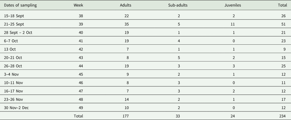
In total, 113 individual lizards (Table 1) were captured 234 times throughout the study. Each individual lizard was captured on average 2 times over the course of the study, with 51.3% of lizards being captured more than once, and a maximum of 9 recaptures per lizard. All lizards were examined for ectoparasites, while 19 faecal samples were collected opportunistically from 17 lizards over the course of the study.
Summary of the number of individual King's skinks captured over the course of the study, with the number of adults, sub-adults and juveniles captured, both overall, and in each week of the study

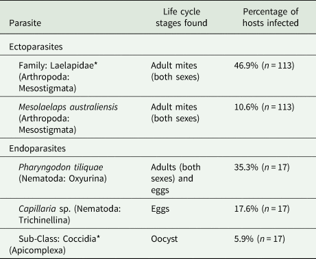
Two ectoparasites and three intestinal parasites were found in this study (Table 2). Additionally, two different mite eggs and one adult cestode were found in the faecal samples but we were not able to classify them further. An undescribed mite species from the family Laelapidae (hereafter termed ‘laelapid mite’) was the most common parasite found in this study, followed by the nematode Pharyngodon tiliquae (Table 2). All other parasites were detected in less than 20% of individuals. Mite loads were generally low, with an average of 2.9 laelapid mites per infested individual (range = 1–10), and 1.3 M. australiensis mites per infested individual (range = 1–2).
Summary of the parasites identified from King's skinks on Penguin Island, including their taxonomy (to the lowest possible taxonomic level), the life cycle stages that were found and the percentage of individual hosts that the parasite was detected in (with an individual host classified as ‘infected’ if the parasite was detected in that host at any point in the study)

Note that two parasites (denoted with an asterisk *) could not be identified to genus level.
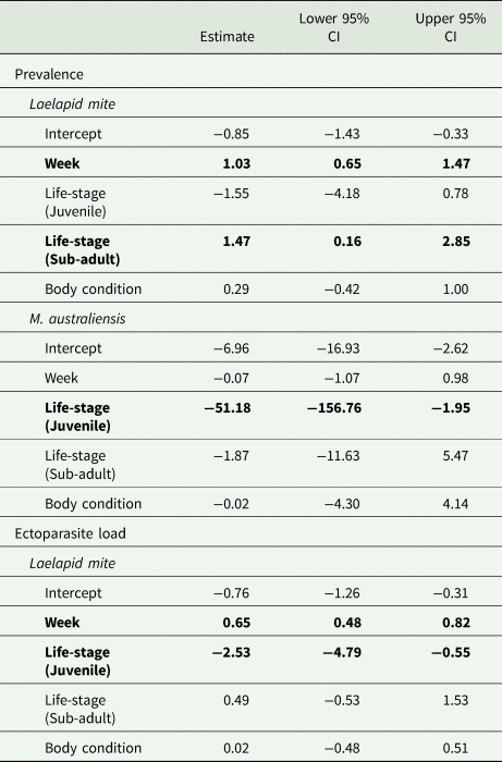
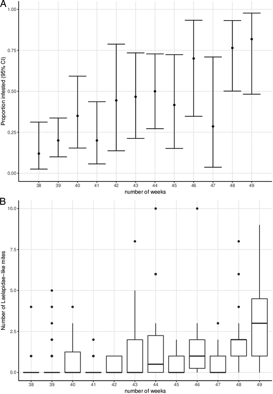
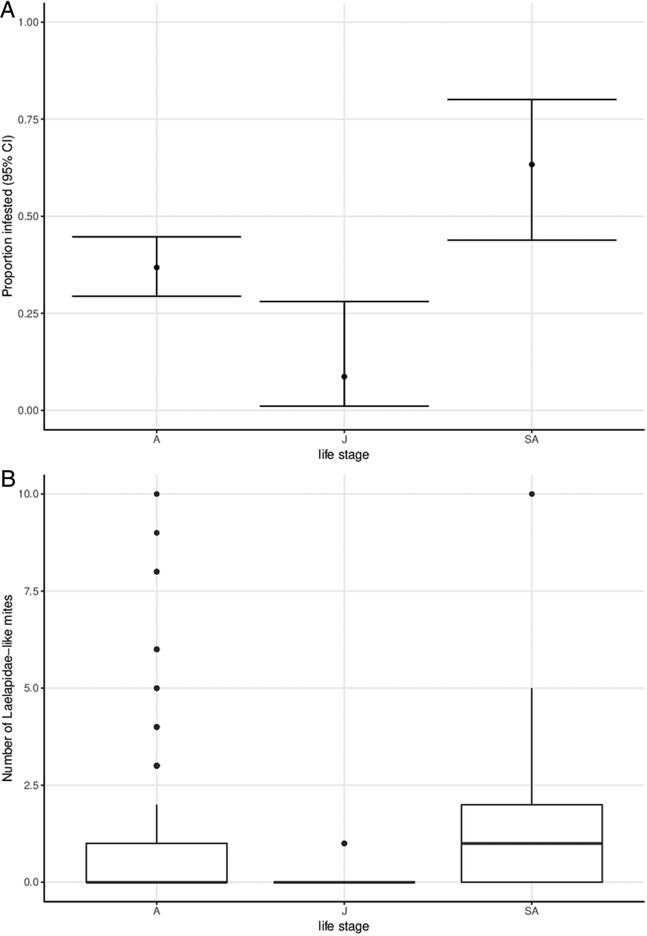
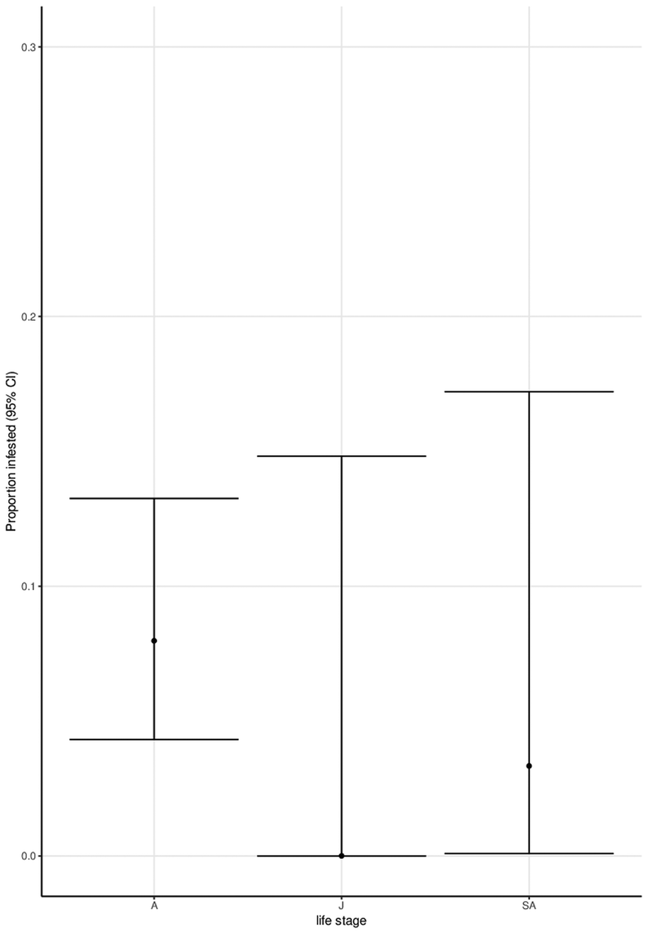
The prevalence and intensity of laelapid mites varied with the week of sampling and the life-stage of the lizard (Table 3). Both the prevalence of laelapid mites (Fig. 1a) and the intensity infesting each lizard (Fig. 1b) increased with time. Sub-adults were more frequently infested by laelapid mites than adults or juveniles (Fig. 2a), and juveniles were infested with fewer mites than adults or sub-adults (Fig. 2b). Mesolaelaps australiensis mites were less common in juveniles than adults (Table 3, Fig. 3). No other factors were associated with patterns of M. australiensis prevalence (Table 3).
A summary of the model estimates from a Bayesian generalized linear mixed model, examining the effects of the week (as a measurement of time), lizard life-stage (adult is the reference category) and body condition on the likelihood of infection with (a) laelapid mites, (b) Mesolaelaps australiensis, and (c) on the number of laelapid mites found infesting King's skinks

Estimates for continuous predictor variables (week and body condition) are standardized and are provided alongside the upper and lower 95% credible intervals. Factors are bolded when the 95% credible intervals exclude zero.
(a) The proportion of King's skinks infested with laelapid mites in each week of the study, with 95% confidence intervals (exact) and (b) boxplots of the number of laelapid mites infesting lizards in each week of the study.

(a) the proportion of King's skinks infested with laelapid mites for each life-stage (A = adults, J = juveniles, SA = sub-adults), with 95% confidence intervals (exact) and (b) boxplots of the number of laelapid mites infesting lizards for each life-stage.

The proportion of King's skinks infested with Mesolaelaps australiensis mites for each life-stage (A = adults, J = juveniles, SA = sub-adults), with 95% confidence intervals (exact).

Among the intestinal parasites, there was only a sufficient sample of infected individuals to examine infection patterns for P. tiliquae. A Pearson's chi-square test revealed that there was a trend (X 2 = 5.4, df = 2, P = 0.07) for juveniles (1 of 2 individuals infected) and sub-adults (3 of 4 individuals infected) to be more commonly infected than adults (2 of 13 individuals infected).
Discussion
This study is the first to describe the parasites infecting a King's skink population. At least five different parasite species were detected in this study, including two ecto-parasitic mites, two nematodes and a gastrointestinal protozoan. This study builds on a previous study (Yang et al., Reference Yang, Brice, Bennett, Eliott and Ryan2013) that examined the parasites infecting a single individual King's skink that had been brought into a wildlife rehabilitation centre. The current study demonstrates that King's skinks are host to a number of parasites on Penguin Island, although it is worth noting this is likely an underestimate since blood samples were not collected in the present study. Similarly, detecting parasites in the feces may not detect all gastrointestinal parasites present in the host, since parasites may be present but not detected if they are not producing eggs.
Two different mite species were found in this study; both from the family Laelapidae. The family Laelapidae is the most morphologically and ecologically diverse group of Mesostigmata and consists of a large number of bloodsucking mites. Most have been found on Australian mammals and only M. australiensis has been recorded on lizards (Domrow, Reference Domrow1988). The laelapid mite discovered in this study was the most abundant, but because it was not classified to genus or species level, it is not possible to predict what other host species it is found on. Mesolaelaps australiensis mites have a wide host range and have been found on Egernia major (Domrow, Reference Domrow1980, Reference Domrow1988) where they were found inside the ear canal, as we found in this study. The fact that this parasite now has been found on two Egernia species and also on the little penguin Eudyptula minor (Domrow, Reference Domrow1988) raises questions about the possible transmission between penguins and King's skinks that coexist on Penguin Island. We know that King's skinks prey on the eggs of little penguins on Penguin Island (Meathrel and Klomp, Reference Meathrel and Klomp1990). During the present study, they were spotted many times sharing the nests of the penguins, making penguin nests an obvious transmission location between the two species.
For both mites, adults of both sexes were found in the ears of the lizards. Very little is known of the lifecycle of Laelapid mites, but typically consists of an egg, hexapod non-feeding larva, and then octopod, protonymph, and deutonymphs, before the adult stage (Radovsky, Reference Radovsky and Houck1994). The fact we only observed adult mites of either species on King's skink could reflect seasonal patterns in the lifecycle of the mites, or that immature stages of the mites occur on other host species. Alternatively, it is possible small immature stages were not visible if they were hiding beneath scales.
Laelapid mites were the most common in the King's skink population, and showed a significant temporal pattern in likely occurrence, increasing in prevalence and abundance over the course of the study. Since mites were removed on each capture to enable identification, the increase in mites in the study reflects an increase in new individual mites entering the lizard population (not just recounting the same individual mites). Mesostigmatid mite lifecycles are often affected by temperature and humidity (Ganjisaffar et al., Reference Ganjisaffar, Fathipour and Kamali2011; Li et al., Reference Li, Huang, Wang and Zhang2015), where warmer temperatures and higher humidity favours faster development in Dermanyssus gallinae up to a point where extreme temperatures or low humidity is lethal (e.g. Nordenfors et al., Reference Nordenfors, Höglund and Uggla1999). Thus, the increase in parasitism observed over the course of our study could reflect changes in development or activity as the temperature range increased during the study towards summer. Alternatively, it could reflect an increase in activity of the lizard host, as has been suggested in other lizard-ectoparasite systems where seasonal increases in parasitism correspond with an increase in lizard activity (e.g. Eisen et al., Reference Eisen, Eisen and Lane2001). As the study was undertaken over approximately 3 months, we do not know whether this increase is sustained over the summer months. Similar temporal patterns were not observed for M. australiensis, although the number of lizards infested was much lower.
Mite infection patterns also varied with lizard life-stage. For the laelapid mites, we found an unusual pattern where sub-adults were more commonly infested than adults or juveniles, and sub-adults and adults were infested by more mites than juveniles. It is possible that sub-adult lizards are more mobile, and perhaps pick up more mites by ranging over a larger area than juveniles or adults. Similar explanations have been suggested to explain differences in ectoparasitism between male and female lizards (Pollock et al., Reference Pollock, Vredevoe and Taylor2012), and relationships between home range size and blood parasite infection (vectored by ticks) have been suggested to relate to increased movement exposing hosts more to ectoparasites in the off-host environment (Bouma et al., Reference Bouma, Smallridge, Bull and Komdeur2007). Mesolaelaps australiensis mites showed a different pattern with respect to life-stage, with adults having a higher prevalence of M. australiensis than juveniles or sub-adults. We found no evidence for a relationship between the body condition of the lizards and infection with either mite species.
Gastrointestinal parasites were less common in the King's skink population, with the nematode P. tiliquae being the most commonly detected. This nematode appears to be specific to reptiles, but has a wide host range, infecting several other species from the Egerniinae lineage of skinks (Thapar, Reference Thapar1925; Adamson, Reference Adamson1984; Hallas et al., Reference Hallas, Bull and Bursey2005). Pharyngodon tiliquae is common in lizards but has not previously been observed in south-western Australia. Unfortunately, our sample size was small, but there was a trend for the higher prevalence in juvenile and sub-adult lizards compared to adults. Capillaria sp. and a coccidian were also detected, but at relatively low levels (<20%) in the population.
This study makes a substantial contribution to our understanding of the host-parasite ecology of the King's skink on Penguin Island. We found that mites are commonly found on these lizards, and a number of other gastrointestinal parasites are less frequently found. This study provides a new host record for both M. australiensis and P. tiliquae, and further demonstrates the presence of coccidian parasites in King's skinks, building on work by Yang et al. (Reference Yang, Brice, Bennett, Eliott and Ryan2013). Given that these lizards are thought to be group-living, this study provides important insights into their parasite fauna, which may have implications for understanding what costs parasites pose to the evolution of group-living in this system.
Acknowledgements
We would like to thank James Barr for assistance with fieldwork, Bruce Halliday who provided advice on mite taxonomy, Aileen Elliot for assistance with laboratory work, and Hugh Jones for help on endoparasite taxonomy. We would like to thank Perth Wildlife Encounters for free transport to Penguin Island, and Erin Clitheroe and staff at the Shoalwater Islands Marine Park (Department of Biodiversity, Conservation and Attractions, Western Australia) for their support of our research and allowing us to use the research quarters on the island. We thank two anonymous reviewers for their constructive feedback of the manuscript.
Financial support
This research was supported by the Murdoch University Small Grant Scheme (awarded to SSG).
Conflict of interest
None
Ethical standards
This research was approved by the Murdoch University Animal Ethics Committee (permit number RW2708/14) and conducted under a Regulation 17 Licence to Take Fauna for Scientific Purposes (licence number SF010261) and a Regulation 4 Written Notice of Lawful Authority (CE004810) issued by the Department of Biodiversity, Conservation and Attractions.