INTRODUCTION
Chagas disease is a chronic illness caused by the flagellate protozoan Trypanosoma cruzi that constitutes a major health problem in the Americas, where about 20 million cases have been reported and an estimated total of 40 million people are at risk of becoming infected. The acute infection is characterized by patent parasite burden and several alterations including intense polyclonal activation of lymphocytes, immune suppression (Minoprio, 2003) and transient thymic aplasia (Taliaferro and Pizzi, 1955; Savino et al. 1989) among others.
In recent years, great effort has focused on the characterization of molecules involved in host cell attachment/invasion by the parasite and in the development of the Chagas disease-associated physiopathology. One representative example is the trans-sialidase (TS), an enzyme expressed by the infective stage that circumvents the parasite inability to perform sialic acid synthesis de novo (Frasch, 2000). The biological relevance of the enzyme activity during the infection can be dissected in (a) that exerted on the parasite surface and/or its close vicinity during the cell-invasion process and (b) the bloodstream disseminated enzyme that acts on the immune system cells (Leguizamón et al. 1999; Mucci et al. 2002). The activity of TS on the cell surface catalyses the transference of sialyl residues from host glycoconjugates to glycosylated molecules, mainly mucins (Acosta-Serrano et al. 2001). In this way, TS enables the parasite to avoid its destruction by serum factors (Tomlinson et al. 1994; Pereira-Chioccola et al. 2000) and also generates sialylated epitopes critically required for the mammalian cell invasion (Schenkman et al. 1992). The enzyme is also involved in release of the parasite from the parasitophorous vacuole (Hall et al. 1992; Rubin-de-Celis et al. 2006). The infective trypomastigote stage does not only express TS on its surface but also releases it into the milieu, leading to its systemic distribution in the bloodstream where it persists due to its association to the repetitive antigen SAPA (Buscaglia et al. 1999; Alvarez et al. 2004). The intravenous administration of active TS to naïve mice induces alterations on the immune system organs mimicking the damage observed during the infection (Leguizamón et al. 1999; Mucci et al. 2002). These abnormalities are derived from the modification of the cell surface pattern by the acquisition of sialyl residues (Mucci et al. 2006).
The 2 main T. cruzi populations show a differential expression of TS activity (Risso et al. 2004). The virulent T. cruzi II parasite strains shed higher amounts of TS that correlates with severe damage to the immune system (Risso et al. 2004). In contrast, lower TS activity is found in the low virulent T. cruzi I strains and no circulating TS activity is detected during the infection; this correlates with the induction of only slight and transient alterations (Risso et al. 2004). TS-neutralizing antibodies are persistently found throughout the chronic phase of the infection (Leguizamón et al. 1994, 1997; Pereira-Chioccola et al. 1994) and seem to circumscribe the enzyme activity in blood to the early steps of the infection both in patients and mice. The antibody-mediated neutralization of the systemic TS activity shed by the highly virulent T. cruzi strains was studied here through the passive transfer of a monoclonal neutralizing antibody (mNtAb) in extreme infection models. Results stress the protective role of these antibodies in preserving the immune system organs from damage during infection and define the activity of a virulence factor from a complex pathogen.
MATERIALS AND METHODS
Mice
Male C3H/HeN, BALB/c and NIH nu/nu mice (60-day-old) were used throughout. Local animal experimentation guidelines were followed.
Trypanosoma cruzi parasites
The highly virulent Tulahuen and RA strains and the Tulahuen-derived clone Q501/3 were used. All the isolates were genotyped as T. cruzi II (Risso et al. 2004) and maintained by serial passages in mice.
In vivo infection procedures
Fifty bloodstream trypomastigotes in PBS-2% (v/v) bovine serum albumin were administered i.p. Parasitaemia was determined in blood samples by counting trypomastigotes in a Neubauer haemocytometer. Mortality was checked daily.
Monoclonal antibodies and passive transference assays
A TS-neutralizing IgG2a-secreting 13G9-34 hybridoma was obtained after immunizing mice with a modified TS that was depleted of spurious epitopes (Pitcovsky et al. 2002; Tribulatti et al. 2005) and grown as ascites in nude mice rendering high neutralizing titres (over 1[ratio ]80000) (Leguizamón et al. 1997). As controls, ascitic fluid containing the isotype-matched BI24 monoclonal antibody (mAb) anti-lumazine synthase from Brucella spp or ascitic fluid from Sp2/0 cells were used. All the ascites were obtained from nude mice injected with the corresponding cells. Mice (at least n=5 per group in all experiments) were passively transferred with mNtAb (10 μl) or control ascitic fluid diluted in PBS every 3 days, from day 7 p.i. The continuous mNtAb presence in serum was confirmed by its ability to prevent the TS-catalysed transference of the sialyl residue from sialyllactose to [14C]-lactose as previously described (Leguizamón et al. 1997) until the end of the assays. Non-infected mice were passively transferred following the same schedules as the experimental groups.
Histology
Organs collected at different times p.i. were fixed in 4% paraformaldehyde in PBS and embedded in paraffin. Tissue sections were stained with haematoxylin-eosin (H.E.) by following standard procedures. Five slices (5 μm thick) of each organ were taken randomly and analysed in detail. The number of apoptotic bodies/400X field was determined by using 0·01 mm grid and expressed as the mean of 30 fields. The morphological criteria used to characterize apoptosis were applied as described elsewhere (Cummings et al. 1997; Harmon et al. 1998). The thymus cortex/medulla index (naive mice=2[ratio ]1) was evaluated as previously described (Risso et al. 2004).
Statistical analysis
The Student's t-test was used.
RESULTS
Parasitaemia and mortality analysis after the passive transfer of TS-neutralizing antibodies
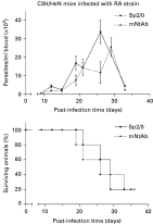
The effect of the mNtAb on systemic TS activity was tested here under extreme experimental conditions. Two parasite strains (RA and Tulahuen) and a Tulahuen-derived clone (Q501/3) belonging to the virulent T. cruzi II lineage (Risso et al. 2004) were used. The BALB/c and C3H/HeN mouse strains that present different susceptibilities to the infection (BALB/c mice being more resistant than C3H/HeN) were assayed. In all cases, an inoculum of 50 bloodstream trypomastigotes induced high values of parasitaemia and mortality (Fig. 1). The mNtAb was passively transferred from day 7 after parasite inoculation, that is, when the infection is established but circulating parasites are still rare. The evolution of parasitaemia and mortality was evaluated. High parasitaemia values (about 4×10⁶ trypomastigotes/ml) were reached by day 20–25 p.i. both in control and mNtAb-transferred C3H/HeN mice when infected with the Q501/3 clone, and no animals survived after 32 (control) or 35 (transferred) days p.i. (not shown). Passively mNtAb transferred C3H/HeN mice that were infected with the RA strain showed a time delay of only 4 days of the parasitaemia peak when compared with control animals (Fig. 1); no differences in mortality rates were found. BALB/c mice infected with the Tulahuen strain and transferred with the mNtAb showed slightly lower parasitaemia values than controls, this was not observed when infected with the Q501/3 clone (not shown). In both BALB/c infection models, parasite levels reached values around 6×10⁶ trypomastigotes/ml by day 25 p.i. and mortality did not show significant differences between mNtAb-transferred and control infected mice (not shown).

Fig. 1. Effect of passive transfer of the mNtAb of TS on Trypanosoma cruzi infection. C3H/HeN male mice were intraperitoneally infected with 50 bloodstream parasites of the RA strain and passively transferred with the mAb from day 7 pi. (A) Parasitaemia. (B) Mortality follow-up. Filled line: animals transferred with ascitic fluid from Sp2/0 cells (n=5). Dotted line: animals transferred with ascitic fluid from 13G9-34 hybridoma mNtAb-secreting cells (n=5).
T. cruzi-induced immune system damage is prevented by the passive transfer of mNtAb
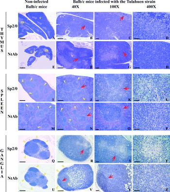
Results from the previous section show the extreme virulence of the models chosen to analyse the effect of the systemic inactivation of TS activity by the passive transference of mNtAb. In these models the absence of protection revealed by similar mortality and parasitaemia is not an unexpected phenomenon since only 1 parasite molecule is blocked. However, the prominent role of TS in the pathogenesis of the infection was highlighted after the histological studies described below which provided clear evidence of immune system tissue preservation by the passive transference of the mNtAb (Figs 2, 3 and Table 1). BALB/c mice were infected with the Tulahuen strain or its derived clone Q501/3 and C3H/HeN animals with the RA strain or Q501/3 clone. In all cases animals were passively transferred with either the mNtAb or the control monoclonal antibodies or ascites. No abnormalities were observed in organs from transferred non-infected control mice.

Fig. 2. mNtAb protects BALB/c mice from Tulahuen strain histological damage. Red arrow indicates successive magnification areas. Thymus. Sp2/0 transferred mice: (B, C) Thymic cortex reduction. (D) Detail showing loss of cellulariry. mNtAb transferred mice: (F, G) preservation of cortex/medulla index. (H) Preservation of histo-architecture organization and cellularity. c, cortex; m, medulla. Spleen. Sp2/0 transferred mice: (J, K) follicular splenic disorganization (yellow arrow) and (L) severe apoptotic level. mNtAb transferred mice: (N) conserved follicular organization (yellow arrow). (O) Detail of preserved follicular architecture. (P) Conserved cellularity within follicles. Ganglia. Sp2/0 transferred mice: (R, S) Severe disorganization with absence of follicles. (T) Detail of cellular depletion. mNtAb transferred mice: (V, Y) Follicular hyperplasia (yellow arrow). (Z) Conserved cellularity within the follicle. (A, E, I, M, Q and U) show non-altered histo-architecture in non-infected transferred BALB/c mice. Yellow arrows indicate normal follicles. Bars: Left and 40X rows: 500 μm; 100X row: 100 μm; 400X row: 50 μm.
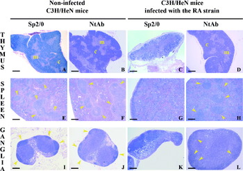
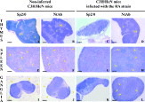
Fig. 3. mNtAb protects C3He/N mice from RA strain histological damage. Thymus. In (C) thymic cortex reduction can be observed in Sp2/0 transferred mice. (D) Shows the preservation of cortex/medulla index in mNtAb transferred mice. c, cortex; m, medulla. Spleen. (G) Shows follicular splenic disorganization in Sp2/0 transferred mice and (H) conserved follicular organization (yellow arrows) in mNtAb transferred mice. Ganglia. (K) Shows severe disorganization with absence of follicles in Sp2/0 transferred mice and (L) follicular hiperplasia (yellow arrows) in mNtAb transferred mice. (A, B, E, F, I and J) show non-altered histo-architecture in non-infected transferred C3H/HeN mice. Yellow arrows indicate normal follicles. Bars: 500 μm.
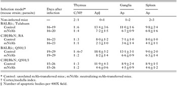
Table 1. Prevention of damage by mNtAb during the infection of BALB/c and C3H/HeN mice with Trypanosoma cruzi II (C/M: cortex/medulla index. Ap: apoptotic bodies per 400X field. For all apoptosis quantification comparisons P<0·001 between neutralizing antibodies groups and their corresponding controls.)

The spleens from infected mice transferred with unrelated antibodies, showed deep disorganization in the tissue architecture. The white pulp was diminished mainly due to the reduction of the B cell areas, where a high number of apoptotic bodies was detected (Figs 2, 3 and Table 1). The T cell peri-arteriolar lymphoid sheath architecture also was altered. Few secondary follicles that always displayed small germinal centres were observed and, in fact, the follicular histo-architecture was often lost after lymphocyte depletion and intense apoptosis (Table 1 and Figs 2 and 3). Diffuse congestion and intense haemorrhage was observed in the red pulp of the spleens from the infected control animals. The macrophage population was strongly increased. In contrast, in the passively mNtAb-transferred mice, the spleen histology was to a large extent conserved and displayed significant diminution of apoptotic bodies (Table 1). The white pulp was increased mainly due to the expansion of the B cell areas with marked hyperplasia, showing the preservation of the follicular structure (Figs 2 and 3). Increased red cell and macrophage presence also was found in the red pulp.
The lymphatic ganglia from infected control mice displayed a reduction of the B cell areas, where small lymphocytes with dense nuclei were found. Only a few follicles remained and an increase in the macrophage population, both in the medullar and subcapsular sinuses, atrophy of the ganglionar cortex and high apoptotic cell numbers were consistently observed (Table 1 and Fig. 2). The absence of follicles together with a reduction in the celullarity is shown in Figs 2 and 3. In contrast, in the mNtAb-passively transferred animals, the damage observed during the infection was prevented because all these alterations were of lower intensity. Conserved general histo-architecture that correlated with a significant reduction of apoptotic body count was recorded (Table 1). Hyperplasia of B cell areas with follicles containing medium-sized and large lymphocytes, and the development of germinative centres in some areas showing mature lymphocyte transformation were observed (Figs 2 and 3). Subcapsular and medullary oedema, with an increased macrophage population, was also found.
The histology of the thymuses from the infected control groups showed severe disorganization with intense cortical thymocyte apoptosis (Table 1) and marked reduction in the cellularity of the cortex leading to altered cortex/medulla indices as described previously (Risso et al. 2004). General thymus histo-architecture remained conserved in the mNtAb-transferred animals, where cortical cellularity and cortex/medulla index were preserved as a result of the reduction of thymocyte apoptosis (Table 1, Figs 2 and 3).
DISCUSSION
A role for anti-TS immunization in protection against the infection has been proposed mainly from results through DNA vaccination approaches. DNA constructions containing TS-encoding sequences (Costa et al. 1998; Fralish and Tarleton, 2003; Mussalem et al. 2006) or TS-derived CD4 and CD8 T cell epitopes (Fujimura et al. 2001) provided evidence that anti-TS cellular immunity is involved in protection against the infection. Reduction of parasitaemia and mortality in mice have also been described as a result of immunization schemes using DNA encoding members of the TS family (ASP-1, ASP-2 and TSA-1) (Garg and Tarleton, 2002; Vasconcelos et al. 2004) and the co-administration of an interleukin-12 coding gene improved the efficacy against lethal T. cruzi infection (Katae et al. 2002).
In contrast, the role of antibodies induced by TS protein family members in the control of T. cruzi infection in the mouse model seems to be more controversial. Their relevance was highlighted by some authors who reported protection by the passive transfer of Fab' fragments or by the incubation of trypomastigotes with purified TS-neutralizing antibodies before infection (Chuenkova and Pereira, 1995; Villalta et al. 1996). However, immunization approaches with naked DNA revealed protection even in the absence of antibodies as evaluated by survival of infection (Fujimura et al. 2001). It has also been reported that a mAb directed to a sialylated epitope of mucin-like glycoproteins decreased parasitaemia but only upon challenge with attenuated (cell culture-derived) trypomastigotes (Franchin et al. 1997).
Here we analysed the role of the induced humoral TS-neutralizing response on the systemic TS activity by transferring a mNtAb to mice infected with highly virulent parasites, which lead to strong tissue damage and 100% mortality with an inoculum of only 50 bloodstream trypomastigotes. These parasites are representative of the lineage II that sheds higher amounts of TS (Risso et al. 2004). These extreme conditions allowed us to highlight the ability of the antibodies to protect the immune system tissues against the TS-induced damage.
The induction of B cell apoptosis in the experimental infection is known in Chagas disease (Zúñiga et al. 2000). In transferred mice the B cell areas were those predominantly protected both in the ganglia and spleen, whereas in the thymus the cortex area was preserved. Our results were consistently reproduced independently of the mouse genetic backgrounds and parasite strains and similar findings were observed along the course of the infection. These results led us to identify the role of the neutralizing antibodies that are induced and persist along the infection (Leguizamón et al. 1994, 1997). Findings also highlight the activity of the bloodstream-borne enzyme on the immune system as a central role for this virulence factor during the early steps of the infection.
As a general rule, seemingly applicable to this model, the resistance to intracellular pathogens relies mainly on the cellular response, meanwhile the neutralization of the circulating toxins or virulence factors as in this case, finds its base in the humoral immune response. The neutralization of the TS circulating activity by the mNtAb was able to prevent the immune system damage even in the extremely virulent models assayed here. The biological activity of TS related to the sialylation of surface parasite molecules that enhances their infectivity could not be prevented by the mNtAb in the infection nests, as suggested by the high parasitaemia and mortality recorded in both control and transferred groups. This might be due to the high rate of TS shedding by T. cruzi II strains (Risso et al. 2004) together with the inaccessibility of the antibody molecules in sufficient amounts to the replication sites. Moreover, the host cell invasion is a complex multi-step process and therefore several alternative invasion strategies might be displayed (Yoshida, 2003). It is very well known that the survival to T. cruzi infection is due to the immune response both in experimental models and patients. Therefore it is somewhat puzzling to find that the immune system damage prevention, although necessary, is not a sufficient condition to survive the infection. This would suggest that the highly virulent parasite strains overthrow the host defences leading to the early mortality recorded.
The TS, a shed virulence factor designed to persist in the circulation, contains in the catalytic domain a cross-reactive epitope network that delays the elicitation of neutralizing antibodies (Pitcovsky et al. 2001, 2002; Alvarez et al. 2001). During the early infection, this delay might allow depression of the immune system by the TS while the parasite is spreading in the host.
The preservation of the immune system tissue by inactivating a defined systemic virulence factor is a major finding in the context of infections caused by organisms with a high level of complexity.
The authors are indebted to Dr E. Mocetti (Hospital Italiano de Buenos Aires) for helpful suggestions and assistance in preliminary assays and Dr Monica Esteva (Instituto Fatala Chaben. Argentina) for providing the Tulahuen parasite strain. The anti-lumazine synthase ascites was kindly provided by Dr G. Giambartolomei (Universidad de Buenos Aires). The critical reading of the manuscript by Dr C. A. Buscaglia is also appreciated. This work was supported by Agencia Nacional de Promoción Científica y Tecnológica (ANPCyT); Consejo Nacional de Investigaciones Científicas y Técnicas (CONICET); Universidad Nacional de San Martín and Universidad de Buenos Aires, Argentina and United Nations Development Programme/World Bank/World Health Organization Special Program for Research and Training in Tropical Diseases. M.G.R. and T.A.P. are fellows and O.C. and M.S.L. are researchers from the CONICET.