Coronary artery anomalies are rare but potentially serious congenital cardiac lesions. A prospective echocardiographic study on asymptomatic children found either an anomalous origin of the left main coronary artery from the right sinus of Valsalva or an anomalous origin of the right coronary artery from the left sinus of Valsalva in 0.17% of patients. Although such lesions might be detected in asymptomatic children, undiagnosed anomalous coronary arteries are a well-recognised cause of sudden cardiac death.Reference Kaushal, Backer and Popescu 1 – Reference Liberthson 4
Case report
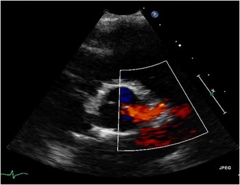
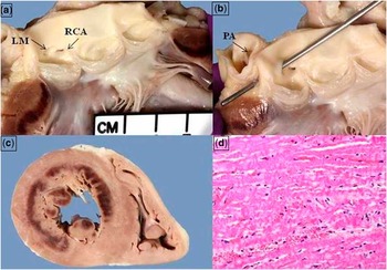
A 5-year-old previously healthy boy presented to an outside hospital with acute-onset abdominal pain and vomiting while playing at summer camp. He experienced syncope and went into a witnessed cardiac arrest – pulseless electrical activity – in the emergency room. Cardiopulmonary resuscitation was immediately commenced and continued for 45 minutes during transfer to our Pediatric Intensive Care Unit for further management. Upon arrival at the intensive care unit, he remained in cardiac arrest, requiring ongoing resuscitation, and was placed on extracorporeal membrane oxygenation. His initial electrocardiograph revealed an idioventricular rhythm. The laboratory tests at admission were significant for a lactate level of 17.0 mmol/L, CK-MB level of 183.9 mcg/L (range 0–7.0), and Troponin level of 7.02 ng/ml (normal range 0–0.059). The transthoracic echocardiogram demonstrated severe global left ventricular dysfunction and apparently normal origins of the coronary arteries (Fig 1). Transoesophageal echocardiography was recommended but was postponed because of patient instability. Unfortunately, within the first few hours, he was observed to have bilateral fixed and dilated pupils, and a computed tomography of the head demonstrated severe global cerebral oedema and evidence of hypoxic ischaemic injury. Given the poor prognosis, the patient's family and physicians decided to withdraw support, and the patient died within 16 hours of presentation. On autopsy, an anomalous origin of the left main coronary artery from the right sinus of Valsalva from a separate slit-like orifice just superior to the right coronary artery orifice – a slightly “high take-off” – was found, with fresh circumferential subendocardial infarction involving the left ventricle and no evidence of an old scar (Fig 2).
The left coronary arteries appear to arise from the left sinus of Valsalva on colour Doppler.

( a ) An anomalous left main coronary artery (LM) arising from the right coronary sinus. ( b ) Probe in LM shows the proximal course between the aorta and pulmonary artery. ( c , d ) Cross-section of the heart and a histological section showing a nearly circumferential reperfusion injury of the left ventricle.

Discussion
Anomalous aortic origin of a coronary artery from the opposite sinus of Valsalva with an interarterial course is a rare congenital anomaly and is the second leading cause of sudden death in older children and young adults after hypertrophic cardiomyopathy.Reference Davis, Cecchin, Jones and Portman 3 – Reference Lytrivi, Wong and Ko 6 Anomalous origin of the left coronary artery from the right sinus of Valsalva has been implicated in numerous case reports and autopsy series in sudden, unexpected cardiac death in young adults, particularly during or immediately after vigorous exercise.Reference Lytrivi, Wong and Ko 6 – Reference Barth and Roberts 9
The pathophysiological mechanism that leads to sudden death in this anomaly is not completely clear; however, the intramural course, acute angle of take-off, systolic stretching, and/or compression of the aberrant coronary between the great arteries during exercise as well as myocardial fibrosis from subclinical ischaemia may lead to ventricular tachycardia and/or fibrillation.Reference Kaushal, Backer and Popescu 1 , Reference Lytrivi, Wong and Ko 6 Most of the sudden deaths from the literature review occurred during or immediately after physical exercise, with as many as 2/3 of the patients being asymptomatic before the final event.Reference Lytrivi, Wong and Ko 6 , Reference Taylor, Byers, Cheitlin and Virmani 10
Improvements in echocardiographic technology and increased vigilance have reduced the rate of false negatives; however, the absence of a “gold standard” by which to measure sensitivity and specificity impairs our ability to judge the technique's success in coronary anomaly diagnosis.Reference Lytrivi, Wong and Ko 6 In many cases, two-dimensional imaging may be misleading in patients with an anomalous aortic origin of a coronary artery that has an intramural course as it can appear to arise normally from the appropriate sinus as it exits the aortic wall. Colour Doppler and multi-plane imaging are particularly useful because these techniques can provide additional information on the direction of flow in the intramural segment. This helps in differentiating whether the anomalous coronary arises from the right or left sinus. We speculate that the relatively high take-off of the left coronary artery from the right coronary sinus in the present case may have made diagnosis by transthoracic echocardiography more difficult. Moreover, the intramural course posteriorly between the aorta and the pulmonary artery may have given the impression that the left coronary artery actually originated from the left sinus when, in fact, this was an illusion created by the intramural course.
Conclusion
We conclude that transoesophageal echocardiographic imaging and possibly other imaging options including computed tomography, magnetic resonance imaging, or coronary angiography should be considered if there is a failure to demonstrate the origin of both proximal left and right coronary arteries from their usual coronary sinuses by transthoracic echocardiography or when there is a high index of clinical suspicion despite apparently normal coronary origins in a young patient with a suspected acute cardiac event.
Acknowledgements
The authors thank Robert Kutys from the cardiovascular pathology department for providing the autopsy images.
Financial Support
This research received no specific grant from any funding agency, commercial or not-for-profit sectors.
Conflicts of Interest
None.