Introduction
The current prevalence of anxiety disorders adds up to 10.4% in Western cultures. Anxiety disorders are among the 10 leading causes of disability worldwide, and among the three most expensive mental disorders in Europe (Olesen et al. Reference Olesen, Gustavsson, Svensson, Wittchen and Jönsson2012; Baxter et al. Reference Baxter, Scott, Vos and Whiteford2013). Features shared by anxiety disorders, such as panic disorder (PD), social anxiety disorder (SAD), or specific phobia, are excessive fear and anticipatory anxiety, physical symptoms, escape and avoidance behaviour, thoughts of current threat, and mental apprehension (Craske et al. Reference Craske, Rauch, Ursano, Prenoveau, Pine and Zinbarg2009). Cues triggering this initial fear response depend on the disorder and are thus disorder-related (American Psychiatric Association, 2000). In the recently published Diagnostic and Statistical Manual of Mental Disorders, 5th revision (DSM-5; American Psychiatric Association, 2013), post-traumatic stress disorder (PTSD) has been classified as a trauma- and stressor-related disorder, distinguishing PTSD from anxiety disorders. As the symptoms for anxiety disorders and trauma- and stressor-related disorders overlap, the classification change of PTSD fuelled intensive discussion (Friedman et al. Reference Friedman, Resick, Bryant, Strain, Horowitz and Spiegel2011; Zoellner et al. Reference Zoellner, Rothbaum and Feeny2011).
Shared behavioural features suggest a dimensional view of anxiety and trauma- and stressor-related disorders. Keeping with recent dimensional approaches such as the Research Domain Criteria (RDoC) project (Cuthbert, Reference Cuthbert2015), studies examining potential transdiagnostic neurobiological alterations that cut across anxiety, and trauma- and stressor-related disorders are needed.
Since the amygdala is linked to fear mechanisms in healthy subjects and in pathological anxiety, it could potentially represent a target structure in a dimensional approach (Rauch et al. Reference Rauch, Shin and Wright2003; Zald, Reference Zald2003; Phelps, Reference Phelps2006; Forster et al. Reference Forster, Novick, Scholl, Watt and Ferry2012). The amygdala is a central processing hub for emotional information. It receives input from sensory processing structures (e.g. thalamus) and higher-order regions (e.g. prefrontal cortex; PFC), and is essential for the detection of relevant emotional information (Phelps & LeDoux, Reference Phelps and LeDoux2005; Sergerie et al. Reference Sergerie, Chochol and Armony2008; Forster et al. Reference Forster, Novick, Scholl, Watt and Ferry2012). The amygdala can be divided into lateral, basal and central components, with the lateral part functioning mainly as an entrance region for sensory information while the central region mediates behavioural and autonomous reactions via various output pathways (LeDoux, Reference LeDoux2007; Janak & Tye, Reference Janak and Tye2015).
Reviews and meta-analyses have drawn an inconsistent picture with regard to amygdala involvement across anxiety disorders during emotional processing (Rauch et al. Reference Rauch, Shin and Wright2003; Etkin & Wager, Reference Etkin and Wager2007; Craske et al. Reference Craske, Rauch, Ursano, Prenoveau, Pine and Zinbarg2009; Shin & Liberzon, Reference Shin and Liberzon2010; Holzschneider & Mulert, Reference Holzschneider and Mulert2011; Fredrikson & Faria, Reference Fredrikson and Faria2013; Duval et al. Reference Duval, Javanbakht and Liberzon2015; Taylor & Whalen, Reference Taylor and Whalen2015). This variable involvement of the amygdala in different anxiety disorders is not well understood. In general, more consistent amygdala activations are reported for SAD and specific phobia and partially PTSD as compared with PD, for example (Etkin & Wager, Reference Etkin and Wager2007; Shin & Liberzon, Reference Shin and Liberzon2010; Duval et al. Reference Duval, Javanbakht and Liberzon2015). However, since designs are only partially comparable, findings are difficult to interpret. Meta-analyses have not yet included PD (Etkin & Wager, Reference Etkin and Wager2007; Fredrikson & Faria, Reference Fredrikson and Faria2013). Furthermore, it is unknown whether subregions of the amygdala are differentially involved when comparing effects across anxiety disorders.
Besides the amygdala, altered activation in anxiety disorders has been observed in the insula, thalamus, anterior cingulate cortex (ACC) and medial PFC (mPFC) (Etkin & Wager, Reference Etkin and Wager2007; Shin & Liberzon, Reference Shin and Liberzon2010; Duval et al. Reference Duval, Javanbakht and Liberzon2015). While reviews and meta-analyses propose a set of brain regions involved in anxiety disorders, findings within each brain region vary between studies. Further, conclusions are limited since they include studies with different tasks, designs, sample characteristics and stimuli that are only partially disorder-relevant. This calls for studies across multiple anxiety disorders that use comparable stimuli, tasks and designs, while controlling for sampling confounds (Etkin & Wager, Reference Etkin and Wager2007; Craske et al. Reference Craske, Rauch, Ursano, Prenoveau, Pine and Zinbarg2009; Duval et al. Reference Duval, Javanbakht and Liberzon2015).
Some studies related to this research question directly compared the neural correlates across different anxiety disorders, using facial expressions (Blair et al. Reference Blair, Shaywitz, Smith, Rhodes, Geraci, Jones, McCaffrey, Vythilingam, Finger, Mondillo, Jacobs, Charney, Blair, Drevets and Pine2008; Killgore et al. Reference Killgore, Britton, Schwab, Price, Weiner, Gold, Rosso, Simon, Pollack and Rauch2014; Pantazatos et al. Reference Pantazatos, Talati, Schneier and Hirsch2014; Fonzo et al. Reference Fonzo, Ramsawh, Flagan, Sullivan, Letamendi, Simmons, Paulus and Stein2015). Both differential amygdala activation [Blair et al. Reference Blair, Shaywitz, Smith, Rhodes, Geraci, Jones, McCaffrey, Vythilingam, Finger, Mondillo, Jacobs, Charney, Blair, Drevets and Pine2008: generalized anxiety disorder (GAD), SAD] and similar amygdala activation across groups (Killgore et al. Reference Killgore, Britton, Schwab, Price, Weiner, Gold, Rosso, Simon, Pollack and Rauch2014: PTSD, PD and specific phobia; Fonzo et al. Reference Fonzo, Ramsawh, Flagan, Sullivan, Letamendi, Simmons, Paulus and Stein2015: GAD, SAD and PD) have been found. General threat-processing studies implementing emotional pictures have not yet compared more than two anxiety disorders. SAD patients and healthy controls (HC) have been found to show greater amygdala responses than GAD patients when confronted with negative pictures (Blair et al. Reference Blair, Geraci, Smith, Hollon, DeVido, Otero, Blair and Pine2012). No differential amygdala activation to negative pictures was found in GAD v. PD patients (Ball et al. Reference Ball, Ramsawh, Campbell-Sills, Paulus and Stein2013). Only one study used disorder-related stimuli (words) with three different anxiety disorders (van den Heuvel et al. Reference van den Heuvel, Veltman, Groenewegen, Witter, Merkelbach, Cath, van Balkom, van Oppen and van Dyck2005). Processing disorder-related words resulted in heightened activation in the right amygdala in obsessive–compulsive disorder and PD patients compared with HC.
Taken together, there are no studies that directly compared neural responses to ecologically valid disorder-specific triggers of anxiety across anxiety disorders. By means of event-related functional magnetic resonance imaging (fMRI), we investigated common and distinct neural correlates of disorder-related scene processing in PD, SAD, dental phobia (DP) and PTSD patients. We used separate, tailor-made stimulus sets displaying situations specifically relating to the core fears of each disorder.
The present study should be able to detect transdiagnostic neurobiological alterations that may cut across anxiety, and trauma- and stressor-related disorders due to high statistical power. Statistical power was increased by a large patient sample, which makes the detection of true effects across diagnoses more likely (Rauch et al. Reference Rauch, Shin and Wright2003; Button et al. Reference Button, Ioannidis, Mokrysz, Nosek, Flint, Robinson and Munafò2013). To additionally increase internal validity, all patients were free of psychiatric medication, and patients with any of the other three disorders as a co-morbid condition were excluded. Based on this methodological stringency we expected to better understand common and diverging brain responses in the amygdala and other brain regions across the four disorders.
Method
Subjects
Patients suffering from PD, SAD, DP or PTSD were recruited via public notices, local paper advertisements and a collaborating out-patient clinic. All PTSD patients suffered from psychopathology after interpersonal violence. HC were drawn from a larger number of screened healthy controls ascertained within the framework of the Collaborative Research Center ‘Fear, Anxiety, Anxiety Disorders’ (TRR SFB 58; http://sfbtrr58.uni-muenster.de/) or were recruited by means of flyers and newspaper advertisements.
Prior to participation, all patients and HC were interviewed by a psychologist using the Structured Clinical Interview for DSM-IV Axis I Disorders (SCID; German version, Wittchen et al. Reference Wittchen, Wunderlich, Gruschwitz and Zaudig1997). Patients fulfilled the criteria of PD, SAD, DP or PTSD according to DSM-IV as main diagnosis and were excluded if they had any of the other investigated disorders as a co-morbid condition (for other co-morbid conditions, see online Supplementary Table S1). HC were free of any psychiatric diagnosis. Further exclusion criteria for patients and HC were psychotropic medication, presence or history of neurological, psychotic or bipolar disorders, drug dependence or abuse within the last 10 years, suicidal ideations, and fMRI contraindications. All participants had normal or corrected-to-normal vision and were right-handed as assessed with the Edinburgh Handedness Inventory (Oldfield, Reference Oldfield1971).
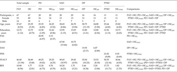
The final patient sample comprised 20 PD patients (16 female), 20 SAD patients (13 female), 16 DP patients (13 female) and 11 PTSD patients (all females). All patients were part of larger disorder-specific studies, with less restrictive sample requirements (Feldker et al. Reference Feldker, Heitmann, Neumeister, Bruchmann, Vibrans, Zwitserlood and Straube2016; Heitmann et al. Reference Heitmann, Feldker, Neumeister, Zepp, Peterburs, Zwitserlood and Straube2016; Neumeister et al. Reference Neumeister, Feldker, Heitmann, Helmich, Gathmann, Becker and Straubein press). In the present study, a same-size HC group, matched for age, gender and education was assigned to each patient group (see Table 1 for sample characteristics), resulting in a specific control group for each patient group. All subjects gave written informed consent. The study conforms to the Declaration of Helsinki and was approved by the ethics committee of the University of Muenster.
Sample characteristics of patient groups and their respective HC groups regarding gender ratio, age, educational attainment and scores in clinical questionnaires a

Data are given as mean (standard deviation) unless otherwise indicated.
HC, Healthy controls; PD, panic disorder patients; SAD, social anxiety disorder patients; DP, dental phobia patients; PTSD, post-traumatic stress disorder patients; PAT, all patients; PAS, Panic and Agoraphobic Scale (Bandelow, Reference Bandelow1997); LSAS, Liebowitz Social Anxiety Scale (Stangier & Heidenreich, Reference Stangier and Heidenreich2005); DAS, Dental Anxiety Scale (Corah, Reference Corah1969); PDS, Post-traumatic Diagnostic Scale (Foa et al. Reference Foa, Cashman, Jaycox and Perry1997); STAI-T, State-Trait-Anxiety-Inventory – Trait version (Laux et al. Reference Laux, Glanzmann, Schaffner and Spielberger1981); BDI, Beck Depression Inventory (Hautzinger et al. Reference Hautzinger, Bailer, Worall and Keller1995).
a PAS, BDI and DAS scores were missing of one HCDP: the clinical interview and screening procedure did not reveal any clinical symptoms. Comparisons: gender differences were calculated by χ2 tests. Other differences were tested with t tests. For demographic data, p < 0.05, no Bonferroni correction was applied in order not to miss possible differences between groups. For questionnaire data, p < 0.05, Bonferroni corrected at p < 0.005. As expected, symptom severity scores of each patient group differed significantly from scores in the respective HC group. Average symptom severity in each patient group was mild to severe as categorized in questionnaires’ manuals (Corah, Reference Corah1969; Bandelow, Reference Bandelow1997; Foa et al. Reference Foa, Cashman, Jaycox and Perry1997; Heimberg et al. Reference Heimberg, Horner, Juster, Safren, Brown, Schneier and Liebowitz1999).
Stimuli
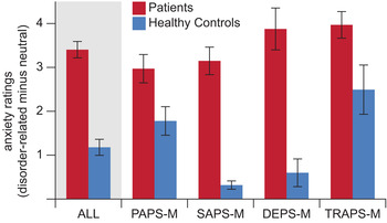
A tailor-made set of 50 disorder-related and 50 neutral scenes was presented to each patient group and the respective HC group, resulting in four different picture sets. Disorder-related scenes tailored to the particular disorders depicted disorder-related feared situations or cues as well as persons suffering from disorder-related symptoms (e.g. close-up of dental treatment procedure for DP). Detailed descriptions of the sets can be found elsewhere: Social Anxiety Picture Set Muenster (SAPS-M; Heitmann et al. Reference Heitmann, Feldker, Neumeister, Zepp, Peterburs, Zwitserlood and Straube2016); Panic-related Picture Set Muenster (PAPS-M; Feldker et al. Reference Feldker, Heitmann, Neumeister, Bruchmann, Vibrans, Zwitserlood and Straube2016); Trauma-related Affective Picture Set Muenster (TRAPS-M; Neumeister et al. Reference Neumeister, Feldker, Heitmann, Helmich, Gathmann, Becker and Straubein press). Similarly, the Dental Phobia Picture Set Muenster (DEPS-M) was developed (for further details, see online Supplementary Table S2). Each patient group and the respective HC group rated all 100 scenes of the respective disorder-related picture set in a separate post-scanning rating session. Each picture was presented for 2 s on a computer screen. A nine-point Likert scale was used to assess valence (1 = very unpleasant, 5 = neutral to 9 = very pleasant), arousal (1 = not arousing to 9 = very arousing) and anxiety (1 = not anxiety inducing to 9 = very anxiety inducing). Each patient group in the current sample rated the disorder-related v. neutral scenes as more anxiety-inducing than the respective HC group. Patient groups did not differ from each other regarding scores in anxiety rating (see Fig. 1 and online Supplementary Table S3), indicating a comparable degree of experienced threat.
Anxiety ratings (disorder-related minus neutral) for patients and healthy controls across picture sets (ALL) and for each set: Panic-related Picture Set Muenster (PAPS-M); Social Anxiety Picture Set Muenster (SAPS-M); Dental Phobia Picture Set Muenster (DEPS-M); Trauma-related Affective Picture Set Muenster (TRAPS-M). Values are means, with standard errors represented by vertical bars. For a colour figure, see the online version.

Experimental task
All subjects participated in the same fMRI experiment, but, as described before, the presented stimulus set depended on the specific disorder. One patient group (e.g. PD) and their respective HC group saw the same stimulus set (here PAPS-M). Another patient group (e.g. SAD) and their respective HC group saw another stimulus set relevant for the disorder (here SAPS-M). During the 8 min 19 s functional run, each of the 50 disorder-related and 50 neutral pictures was presented once in an event-related design. Neutral and disorder-related pictures were presented in a random sequence, optimized and counterbalanced using the Optseq algorithm (http://www.surfer.nmr.mgh.harvard.edu/optseq/), which also provides temporal jitter to increase signal discriminability (Dale et al. Reference Dale, Greve and Burock1999). Each picture was presented for 800 ms. A white fixation cross was presented between two stimuli, for a jittered time window of 1280–15 320 ms (mean = 3915 ms). To keep participants’ attention and gaze towards the stimulus, they were instructed to press a button with their right index finger whenever a blurred picture occurred. Five pictures [originally EmoPicS (Wessa et al. Reference Wessa, Kanske, Neumeister, Bode, Heissler and Schönfelder2010), blurred with Adobe Photoshop CS6 (version 13.0.1, Adobe Sytems Inc., USA)] were randomly presented over the course of the experiment. These five trials were modelled as nuisance regressors in the fMRI analysis.
fMRI acquisition and analysis
Blood oxygenation level-dependent (BOLD) responses and structural brain information were recorded using a 3 Tesla magnetic resonance scanner (‘Magnetom PRISMA’; Siemens, Germany) and a 20-channel Siemens Head Matrix Coil. A high-resolution T1-weighted MPRAGE anatomical volume with 192 slices was recorded for anatomical localization. For functional data a run of 255 volumes was conducted using a T2*-weighted echo-planar sequence (echo time = 30 ms, flip angle = 90°, matrix = 92 × 92 voxels, field of view = 208 mm2, repetition time = 2080 ms). Each volume consisted of 36 axial slices (thickness = 3 mm, gap = 0.3 mm, in plane resolution = 2.26 mm × 2.26 mm). To minimize susceptibility artifacts in inferior parts of anterior brain areas, the volumes were tilted approximately 20° from the anterior commissure/posterior commissure line. A shimming field was applied before functional imaging to further reduce external magnetic field inhomogeneities. fMRI data were pre-processed and analysed with BrainVoyager QX software (version 2.4; Brain Innovation, The Netherlands) and Matlab (version 8.2, The MathWorks Inc., USA). The first 10 volumes of each run were discarded from analysis to ensure steady-state tissue magnetization. First, all volumes were realigned to the first volume, to minimize artifacts due to head movements. No participant showed excessive head movement (>1 voxel). Further data pre-processing steps comprised spatial (6 mm full-width half-maximum isotropic Gaussian kernel) as well as temporal smoothing (high-pass filter: 10 cycles in time course; low-pass filter: 2.8 s; linear trend removal). The anatomical and functional images were co-registered and normalized to Talairach space (Talairach & Tournoux, Reference Talairach and Tournoux1988). Volumes were resampled to a voxel size of 2 × 2 × 2 mm, and slice-time correction was applied. Multiple linear regressions modelling the signal time course at each voxel were calculated with adjustment for autocorrelation. The expected BOLD signal change for each predictor was modelled with a canonical double γ haemodynamic response function. Predictors of interest were the two stimulus types: disorder-related and neutral scene. In the first step, voxelwise statistical maps were generated and the relevant planned contrasts of predictor estimates (β weights) were computed for each individual. β Maps for differential activation (disorder-related minus neutral) per person were then exported to Matlab. In the latter step, a group analysis of these individual contrasts was performed.
The region of interest was the bilateral amygdala (1 mm dilated), for which local information was derived from the Automated Anatomical Labeling atlas included in the Wake Forest University pick atlas (Tzourio-Mazoyer et al. Reference Tzourio-Mazoyer, Landeau, Papathanassiou, Crivello, Etard, Delcroix, Mazoyer and Joliot2002; Maldjian et al. Reference Maldjian, Laurienti, Kraft and Burdette2003). The obtained Montreal Neurological Institute (MNI) coordinates were converted to Talairach space in Matlab using the ICBM-152 routine proposed by Lancaster et al. (Reference Lancaster, Tordesillas-Gutiérrez, Martinez, Salinas, Evans, Zilles, Mazziotta and Fox2007). Obtained peak coordinates were verified with the Mai atlas (Mai et al. Reference Mai, Assheur and Paxinos2004). An a priori-defined whole-brain mask was used to mask out non-brain tissue. The watershed algorithm of Neuroelf (v0.9c; http://neuroelf.net/; i.e. the splitclustercoords function) was used to assess local maxima of clusters.
For statistical analyses, a cluster-based permutation (CBP) approach was used, as often suggested (Bullmore et al. Reference Bullmore, Suckling, Overmeyer, Rabe-Hesketh, Taylor and Brammer1999; Hayasaka & Nichols, Reference Hayasaka and Nichols2004; Maris & Oostenveld, Reference Maris and Oostenveld2007; Kriegeskorte et al. Reference Kriegeskorte, Simmons, Bellgowan and Baker2009; Eklund et al. Reference Eklund, Nichols and Knutsson2016). CBP approaches require no assumptions about the test statistic distribution and have recently been shown to be more valid than classical parametric fMRI analyses, and offer precise control of the false discovery rate (Eklund et al. Reference Eklund, Nichols and Knutsson2016). In the first step, we investigated the group effect (all patients v. all HC) across disorders using the differential β values (disorder-related v. neutral pictures). In the second step, differential effects were investigated by pairwise group (patients, HC) x stimulus set (PAPS-M, SAPS-M, DEPS-M, TRAPS-M) interactions. The cluster-level α was Bonferroni-corrected to adjust for repeated comparisons.
All permutation tests were performed with 1000 permutations (Bullmore et al. Reference Bullmore, Suckling, Overmeyer, Rabe-Hesketh, Taylor and Brammer1999; Maris & Oostenveld, Reference Maris and Oostenveld2007). For each permutation, the individual β maps (including the emotion effect: disorder-related minus neutral) were randomly assigned without replacement to one of the eight experimental groups. Voxel threshold was set at p voxel < 0.005 to balance between type I and type II error type (Lieberman & Cunningham, Reference Lieberman and Cunningham2009). Cluster mass was calculated by adding all F values in neighbouring significant voxels. The cluster mass observed in the contrast of interests was compared with the distribution of the maximal cluster mass observed in each of the 1000 permutations. Clusters masses larger or equal to the 95th percentile of the permutation distribution were considered as statistically significant clusters (i.e. p cluster < 0.05).
Correlational analyses were conducted to account for dimensional effects between amygdala activation clusters of the emotion effect (disorder-related minus neutral) and scene-induced anxiety, depressive symptoms and trait anxiety across all patients. β Weights of the emotion effect for each patient were normalized relative to the β weights of the respective HC group. The mean β weight of the emotion effect of the respective HC group was subtracted from each patient's β value and this difference was divided by the standard deviation of β weights in the respective HC group. The resulting normalized β weights were then correlated with the anxiety-rating scores (disorder-related minus neutral) across all patients, and with Beck Depression Inventory (BDI) and State-Trait Anxiety Inventory – trait version (STAI-T) scores. Bonferroni correction was applied considering the number of significant clusters.
Results
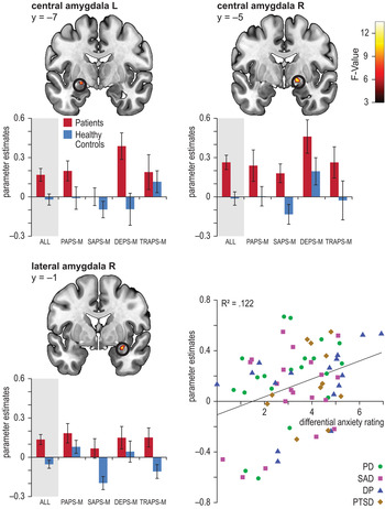
Analysis of the main effect of group showed that all patients compared with all HC showed a stronger emotion effect (disorder-related > neutral) in the left central amygdala (peak voxel Talairach coordinates: x = − 23, y = −7, z = −10, k: 10 voxels, average F: 9.112, maximal F: 10.03, p < 0.05 corrected), right central amygdala (peak voxel Talairach coordinates: x = 15, y = −5, z = −6, k: 16 voxels, average F: 9.763, maximal F: 12.213, p < 0.05 corrected) and right lateral amygdala (peak voxel Talairach coordinates: x = 31, y = −1, z = −16, k: 14 voxels, average F: 9.62, maximal F: 13.42, p < 0.05 corrected). There were neither significant effects for the reversed comparison (HC > patients), nor any differential effects between patient groups in the bilateral amygdala. The size of the emotion effect in the right lateral amygdala cluster correlated significantly with anxiety ratings (disorder-related minus neutral) across all patients (r = 0.349, p = 0.002, see Fig. 2). No cluster in the amygdala correlated with BDI and STAI-T. Correlations were considered as statistically significant if p < 0.016 (Bonferroni-corrected for the three significant clusters in the amygdala).
Shared brain activation (disorder-related minus neutral) in the bilateral amygdala. Bar graphs are shown for patients and healthy controls across picture sets (ALL) and for each set: Panic-related Picture Set Muenster (PAPS-M); Social Anxiety Picture Set Muenster (SAPS-M); Dental Phobia Picture Set Muenster (DEPS-M); Trauma-related Affective Picture Set Muenster (TRAPS-M). Scatterplot displays correlation of activation in the right lateral amygdala and anxiety ratings (disorder-related minus neutral). L, Left; R, right; PD, panic disorder; SAD, social anxiety disorder; DP, dental phobia; PTSD, post-traumatic stress disorder. Values are means, with standard errors represented by vertical bars. All values displayed at p < 0.05 (corrected).

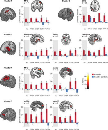
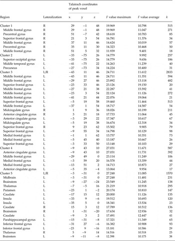
Additionally, whole-brain analyses of the main effect of group yielded significant clusters in middle, medial and superior frontal regions, dorsal and pregenual ACC, midcingulate cortex, thalamus, insula, brainstem and occipital lobe (see Table 2 and Fig. 3). There were no significant effects for the reversed comparison (HC > patients) nor any differential effects between patient groups in the whole-brain analysis. Three whole-brain clusters correlated significantly with scene-induced anxiety across all patients (cluster 1: r = 0.389, p = 0.001; cluster 3: r = 0.419, p < 0.001; cluster 5: r = 0.294, p = 0.008) and two clusters correlated with BDI scores (cluster 4: r = 0.338, p = 0.003; cluster 5: r = 0.455, p < 0.001). Correlations were considered as statistically significant if p < 0.01 (Bonferroni-corrected for the five significant clusters).
Shared brain activation (disorder-related minus neutral) across disorders as revealed in whole-brain analysis. Each row presents results for one cluster (whole cluster presented in three dimensions). Selected local maxima (two dimensions) are presented with corresponding bar graphs. Bar graphs are shown for patients and healthy controls across picture sets (ALL) and for each set: Panic-related Picture Set Muenster (PAPS-M); Social Anxiety Picture Set Muenster (SAPS-M); Dental Phobia Picture Set Muenster (DEPS-M); Trauma-related Affective Picture Set Muenster (TRAPS-M). MFG, Middle frontal gyrus; SOG, superior occipital gyrus; dACC, dorsal anterior cingulate cortex; mPFC, medial prefrontal cortex; pgACC, pregenual anterior cingulate cortex. Values are means, with standard errors represented by vertical bars. All values displayed at p < 0.05 (corrected). For illustration purposes MRIcroGL was used (http://www.mccauslandcenter.sc.edu/mricrogl/).

Significant hyperactivations for disorder-related v. neutral scenes across all patients relative to healthy controls revealed by whole-brain analysis in five clusters (p ⩽ 0.05 corrected) a

k, Number of voxels; R, right; L, left.
a The watershed algorithm of Neuroelf (v0.9c; http://neuroelf.net/, i.e. the splitclustercoords function) was used to assess local maxima of clusters.
Discussion
The aim of the present study was to investigate neural correlates of disorder-related threat processing across PD, SAD, DP and PTSD, with all patients free of psychiatric medication. To trigger disorder-related visual processing, we presented ecologically valid stimuli tailor-made for each disorder. Compared with HC, enhanced neural correlates of disorder-related processing across PD, SAD, DP and PTSD were found in distinct clusters in the bilateral amygdala. Emotion effects (always referring to disorder-related > neutral) in the lateral amygdala correlated significantly with subjective level of stimulus-induced anxiety across all patients. Whole-brain analysis yielded further common neural correlates in middle, medial, superior frontal and (para-)limbic regions, in the brainstem and in the occipital lobe, encompassing the most prominent circuits associated with anxiety (Davidson, Reference Davidson2002; Etkin, Reference Etkin2010; Sylvester et al. Reference Sylvester, Corbetta, Raichle, Rodebaugh, Schlaggar, Sheline, Zorumski and Lenze2012; Duval et al. Reference Duval, Javanbakht and Liberzon2015; Tovote et al. Reference Tovote, Fadok and Lüthi2015). The present analyses yielded no differential effects between patient groups.
The strong emotion effect in the bilateral central and right lateral amygdala across all patients v. HC suggests an involvement of the amygdala in disorder-related threat processing common to all disorders tested here. The amygdala is considered a key element in the human alarm system and has been linked to concepts of salience, attention and vigilance (Davis & Whalen, Reference Davis and Whalen2001; Phelps & LeDoux, Reference Phelps and LeDoux2005). Facilitating attention to, and perception of, incoming emotional information, the amygdala can be considered a relevance detector (Phelps & LeDoux, Reference Phelps and LeDoux2005; Sergerie et al. Reference Sergerie, Chochol and Armony2008; Janak & Tye, Reference Janak and Tye2015). The lateral amygdala, described as the interface of sensory input from the cortex and thalamus, functions as a ‘gatekeeper of the amygdala’ (LeDoux, Reference LeDoux2007, p. R869), assigning emotional value to stimuli and relaying information to the central nucleus of the amygdala via direct and indirect interconnections (Pitkänen et al. Reference Pitkänen, Savander and LeDoux1997; Ball et al. Reference Ball, Rahm, Eickhoff, Schulze-Bonhage, Speck and Mutschler2007; LeDoux, Reference LeDoux2007; Fujieda et al. Reference Fujieda, Koganezawa, Ide, Shirao and Sekino2015). The central nucleus is considered important for vigilance potentiation in response to relevant stimuli and control of bodily reactions. The latter is, for example, based on its projections to the brainstem, which in turn evokes autonomic threat responses and initiates the release of stress hormones (Kim et al. Reference Kim, Loucks, Palmer, Brown, Solomon, Marchante and Whalen2011; Shin, Reference Shin and Ferry2012). Furthermore, the emotion effect in the lateral amygdala correlated with the subjective level of stimuli-induced anxiety across all patients, which fits with the lateral amygdala's function as a gateway for salient sensory information, initiating further processes that result in behavioural and autonomic fear expression. Emotion effects in the amygdala were correlated with scene-induced anxiety but correlated neither with symptoms of depression nor with trait anxiety. This underlines the amygdala's specific role in the weighting of disorder-related information and suggests scene-induced anxiety to be most closely related to emotion effects in the amygdala. The present amygdala findings might also underline the high emotional value of the disorder-related tailor-made stimuli for each disorder and thereby be the neural signature of stimulus threat relevance. Of note, neural correlates of emotion effects are based on implicit stimulus processing while post-scanning rating data rely on explicit emotional processing. Thus, correlational data reveal a relationship of implicit and explicit processing.
Altered amygdala activation has not been consistently reported in studies that focus on one disorder only (Etkin & Wager, Reference Etkin and Wager2007 for PD; Shin & Liberzon, Reference Shin and Liberzon2010 for PTSD; Del Casale et al. Reference Del Casale, Ferracuti, Rapinesi, Serata, Piccirilli, Savoja, Kotzalidis, Manfredi, Angeletti, Tatarelli and Girardi2012 for specific phobia; Schienle et al. Reference Schienle, Scharmüller, Leutgeb, Schäfer and Stark2012 for specific phobia; Ipser et al. Reference Ipser, Singh and Stein2013 for specific phobia; Brühl et al. Reference Brühl, Delsignore, Komossa and Weidt2014 for SAD). These inconsistencies have been attributed to a wide range of study features: block- v. even-related designs (Caseras et al. Reference Caseras, Giampietro, Lamas, Brammer, Vilarroya, Carmona, Rovira, Torrubia and Mataix-Cols2010), task properties (Straube et al. Reference Straube, Mentzel and Miltner2006), possible habituation effects (Taylor et al. Reference Taylor, Liberzon, Fig, Decker, Minoshima and Koeppe1998; Fischer et al. Reference Fischer, Wright, Whalen, McInerney, Shin and Rauch2003), use of psychiatric medication (Brühl et al. Reference Brühl, Delsignore, Komossa and Weidt2014) or fine differences in characteristics of emotional stimuli (Zald, Reference Zald2003; Carlson et al. Reference Carlson, Greenberg, Rubin and Mujica-Parodi2011). Furthermore, differences between disorders have been suggested, with less frequent amygdala and insula hyperactivation in PD and GAD than in PTSD, SAD and specific phobia (Etkin & Wager, Reference Etkin and Wager2007; Shin & Liberzon, Reference Shin and Liberzon2010; Duval et al. Reference Duval, Javanbakht and Liberzon2015). However, to investigate true differences between disorders, it is necessary to control for variables such as task, stimulation procedure, imaging modality and sample characteristics. The present study used this approach and revealed a threat effect across disorders in the amygdala in response to highly relevant disorder-related pictures.
Besides amygdala findings, whole-brain analysis yielded large emotion effects in middle, medial and superior frontal regions and in the pregenual and dorsal ACC and midcingulate cortex when comparing all patients with HC. This common activation in patients fits with earlier studies that reported co-activation of the mPFC and ACC, and might mirror the shared and altered mechanism of generation, evaluation and regulation of emotion across PD, SAD, DP and PTSD (Ochsner & Gross, Reference Ochsner and Gross2005; Kober et al. Reference Kober, Barrett, Joseph, Bliss-Moreau, Lindquist and Wager2008; Etkin et al. Reference Etkin, Egner and Kalisch2011). It could also constitute a neural correlate of shared phenomenological features among patients such as misinterpretation of ambiguous stimuli, over-interpretation of threat signals, and conscious pathological threat appraisal (Raczka et al. Reference Raczka, Gartmann, Mechias, Reif, Büchel, Deckert and Kalisch2010; Maier et al. Reference Maier, Szalkowski, Kamphausen, Perlov, Feige, Blechert, Philipsen, van Elst, Kalisch and Tüscher2012; Kalisch & Gerlicher, Reference Kalisch and Gerlicher2014).
(Para-)limbic emotion effects in whole-brain analysis were for example revealed in the thalamus and insula. As the thalamus is associated with initial processing and relaying sensory information, the increased emotion effect across patients in the present study might reflect elevated visual processing of disorder-related scenes, and thus indicate alterations in early phases of fear processing (Jones, Reference Jones2003; LeDoux, Reference LeDoux2003). The insula is assumed to mediate alertness and awareness to salient multimodal sensory signals (Critchley et al. Reference Critchley, Wiens, Rotshtein, Öhman and Dolan2004; Paulus & Stein, Reference Paulus and Stein2006; Nagai et al. Reference Nagai, Kishi and Kato2007; Craig, Reference Craig2009; Sterzer & Kleinschmidt, Reference Sterzer and Kleinschmidt2010). The integration of these signals goes hand in hand with interoceptive processing, which provides the basis for the subjective experience of feelings representing a crucial process in anxiety disorders (Critchley et al. Reference Critchley, Wiens, Rotshtein, Öhman and Dolan2004; Nagai et al. Reference Nagai, Kishi and Kato2007; Menon & Uddin, Reference Menon and Uddin2010). In line with this, the reported elevated emotion effect during disorder-related processing in the insula might be a correlate of increased interoceptive processing, a mechanism shared across PD, SAD, DP and PTSD, although specific differences with respect to interoception may still exist between these disorders.
Whole-brain analysis also yielded emotion effects in the brainstem, suggesting alterations in this structure to be a correlate of disorder-related processing across PD, SAD, DP and PTSD. The brainstem, considered the heart of the ‘stress circuitry’ and part of the neuroendocrine stress axis, is thought to play a major role in emotional stress responses (Itoi & Sugimoto, Reference Itoi and Sugimoto2010). Brainstem activation is often addressed in PD literature, since PD is particularly characterized by an exaggerated bodily fear reaction during panic attacks. However, bodily fear symptoms evoked by altered autonomic activity and neuroendocrine functioning play a role in all the investigated anxiety disorders (e.g. during a performance situation in SAD) (Sullivan et al. Reference Sullivan, Coplan, Kent and Gorman1999). It is not yet fully understood how brainstem structures modify the perception and processing of stressful and emotional events. Our finding suggests that disorder-related stimulus processing targets a shared mechanism based on brainstem functioning most probably related to changes in the homeostatic alarm system.
The finding of increased activation to threat across disorders in the occipital lobe supports the idea of alterations in sensory processing areas (Sehlmeyer et al. Reference Sehlmeyer, Schöning, Zwitserlood, Pfleiderer, Kircher, Arolt and Konrad2009; de Carvalho et al. Reference de Carvalho, Rozenthal and Nardi2010; Del Casale et al. Reference Del Casale, Ferracuti, Rapinesi, Serata, Piccirilli, Savoja, Kotzalidis, Manfredi, Angeletti, Tatarelli and Girardi2012; Ipser et al. Reference Ipser, Singh and Stein2013; Brühl et al. Reference Brühl, Delsignore, Komossa and Weidt2014), and the notion that basic, initial disorder-related processing is a dysfunctional mechanism shared across anxiety disorders in response to visual threat.
Remarkably, our study which used tailor-made disorder-related stimuli for each disorder and the same task across all disorders revealed no differential effects between disorders. We interpret the absence of statistically significant differential effects and the presence of shared effects as neuronal correlates of phenomenological aspects common to PD, SAD, DP and PTSD, rather than as correlates of disorder-specific aspects, such as the content of concerns (e.g. fear of negative evaluation in SAD v. the fear of bodily symptoms in PD) or feared situations (e.g. trauma-related situations in PTSD v. dental treatment in DP). Speaking in terms of RDoC, disorder-related visual processing might thus be regarded an intermediate phenotype across the four disorders (Cuthbert, Reference Cuthbert2014).
There are no previous studies that investigated responses to disorder-related visual threat across multiple anxiety disorders. Reviews and meta-analyses have drawn an inconsistent picture regarding neural circuitries in anxiety-related disorders (Rauch et al. Reference Rauch, Shin and Wright2003; Etkin & Wager, Reference Etkin and Wager2007; Craske et al. Reference Craske, Rauch, Ursano, Prenoveau, Pine and Zinbarg2009; Shin & Liberzon, Reference Shin and Liberzon2010; Holzschneider & Mulert, Reference Holzschneider and Mulert2011; Fredrikson & Faria, Reference Fredrikson and Faria2013; Duval et al. Reference Duval, Javanbakht and Liberzon2015; Taylor & Whalen, Reference Taylor and Whalen2015). Meta-analyses including SAD, specific phobia and PTSD studies suggested a potentially shared neural basis of disorders, but also noted differences between disorders (Etkin & Wager, Reference Etkin and Wager2007; Fredrikson & Faria, Reference Fredrikson and Faria2013). Taking large heterogeneity between included studies into account, caution is warranted when drawing comparative conclusions across disorders. As meta-analyses have not included PD patients (Etkin & Wager, Reference Etkin and Wager2007; Fredrikson & Faria, Reference Fredrikson and Faria2013), the present study contributes substantially to the knowledge about neural correlates of emotional processing across multiple psychiatric disorders.
Our study controls for task, design, procedure and operationalization of disorder-related processing. Due to its large sample size (n = 134) of matched patients and HC, our study has high statistical power, which makes the detection of reliable neural effects more likely than in studies with smaller sample sizes. However, caution is warranted since patient samples differ in size, with the DP (n = 16) and PTSD (n = 11) patient samples being rather small. The small size of the PTSD sample is due to the difficulty in recruiting unmedicated PTSD patients. Thus, to increase sensitivity for differential effects, future studies should implement larger samples within each disorder. Furthermore, our PTSD sample comprised only women suffering from one specific (but frequent) type of trauma. The present study uses four novel disorder-related stimulus sets, which we deem a fruitful approach also for further studies to target more disorder-relevant psychological processes (e.g. tasks related to automatic processing, interference, attentional bias).
In conclusion, using disorder-related stimuli high in ecological validity, the same procedures and task, implementing CBP statistics and comparing a large sample of medication-free PD, SAD, DP and PTSD patients with matched HC, we found evidence for shared neural correlates, including the bilateral amygdala, wide frontal and (para-)limbic regions as well as the brainstem and occipital lobe. Most strikingly, the elevated emotion effect in the lateral amygdala correlated with subjective levels of anxiety, underlining the central role of the amygdala in the pathology of all four disorders. Our findings suggest that basic mechanisms are shared across disorders, despite their different diagnostic profiles.
Supplementary material
The supplementary material for this article can be found at https://doi.org/10.1017/S0033291716002634
Acknowledgements
The present study was supported by research grants awarded by the German Research Society (Deutsche Forschungsgemeinschaft, DFG; SFB/TRR 58: C06, C07).
Declaration of Interest
None.