

Introduction
The use of vitrification as a simple, viable and promising cryopreservation technique, even as a technology, has been restricted to a very few human and domestic animal embryology laboratories (Kuwayama, Reference Kuwayama2007). This limited practice may be attributed to the lack of consistent results achieved post-warming including survivability, in vitro maturation, fertilization and embryonic developmental rates (Liebermann & Tucker, Reference Liebermann and Tucker2002, Reference Liebermann and Tucker2006; Kuwayama, Reference Kuwayama2007). Different vitrification protocols have been used to cryopreserve bovine embryos or oocytes. Several different studies, to date, have established the obvious fact that the minimum volume vitrification methods avoid two major obstacles associated with the cryopreservation process, i.e. ice crystal formation and chilling injury (Chian et al., Reference Chian, Kuwayama, Tan, Tan, Kato and Nagai2004; Vajta & Kuwayama, Reference Vajta and Kuwayama2006; Kuwayama, Reference Kuwayama2007; Morato et al., Reference Morato, Izquierdo, Paramio and Mogas2008; Hajarian et al., Reference Hajarian, Wahid, Rosnina, Daliri, Dashtizad, Mirzapour, Yimer, Bukar, Iswadi and Abas Mazni2011). Different cryodevices have been used for efficient oocyte vitrification such as cryotop, open-pulled straw (OPS), electron microscopy grid (EMG) and cryoloop; the cryotop device is the most recent minimum volume vitrification approach (<1 μl). This reduction of vitrification solution volume produces exceedingly high cooling/warming rates, consequently reducing the danger of zone temperature (15–5°C), and avoidance of zona pellucida and embryo fracture damage (Kuwayama, Reference Kuwayama2007; Hajarian et al., Reference Hajarian, Wahid, Rosnina, Daliri, Dashtizad, Mirzapour, Yimer, Bukar, Iswadi and Abas Mazni2011). Accordingly, cryotop technology has been the most efficient tool for preserving oocyte fertilization capacity after vitrification (Morato et al., Reference Morato, Izquierdo, Paramio and Mogas2008; Succu et al., Reference Succu, Bebbere, Bogliolo, Ariu, Fois, Leoni, Berlinguer, Naitana and Ledda2008; Hajarian et al., Reference Hajarian, Wahid, Rosnina, Daliri, Dashtizad, Mirzapour, Yimer, Bukar, Iswadi and Abas Mazni2011). Most previous studies have only evaluated the quality of in vitro embryos produced from vitrified⁄warmed oocytes by their structural and morphological attributes, but more thorough assessments are needed. To achieve an optimum and productive procedure for both mammalian oocytes or embryos, further information on molecular and biochemical events induced by the vitrification process needs to be screened, monitored and discussed carefully (Succu et al., Reference Succu, Bebbere, Bogliolo, Ariu, Fois, Leoni, Berlinguer, Naitana and Ledda2008; Habibi et al., Reference Habibi, Farrokhi, Moreira da Silva, Bettencourt, Bruges-Armas, Amidi and Hosseini2010).
Recently, it has been demonstrated that the study of the relative abundance of developmentally important gene transcripts from in vitro produced embryos after cryopreservation could efficiently reflect the success of the vitrification process. Moreover it could open the door to large amounts of molecular data, principally when compared with their in vivo produced counterparts (Stinshoff et al., Reference Stinshoff, Wilkening, Hanstedt, Bruning and Wrenzycki2011). So, there is still a need to conduct studies that involve the most efficient vitrification elements from cryoprotectants to cryodevices. There is an urgent need to correlate the success of this process with gene expression status in embryos produced after oocyte vitrification⁄warming.
Therefore, the aim of the present study was to evaluate the efficiency of immature bovine cumulus–oocyte complex (COC) vitrification using two of the most common cryoprotectants: 1,2-propanediol (PROH) or dimethylsulphoxide (DMSO) in combination with ethylene glycol (EG) using the cryotop method and by measurement of post-warming survivability and subsequent in vitro developmental competence. In addition, the expression patterns of some genes related to embryonic development were monitored, namely connexin 43 (Cx43), cadherin-1 (CDH1), DNA methyltransferase 1 (DNMT1) and heat shock 70 kDa protein 14 (HSPA14).
Materials and methods
Chemicals
All the chemicals and reagents used in this study were obtained from Sigma-Aldrich (St. Louis, MO, USA) unless stated otherwise.
Ovaries
Ovaries (n = 70) were obtained in five trials at a local abattoir from adult animals, trimmed of adhering tissue and transported to the laboratory in Dulbecco's phosphate-buffered saline (DPBS) at temperatures ranging from 34 to 37°C within 2 h post-slaughtering. Upon arrival at the laboratory, all ovaries were rinsed in 70% alcohol and washed with fresh DPBS.
Immature oocytes collection
Cumulus–oocytes complexes were collected by aspiration from antral follicles (2–8 mm diameter) with an 18-gauge needle. Good quality COCs, based on their morphological appearance, for which oocytes must be covered by at least four layers of compacted cumulus cells and show evenly granulated ooplasm, were assigned randomly to the non-vitrified control (fresh COCs) and cryotop vitrification groups.
Experimental design
Two experiments were conducted successively: (1) to evaluate the developmental competence of vitrified⁄warmed bovine immature cumulus–oocyte complexes (COCs) in vitro; and subsequently (2) to study of gene expression patterns of Cx43, CDH1, DNMT1 and HSPA14 from in vitro produced embryos at different embryonic developmental stages.
Experiment 1: In vitro embryo production after oocytes vitrification using the cryotop method
Vitrification/warming of immature oocytes
COCs were vitrified according to the protocol used for cattle by Cetin & Bastan (Reference Cetin and Bastan2006) and assayed as for mice by Habibi et al. (Reference Habibi, Farrokhi, Moreira da Silva, Bettencourt, Bruges-Armas, Amidi and Hosseini2010), with some modifications. Briefly, groups of 4–6 immature oocytes were initially equilibrated for 5 min in holding medium (TCM-199 medium with HEPES, 20% fetal bovine serum (FBS) and 50 μg/ml gentamycin) supplemented with either 1 M PROH or DMSO and 7.5% EG. After achieving equilibration, oocytes were transferred to the vitrification solution (2 M PROH or DMSO in TCM-199 HEPES medium with 15% EG, 0.5 M sucrose, 20% FBS and 50 μg/ml gentamycin) for 30 s. All vitrification processes were performed at room temperature. The oocytes were then loaded on a cryotop with less than 1 μl vitrification solution and plunged immediately into liquid nitrogen (LN2) for storage.
For warming, the cryotops were directly inserted into 1 ml of warming medium (TCM-199 HEPES medium supplemented with 20% FBS) and 0.5 M of sucrose at 37°C for 1 min. COCs were then sequentially rehydrated in the warming medium, in stepwise serial dilution (0.25, 0.1 and 0 M of sucrose for 5, 5 and 10 min respectively) at 37°C. Post-warming, all collected oocytes were counted and only good morphological oocytes with an intact granular ooplasm and undamaged cumulus cells (Santos et al., Reference Santos, Tharasanit, Van Haeften, Figueiredo, Silva and Van den Hurk2007) were classified as surviving COCs and subjected to in vitro maturation. Oocytes that showed a shrunken or irregular dark ooplasm and damaged cumulus cells post warming were considered to be not surviving (Borges et al., Reference Borges, Silva, Futino, Rocha-Junior, Amorim, Bao and Lucci2009; Sharma et al., Reference Sharma, Dubey and Chandra2010).
In vitro oocytes maturation, fertilization and embryonic development
Fresh COCs (control group) and survived COCs post warming (vitrified groups) were washed in TCM-199 HEPES medium supplemented with 2% FBS, 0.3 mg/ml glutamine, 50 μg/ml gentamycin and 20 μg/ml nystatin and matured in TCM-199 with HEPES supplemented with 10% FBS, 5 μg/ml of FSH-LH (Stimufol, Belgium), 1 μg/ml estradiol-17β, 0.15 mg/ml glutamine, 22 μg/ml Na-pyruvate, 50 μg/ml gentamycin and 20 μg/ml nystatin. After 24 h of maturation under 5% CO2 in air in a humidified atmosphere at 38.5°C, only oocytes with cumulus expansion were considered to be mature and subjected to fertilization. Matured oocytes were placed for insemination in Tyrode's albumin lactate pyruvate (TALP) medium according to Faheem et al. (Reference Faheem, Carvalhais, Chaveiro and Moreira da Silva2011). Briefly, thawed semen were washed in IVF-TALP medium supplemented with 10 μg/ml heparin, 6 mg/ml bovine serum albumin (BSA, essential fatty acid-free), 22 μg/ml Na-pyruvate, 50 μg/ml gentamycin and 20 μg/ml of nystatin. Then, the sperm concentration was adjusted to 1 × 10⁶ sperm/ml. Oocytes and sperm were co-cultured for 22–24 h at 38.5ºC in 5% CO2 in air. Presumptive embryos were denuded by vortexing and washed in culture medium consisting of TCM-199 with HEPES and supplemented with 3 mg/ml BSA (Fr. V), 22 μg/ml Na-pyruvate, 10 μl/ml NEAA (MEM, non-essential amino acids), 20 μl/ml EAA (BME, essential amino acids), 50 μg/ml gentamycin and 20 μg/ml of nystatin. After washing, the embryos were uploaded onto a monolayer of granulosa cells for further developmental culture in an incubator at 38.5ºC in 5% CO2 in air. Cleavage rate and early embryonic development (2-cell and 4-cell stages) were determined microscopically (×100 magnification) after 3 days of fertilization (day 0), while later embryonic development from morula and blastocyst stages were evaluated at day 8 of fertilization. Embryos at different stages of development were stored for further analysis.
Granulosa cells preparation
The co-culture monolayers were prepared on the day of oocyte maturation, in which granulosa cells were aspirated from bovine follicles (2–8 mm) according to Khatir et al. (Reference Khatir, Anouassi and Tibary2004) with some modifications. Briefly, follicular fluid containing granulosa cells was centrifuged at 200 g for 10 min, then the pellet resuspended in the granulosa culture medium (TCM-199 without HEPES containing 10% FBS, 50 μg/ml gentamycin and 20 μg/ml of nystatin). The clumps of cells were mechanically broken. Cell concentration was adjusted to 1 × 10⁶ cells/ml. Droplets of the cell suspension (50 μl) were cultured under 5% CO2 in air at 38.5ºC. Before placing the embryos in co-culture and every 48 h, half of the medium was replaced.
Experiment 2: Evaluation of gene expression
Sample preparation, RNA extraction and cDNA synthesis
Total RNA extraction
After embryonic development, all embryos were collected and divided separately according to their developmental stages (2-cell, 4-cell, morula and blastocyst stages), washed three times extensively, to avoid the presence of any co-culture cell, with diethylpyrocarbonate (DEPC)-treated water and stored as pools of 15 embryos at –80ºC. From the same number of bovine embryos, divided into control (non-vitrified group) and PROH- and DMSO-treated groups (vitrification groups), total RNA was extracted with Trizol reagent following the manufacturer instructions (Life Technologies, Inc., Carlsbad, CA, USA) and using the extraction modifications from bovine embryos described by Pavani (Reference Pavani2012). Samples were dissolved in 12 μl of DEPC water and quantified by the average of triplicate spectrophotometric readings at 260 nm using NanoVue™ equipment (GE Healthcare). The purity of total RNA was confirmed by the A260/280 nm ratio. The integrity of RNA samples was measured by electrophoresis and 1% agarose gel.
Single-strand cDNA synthesis
Three micrograms of total RNA was reverse transcribed using an oligo(dT)12–18 (1.0 μl or 500 ng) primer. The reaction was performed in a 20 μl final volume containing 1× reaction buffer (250 mM Tris–HCl at pH 8.3, 250 mM KCl, 20 mM MgCl2, 50 mM DTT), 20 U of RiboLock™ RNase Inhibitor, 20 mM of dNTP mix and 200 U of RevertAid™ H Minus M-MuLV reverse transcriptase (Fermentas Life Science). cDNAs were synthesized at 42°C for 60 min following the inactivation of the transcriptase by heating at 70°C for 5 min. To remove mRNA templates, samples were treated with 1 U RNase H enzyme at 37°C for 30 min.
Specific primer design
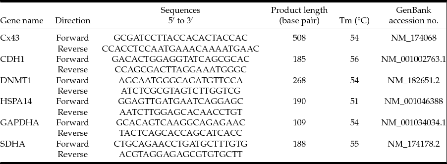
In order to construct standard curves and perform the relative quantification of gene expression in real time, specific primers to four desired genes and two reference genes (shown in Table 1) were designed using Prime3 software (Rozen & Skaletsky Reference Rozen and Skaletsky2000) and synthesised by Invitrogen (Life Technologies Corporation, Europe) from sequences described in the GenBank database (www.ncbi.nlm.nih.gov).
Oligonucleotide primers used for quantitative real-time polymerase chain reactions (qPCR)

Standard curves
In order to determine PCR efficiency and plot calibration curves, five-fold serial dilutions of cDNA were made and amplified using primers to both desired and reference genes. The Ct versus Log10 cDNA dilution was plotted to determine the slope of the line. PCR efficiency was then calculated by the equation m = –(1/logE) where m is the slope of the line and E is the efficiency.
Quantitative real-time polymerase chain reaction (qPCR)
Real-time PCRs were performed using the ABI Prism 7500 (PE Applied Biosystems) in 96 micro-well plates and using the Thermo Scientific Absolute Blue QPCR SYBR Green Low ROX Mix (Thermo Scientific ABgene® UK). All samples, including the external standards and non-template control, were run in triplicate. Reaction conditions were established through a series of preliminary optimization experiments including calibration curves. Each 25-μl final volume reaction contained 1× Blue QPCR SYBR Low ROX (which includes Thermo-Start™ DNA polymerase and 3 mM of MgCl2 in addition to blue dye and ROX dye), 1 μM of each forward and reverse primers, water and template cDNA. Template cDNA corresponded to the 2-cell, 4-cell, morula and blastocyst stage samples from control, PROH- and DMSO-treated groups. Water was included as a non-template control to confirm the absence of contamination in the reaction mixture. The reaction was initiated by activation of Thermo-Start™ DNA polymerase at 95°C for 15 min, followed by 40× three-step amplification cycles consisting of 15 s denaturation at 95°C, 30 s at 54°C and 30 s at 72°C. A final dissociation stage was run to generate a melting curve for verification of amplification product specificity. Quantification was carried out using the comparative cycle threshold (Ct) method; with the results expressed in relation to endogenous reference genes and a control group.
In this experiment the reference genes were GAPDH and SDHA, presenting little variation in mRNA levels during the development of bovine blastocysts (Goossens et al., Reference Goossens, Van Poucke, Van Soom, Vandesompele, Van Zeveren and Peelman2005). The studied genes were Cx43, CDH1, DNMT1 and HSPA14.
Data analysis
Embryonic development data were analysed by one-way analysis of variance (ANOVA) and expressed as mean ± standard error of the mean (SEM) calculated from the survived oocytes for each group. Comparisons were considered to be significantly different if P < 0.05. All analyses were performed using the SPSS Statistics 17.0 program (SPSS Inc., Chicago, Illinois, USA).
For gene analyses, amplicon sizes and specificity of products generated were confirmed by dissociation curves after running amplification cycles. The relative expression ratio of a target gene was determined from the real-time PCR efficiency (E). For each gene, cDNA dilution curves were generated and used to calculate the individual real-time PCR efficiencies by E = 10(–1/slope) – 1. Relative gene expression analysis was performed using the Relative Expression Software Tool (REST©) with the Pair Wise Fixed Reallocation Randomization Test© method (Pfaffl et al., Reference Pfaffl, Horgan and Dempfle2002) with the calculated E values introduced. The relative expression values (Ct) are presented as mean ± SEM. For comparisons between genes, data from fresh embryos (control group) at the same embryonic stage were used as calibrator group for relative gene expression analysis. The mRNAs levels of the Cx43, CDH1, DNMT1 and HSPA14 genes were compared between in vitro produced bovine embryos at 2-cell, 4-cell, morula and blastocyst stages using GAPDH and SDHA as reference genes for normalization.
Results
Experiment 1
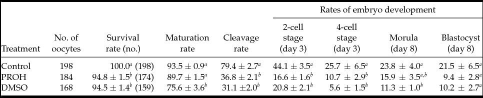
Oocyte viability results after warming, as well as their in vitro competence to develop up to the blastocyst stage, are shown in Table 2. As observed, the viability of COCs vitrified with either PROH or DMSO was comparable, and achieved similar survival rates post warming (94.8 ± 1.5 and 94.5 ± 1.4%, respectively), but were however significantly lower than that of fresh COCs.
Oocytes survival rate and their subsequent in vitro development after vitrification using different cryoprotectants

a,b Numbers in the same column with different letters (a,b,c) differ significantly at P < 0.05.
Data are expressed as mean ± standard error of the mean (SEM).
DMSO, dimethylsulphoxide; PROH, 1,2 propanediol.
All the percentages were based on the number of survived oocytes.
Days 3 and 8 were based on the day of fertilization (day 0).
For maturation rate, assayed by cumulus–oocyte expansion, there was no significant difference between the fresh group and oocytes vitrified using PROH (93.5 ± 0.9 and 89.7 ± 1.5%, respectively); the oocytes vitrified using DMSO had a lower (P < 0.05) maturation rate (75.6 ± 3.6%) compared with the fresh and PROH-treated groups.
The percentage of cleaved embryos did not differ among the two vitrification groups (36.8 ± 2.1 and 31.1 ± 2.0% for PROH and DMSO, respectively), however when compared with the fresh group (79.4 ± 2.7%), both vitrification groups had lower (P<0.05) cleavage rates.
Early embryonic developmental rates at the 2-cell and 4-cell stages were significantly higher in the fresh group compared with both vitrified groups, whereas between PROH- and DMSO-treated groups there were no differences observed for these stages. At the morula stage, although there were no significant differences between the two vitrification groups, the DMSO-treated group had a lower (P<0.05) morula rate compared with the fresh oocyte group. At the blastocyst stage, there was no significant differences between fresh oocytes and both vitrified groups.
Experiment 2
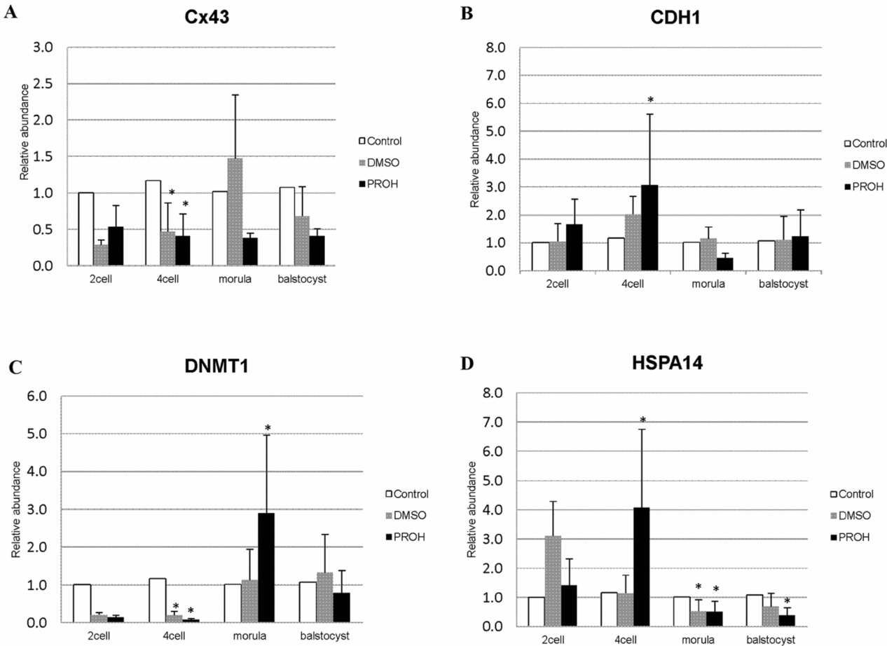
The relative abundances of Cx43, CDH1, DNMT1 and HSPA14 transcripts at the 2-cell, 4-cell, morula and blastocyst stages after immature bovine oocyte vitrification either with PROH or DMSO are depicted in Fig. 1.
Relative abundance of developmentally important genes in fresh and vitrified embryos with PROH or DMSO: (A) Cx43 (Connexin 43); (B) CDH1 (cadherin 1); (C) DNMT1 (DNA methyltransferase1); and (D) HSPA14 (heat shock 70 kDa protein 14) at 2-cell, 4-cell, morula and blastocyst stages of embryonic development.

The relative expression levels of Cx43 (Fig. 1 A) were significantly (P<0.05) down-regulated in both groups of cryoprotectants at the 4-cell stage, while for the other embryonic developmental stages (2-cell, morula and blastocyst), no differences could be observed when compared with the control group (for PROH: P = 0.344, 0.66 and 0.838; for DMSO: P = 0.49, 0.335 and 0.514 for each embryonic stage, respectively).
Although the pattern of CDH1 expression (Fig. 1 B) did not differ throughout the 2-cell, morula and blastocyst stages from both cryoprotectants groups (PROH: P = 0.336, 0.66 and 0.332; DMSO: P = 0.66, 0.83 and, 0.662, respectively) compared with the control group, expression values for embryos derived from the PROH group were significantly up-regulated at the 4-cell stage.
DNMT1 transcript levels (Fig. 1 C) were down-regulated (P < 0.05) in both groups of cryoprotectants at the 4-cell stage compared with the control group. Nevertheless, the PROH-treated group showed significantly up-regulation of DNMT1 expression levels (P < 0.05) at the morula stage. At the 2-cell stage, there were no significant differences for both cryoprotectants groups (P = 0.336 and 0.51 for PROH and DMSO groups, respectively).
For HSPA14, (Fig. 1 D), while significant up-regulation (P < 0.05) of HSPA14 expression levels was seen at the 4-cell stage from the PROH-treated group compared with the control, lower expression levels (P < 0.05) were found throughout the subsequent embryonic morula and blastocyst stages from both groups of cryoprotectants and the PROH-treated group, respectively.
At the 2-cell stage, interestingly, no statistical differences were observed in relative gene expression of all genes studied from both groups of cryoprotectants (Fig. 1).
Discussion
The present study was designed to investigate the influence of vitrification on immature bovine oocytes using cryotop technology by measuring their post-warming survivability and capacity to undergo normal in vitro embryonic development. Consequently and throughout the embryonic developmental stages, four developmentally important genes that are concerned functionally with gap junction communications (Cx43), compaction and cavitation (CDH1), methylation (DNMT1) and cellular stress and thermotolerance (HSPA14), were analysed for their pattern of expression.
The present study, using a cryotop device, resulted in high rates of post-warming oocyte survival, whether from the PROH- or DMSO-treated groups, mostly similarly in both cryoprotectants groups. Our results are in agreement with Morato et al. (Reference Morato, Izquierdo, Paramio and Mogas2008), who found similar survivability rates (94.5%) when using bovine matured oocytes, but higher than that found by Hajarian et al. (Reference Hajarian, Wahid, Rosnina, Daliri, Dashtizad, Mirzapour, Yimer, Bukar, Iswadi and Abas Mazni2011) using immature bovine oocytes (90.34 ± 2.5%) and a cryotop device. In regard to cryoprotectant type, Chian et al. (Reference Chian, Kuwayama, Tan, Tan, Kato and Nagai2004) found that PROH provided an optimal vitrification condition for mature bovine oocytes, resulting in relatively higher survival rates (97.6 ± 4.2%) compared with the addition of DMSO (91.8 ± 6.7%). In contrast, according to Kuwayama (Reference Kuwayama2007) and Dutta et al. (Reference Dutta, Dev and Raj2013), an equal proportion of DMSO and ethylene glycol was the most efficient combination associated with a two-step equilibration method. Based on oocyte survivability, our results could indicate that the method of action of both cryoprotectants, to preserve cells or oocytes, may be similar. However our in vitro maturation results, assayed by cumulus–oocyte expansion, showed an inconsistency in the performance of the two cryoprotectants, and demonstrated that there was a significant defect in oocyte maturation after vitrification when using DMSO compared with PROH (75.6 ± 3.6 and 89.7 ± 1.5%, respectively). Gurtovenko & Anwar (Reference Gurtovenko and Anwar2007) and Varghese et al. (Reference Varghese, Nagy and Agarwal2009) found that the integrity of the cell membrane with its functional ligand receptor could be adversely affected by DMSO, leading to incomplete maturation, as the maturation process of frozen–thawed immature oocytes requires a proper interaction between the oocytes and their receptors. Interestingly, similar oocyte cleavage rates were observed between the two cryoprotectant groups. In an attempt to clarify this observation, this could be attributed to the presence of cumulus cells around the oocytes prior to freezing, and which play an important role in reducing the adverse effects of DMSO on the oocytes (Johnson & Pickering, Reference Johnson and Pickering1987; Chian et al., Reference Chian, Kuwayama, Tan, Tan, Kato and Nagai2004). On the other hand, the DMSO mechanism of action may impair the ability of cumulus cells to expand during the cytoplasmic maturation process, probably without affecting the nuclear maturity of the oocytes (Cetin & Bastan, Reference Cetin and Bastan2006; Ebrahimi et al., Reference Ebrahimi, Valojerdi, Eftekhari-Yazdi and Baharvand2010; Hajarian et al., Reference Hajarian, Wahid, Rosnina, Daliri, Dashtizad, Mirzapour, Yimer, Bukar, Iswadi and Abas Mazni2011). Accordingly, as our results showed, the nuclear maturity of the oocytes was not affected resulting in a comparable cleavage rate between DMSO- and PROH-treated groups even if the maturation rate was statistically different.
When in vitro embryonic developmental rates, from morula and blastocyst stages, were calculated based on the number of matured oocytes, no statistical differences were observed between fresh oocytes (25.4 ± 4.2 and 22.9 ± 6.9%, respectively) and both vitrification groups (17.7 ± 4.0, 10.4 ± 3.1% and 14.9 ± 1.1, 14.2 ± 4.3% for PROH and DMSO treatments, respectively). These results are in agreement with our previous finding using DMSO as a cryoprotectant for ovarian tissue cryopreservation (Faheem et al., Reference Faheem, Carvalhais, Chaveiro and Moreira da Silva2011), and stating that the impact of the cryopreservation process did not exceed the maturation stage and that the oocytes able to be matured after cryopreservation had the same competence to develop in vitro as fresh matured oocytes. Moreover, when calculated from the survived oocyte numbers, the same trend of embryonic development was observed between the fresh oocytes and the oocytes derived from the PROH-treated group, while the DMSO-treated showed significant difference at the morula stage compared with the fresh oocyte group.
Molecular analysis, thereafter, was used to assess the success of the vitrification process using either PROH or DMSO with the cryotop method, and by detecting the expression patterns of Cx43, CDH1, DMNT1 and HSPA14 at the 2-cell, 4-cell, morula and blastocyst stages of embryonic development, compared with the corresponding expression levels from the control group.
As gap junctions are required during preimplantation embryonic development (Houghton, Reference Houghton2005), the functional significance of Cx43 expression probably appears at the 8-cell stage of embryonic development, virtually when cell adhesion between blastomeres occurs during compaction in mammalian embryos (murine: Goodall & Johnson, Reference Goodall and Johnson1984; Fleming et al., Reference Fleming, Sheth and Fesenko2001). Moreover, Ogawa et al. (Reference Ogawa, Oyamada, Mori, Mori and Shimizu2000) found that levels of the phosphorylated form of Cx43 started to increase from the 8-cell stage onwards. Our results revealed that, at the 4-cell stage, both cryoprotectants groups showed a significant Cx43 down-regulation, assuming that the influence of the vitrification process could be responsible for this low expression more than the individual cryoprotectant effect itself. Noteworthily, this influence might be increased in coincidence with maternal-zygotic transition as the Cx43 transcripts for in vitro produced embryos were generated during this embryonic stage (Houghton, Reference Houghton2005).
However, the embryos derived from the DMSO-treated group showed, to some extent, higher expression levels of Cx43 at the morula stage compared with the same embryo stage derived from the PROH-treated group, although this difference did not improve in the rate of morula development in the DMSO group. One can speculate that Cx43, functionally, might not be the only gene responsible for embryonic competence to develop to the morula stage under in vitro conditions.
The cell adhesion molecule E-cadherin, also known as CDH1 (cadherin 1) or uvomorulin (Yang et al., Reference Yang, Wang, Zuo, Hao, Lu, Liu, Xu, Lu and Lu2012), is responsible for the cell adhesion of epithelial cells and mediates the compaction process of preimplantation embryos (Tesfaye et al., Reference Tesfaye, Lonergan, Hoelker, Rings, Nganvongpanit, Havlicek, Besenfelder, Jennen, Tholen and Schellander2007; Modina et al., Reference Modina, Leoni, Lodde, Naitana, Pirani, Succu, Berlinguer and Luciano2010). Furthermore, CDH1 was found to be the major functional gene controlling the embryonic compaction process as, experimentally, antibodies that block cadherin1 cause decompaction to early embryo stages (Hyafil et al., Reference Hyafil, Morello, Babinet and Jacob1980; Vestweber & Kemler, Reference Vestweber and Kemler1984; Riethmacher et al., Reference Riethmacher, Brinkmann and Birchmeier1995). In the present study, the pattern of CDH1 expression was apparently increased from the 2- to 4-cell stage for both groups of cryoprotectants, however a higher level of expression was found to be significantly confined to the PROH-treated group at the 4-cell stage. This reflects the competence of PROH-treated group cells in increasing CDH1 expression levels before the onset of morula compaction. This predominantly, in turn was involved in protein synthesis required for embryo programming for compaction (Riethmacher et al., Reference Riethmacher, Brinkmann and Birchmeier1995). Subsequently, the higher expression level of CDH1 in the PROH-treated group before compaction process could be the possible reason for the relatively higher morula rate shown in the PROH-treated group over the DMSO-treated group. Yet, it may be of maternal origin, as CDH1 can be synthesised and accumulated during oogenesis and oocyte maturation (Fleming et al., Reference Fleming, Sheth and Fesenko2001; Modina et al., Reference Modina, Leoni, Lodde, Naitana, Pirani, Succu, Berlinguer and Luciano2010). At the morula stage, the level of CDH1 expression decreased markedly in the PROH-treated group, and then increased at the blastocyst stage. As bovine embryos begin to synthesis their own mRNA at the 8–16-cell stage (Telford et al., Reference Telford, Watson and Schultz1990), the cause for this increase observed from morula to blastocyst stage can be probably explained due to zygotic genome activation.
DNMT1 is the most abundant form of DNA methyltransferase (DNMT1, DNMT2 and DNMT3) in mammalian cells and the main methyltransferase (Shi & Wu, Reference Shi and Wu2009). Its role, functionally, is to maintain DNA methylation patterns.
Mammalian preimplantation development represents a critical stage for the establishment of the epigenome and, as DNA methylation is one of the main epigenetic events, the genomic methylation pattern is stripped and then reasserted anew during the epigenetic reprogramming mechanism (Golding & Westhusin, Reference Golding and Westhusin2003). In agreement with our observed expression pattern for DMNT1, Dean et al. (Reference Dean, Santos, Stojkovic, Zakhartchenko, Walter, Wolf and Reik2001) demonstrated that at the zygote 1-cell stage, diminished methylation patterns were observed during the first cleavage-stage divisions, mostly from the maternal genome as the paternal genome undergoes genome-wide demethylation only hours after fertilization. Noteworthy, this loss of methylation is consistent with a passive demethylation mechanism by the absence or exclusion of DNMT1 from the nucleus and thereafter substantial de novo methylation occurs at the 8–16-cell stage in bovine embryos. In regard to this remarkable theory, similar dynamics of DNMT1 expression were monitored after the vitrification process during the first stages of divisions, although it was significantly down-regulated for both groups of cryoprotectants at the 4-cell stage. The high expression level of DNMT1 found at the morula stage in the PROH-treated group might reveal the earlier de novo methylation in the embryos derived from the PROH- rather than the DMSO-treated group.
The expression of heat shock proteins is induced by stress factors and their transcript level reflects both cell response and stress intensity (Neuer et al., Reference Neuer, Spandorfer, Giraldo, Dieterle, Rosenwaks and Witkin2000). In the present study, the pattern of HSPA14 expression during the first embryonic divisions, including 2- and 4-cell stages, was relatively up-regulated. Interestingly, data were inconsistent between the two groups of cryoprotectants, revealing: (1) the stress intensity was relatively restricted to the first stages of divisions; and (2) each type of cryoprotectants has an intrinsic role in controlling HSPA14 expression in response to stress. From the 2- and 4-cell stages and alongside the mentioned role of PROH in increasing the expression of CDH1 required for embryonic compaction, PROH effectively strengthens embryonic resistance to thermal stress by raising HSPA14 expression levels. This HSPA14, in turn, plays a protective role within a cell preventing protein degradation as a result of exposure to stress (Levy, Reference Levy2001). Consequently, the potential impact of PROH as a cryoprotectant could be elucidated through the mechanism of increasing both CDH1 and HSPA14 expression for in vitro produced embryos after vitrification.
Conclusion
The present study revealed the effectiveness of the cryotop method for vitrification of immature bovine oocytes in terms of embryonic developmental competence as well as at the molecular level. Post-warming oocyte survivability, embryo cleavage and in vitro embryonic developmental rates achieved comparable results between embryos derived from either the PROH- or DMSO-treated groups, nevertheless, PROH-treated group showed significant advantage as indicated by in vitro oocyte maturation rate. Concerning the molecular analysis, at the 4-cell stage of embryonic development most of the genes under study showed an inconsistent pattern of expression either in the PROH- or DMSO-treated group, presenting significantly down-regulation of Cx43 and DNMT1 levels for both groups of cryoprotectants, but up-regulation of CDH1 and HSPA14 levels in the PROH-treated group. Noteworthily, these differences in gene expression at the morula and blastocyst stages seemed to be limited and were confined mostly to down-regulation of HSPA14 expression levels.
Acknowledgements
The authors would like to thank Sandra Benites and Margarida Andrade for their skilled technical assistance in real-time PCR.
Financial support
The first author is supported by DRCT (Direcção para a Ciência e Tecnologia–Azores, Portugal) Grant BD M3.1.3/F/003/2009. CITA-A is fully acknowledged.
Conflict of interest statement
None