Advanced chronic heart failure is associated with high morbidity and mortality. It presents with reduced systolic and diastolic function, increased arterial resistance, and arterial–ventricular uncoupling, accompanied by excessive sympathetic activation. Reference Talan, Ahmet, Xiao and Lakatta1 These changes at the cellular and molecular levels in the myocardium cause remodelling in patients with chronic heart failure. Such patients can be treated with a variety of drugs to prevent further remodelling following heart failure. However, if remodelling has progressed to an uncompensated state that is refractory to inotropic support, clinicians must consider mechanical ventricular support or heart transplantation. Reference Heerdt, Holmes and Cai2
Left ventricular assist devices enable recovery from severe heart failure and can facilitate the transition to transplantation. There is substantial evidence that mechanical ventricular unloading can help to induce the structural reverse remodelling associated with functional recovery, permitting explantation of the left ventricular assist device. However, recovery from severe heart failure following this is found in only 5–24% of patients, possibly because prolonged left ventricular assist device support can result in unloading in the ventricle, potentially causing apoptosis and irreversible changes in the myocardium. Reference Schena, Kurimoto and Fukada3–Reference Brinks, Giraud and Segiser5 To reduce apoptosis in the myocardium during mechanical unloading, various drugs have been investigated in animal studies. The β2-agonist clenbuterol has been tested in a rat model of mechanical unloading. Reference Tsuneyoshi, Oriyanhan and Kanemitsu4,Reference Soppa, Lee and Stagg6,Reference Navaratnarajah, Siedlecka and Ibrahim7 Moreover, the β1-blocker metoprolol has been investigated in various studies and has been shown to have a positive effect on apoptosis prevention in rat hearts subjected to mechanical unloading. Reference Navaratnarajah, Siedlecka and Ibrahim7,Reference Ahmet, Krawczyk and Zhu8
For treating cases of heart failure, the nonselective β-blocker carvedilol is a representative drug that has cardioprotective effects similar to those of metoprolol. Reference Li, Chen and Gao9 However, no study has compared carvedilol and metoprolol with regard to their impact on the mechanically unloaded heart. A heterotopically transplanted heart in a rat is in a mechanically unloaded state similar to that induced by left ventricular assist devices. Reference Schena, Kurimoto and Fukada3,Reference Tsuneyoshi, Oriyanhan and Kanemitsu10 Therefore, in host and unloaded rat hearts, we investigated the ability of these drugs to induce beneficial changes in histology, myocardial apoptosis, and RNA expression levels, as well as myocardial changes throughout the course of mechanical unloading.
Materials and methods
Experimental design
Eight-week-old male Lewis rats (weighing 250–350 g; Koatech, Seoul, Republic of Korea) were used in this study. In all, 30 rats underwent heterotopic heart transplantation (see below). The transplant-receiving rats were randomly divided into three groups: a control group that received no medication, a treatment group that received carvedilol once daily at a dose of 15 mg/kg body weight for 2 and 4 weeks, and a treatment group that received metoprolol once daily at a dose of 50 mg/kg body weight for 2 and 4 weeks. In all three groups, host and unloaded hearts were investigated after 2 and 4 weeks (n = 5 at each time point for each group). All experimental procedures were conducted according to Pusan National University’s guidelines for animal care.
Heterotopic heart transplantation
Heterotopic heart transplantation was performed as follows: Lewis rats were anaesthetised with pentobarbital sodium (120 mg/kg) via intraperitoneal injection. In the donors, a butterfly incision was made in the thoracic cage by resecting the ribs at the bilateral axillary lines after the incision in the diaphragm and the rib cage was retracted upwards. The right and left superior caval veins, including the azygos vein, were ligated and cut distally. The thymus gland was dissected from the major blood vessels, and the aorta was cut at the innominate artery. The hearts and lungs were perfused using 3–4 ml of sterile saline via the inferior caval vein, after which the vein was ligated and cut distally. The pulmonary artery was then cut at the bifurcation, and the pulmonary veins were ligated together and cut distally. The heart was removed from the donor rat and stored in cold saline. For transplantation, the aorta and inferior caval veins were separated from each other after placement in the abdomen of the host rat and clamped with a single clamp.
A venotomy was performed at the 7–10 o’clock position and flushed with sterile saline. An adventectomy was performed on the side of the aorta, and an elliptical excision of the aortic wall was made and flushed with saline. The graft heart was placed in the right lower abdomen of the rat; an end-to-side aorto-aortic anastomosis was made using 9-0 or 10-0 microsutures in a continuous fashion. The pulmonary artery was then anastomosed end-to-side to the host’s caval vein using 8-0 microsutures. The clamp was slowly released to restore the cardiac rhythm. Gentle pressure with small gauze pads on the suture lines for 2 or 3 minutes controlled most of the oozing. If gaps or tears were present, the aorta and caval veins were clamped again and mended. Reference Lee11 The transplanted hearts exhibited continued contraction because of flow from the coronary arteries; blood through the coronary sinus passed through the right ventricle and entered the host’s inferior caval vein, but the left ventricle was completely unloaded. The total procedure time was < 30 minute, and the surgical mortality rate was <5%.
Pathology studies
The left ventricular myocardium was transversely sliced into sections (2 mm in diameter) at the base of the papillary muscle and then fixed in 10% buffered formalin. The remaining left ventricular myocardium was frozen at −80 °C until analysis. Transverse sections of left ventricular myocardium were stained with haematoxylin and eosin to measure myocyte size. Mean myocyte diameter was calculated by measuring 50 cells, magnified by a factor of 400. Cells needed to have a visible nucleus, with preserved membranes.
A terminal deoxynucleotidyl-transferase-mediated dUTP nick end-labelling assay was used to evaluate the degree of apoptosis according to the manufacturer’s protocol (Takara Korea Biomedical Inc.). Terminal deoxynucleotidyl-transferase-mediated dUTP nick end-labelling positive cells were counted in a blinded manner (300–400 cells) in 50 fields. Other transverse sections were stained with Masson’s trichrome stain to determine the degree of myocardial fibrosis. All slide images were processed with CaseViewer (3DHISTECH Ltd; https://www.3dhistech.com), and the percentage of fibrosis was determined.
Analysis of mRNA expression levels
Total mRNA was extracted from frozen left ventricle samples with TRIzol reagent (Thermo Fisher Scientific Inc.) and subjected to reverse transcription, and cDNA was produced using the Superscript™ II reverse-transcription quantitative real-time polymerase chain reaction system (Invitrogen, Karlsruhe, Germany) according to the manufacturer’s recommendations for oligo(dT) 20 primed cDNA synthesis. cDNA synthesis was performed with 500 ng of RNA at 42 °C. Finally, cDNA was diluted 1:2 prior to use for quantitative polymerase chain reaction, which was performed in an ABI PRISM 7900HT Sequence Detection System (Applied Biosystems, Foster City, CA, United States of America) in 384-well microtiter plates with a final volume of 10 μL. The optimum reaction conditions were obtained with 5 μl of Universal Master Mix (Applied Biosystems) containing dNUTPs, MgCl2, reaction buffer, Ampli Taq Gold, 90 nM of primer, and 250 nM of fluorescently labelled TaqMan probe. Finally, 2 μl of template cDNA was added to the reaction mixture. The primer/TaqMan probe combinations were designed for each target sequence. Amplifications were performed starting with a 10 minute template denaturation step at 95 °C, followed by 40 cycles at 95 °C for 15 s and at 60 °C for 1 min. All samples were amplified in triplicate, and data were analysed using Sequence Detector software (Applied Biosystems).
The nucleotide sequences for quantitative polymerase chain reaction primers were as follows: caspase-3 forward, 5′–AATTCAAGGGACGGGTCATG–3′, and reverse, 5′–GCTTGTGCGCGTACAGTTTC–3′; SERCA2a forward, 5′–CCTATGCACCCATTGGAG–3′, and reverse, 5′–ACAAGCCCGTCATACTGATG–3′; β1-adrenoreceptor forward, 5′–TCTGTGAGCTCTGGACTTCG–3′, and reverse, 5′–ATGACACACAGGGTCTCGAT–3′; and β2-adrenoreceptor forward, 5′–TTCGAGCGACTACAAACCGT–3′, and reverse, 5′–ATATGACTGGCCCCAAAAGG–3′. Rat glyceraldehyde 3-phosphate dehydrogenase was used as an endogenous control.
Statistical analysis
The left ventricle weight and data from pathology studies are expressed as the mean ± standard error of the mean. Paired data between the host and unloaded hearts were analysed using Wilcoxon signed-rank tests, and unpaired data were compared with controls using the nonparametric Mann–Whitney U-test. Statistical analyses were performed using IBM SPSS Statistics 21 for Windows (IBM Corp., Armonk, NY, United States of America). p Values <0.05 were considered statistically significant. Data from the mRNA analysis were analysed using the comparative C T method for relative quantification. The difference or delta (Δ) C T value was determined by subtracting the average endogenous control C T value from the individual C T value of the target gene. The ΔΔC T value was determined by subtracting the ΔC T of the control sample from the individual ΔC T value of the test sample. The fold change of each test sample was relative to control by finding the value of 2ΔΔCT. Fold changes in each group were compared with the control values after 2 weeks. Fold changes >2 or <0.5 were considered statistically significant.
Results
As shown in Table 1, the myocyte diameter in the unloaded hearts was significantly smaller than that in the host hearts in the carvedilol and metoprolol groups after 2 and 4 weeks. The myocyte diameter was also smaller in the unloaded hearts than in the host hearts in the control group after 4 weeks, but not after 2 weeks. In unloaded hearts, no differences in myocyte diameter were observed between the experimental and control groups. There were no differences between the host and unloaded hearts in any of the three groups.
Myocyte diameters in different treatment groups

* p < 0.05 versus host heart.
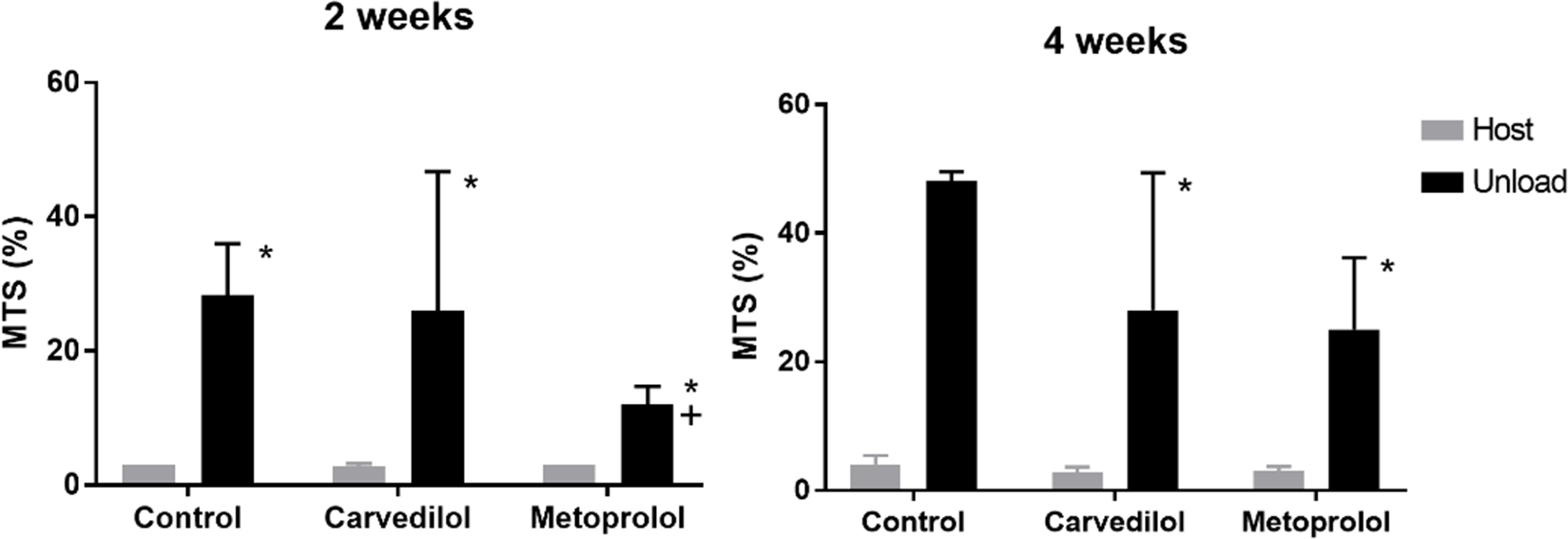
We analysed myocardial fibrotic areas stained with Masson’s trichrome. The host hearts in all groups exhibited differences in fibrotic area. The myocardial fibrotic areas in the unloaded hearts of the control, carvedilol, and metoprolol groups were 28.3 ± 7.6%, 26.0 ± 20.7%, and 12.0 ± 2.7%, respectively, after 2 weeks, and 48.0 ± 13.5%, 28.0 ± 21.3%, and 25.0 ± 11.1%, respectively, after 4 weeks. The proportion of fibrotic area in the unloaded hearts in the metoprolol group was smaller than that in the control group after 2 weeks, as shown in Figure 1.
Analysis of fibrosis area using Masson’s trichrome staining in all three groups at 2 and 4 weeks after transplantation. All values are represented as the mean ± SEM. *p < 0.05 versus host hearts in the same treatment group; +p < 0.05 versus unloaded hearts in the control group. MTS = Masson’s trichrome stain.

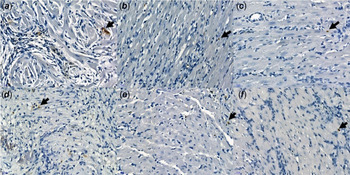
Figure 2 illustrates apoptosis as visualised with terminal deoxynucleotidyl-transferase-mediated dUTP nick end-labelling staining in all three groups. The unloaded hearts in the carvedilol and metoprolol groups 2 weeks after transplantation exhibited decreases in the proportion of terminal deoxynucleotidyl-transferase-mediated dUTP nick end-labelling positive cells, but this difference was not statistically significant (p = 0.071, control versus metoprolol after 2 weeks). The unloaded hearts in the carvedilol and metoprolol groups 4 weeks after transplantation showed no significant differences in the proportion of terminal deoxynucleotidyl-transferase-mediated dUTP nick end-labelling positive cells.
TUNEL staining in all three groups. Arrows (black) point to TUNEL-positive cells. a, b, and c represent the control, carvedilol, and metoprolol groups at 2 weeks after transplantation, respectively; d, e, and f represent the control, carvedilol, and metoprolol groups at 4 weeks after transplantation, respectively. TUNEL = terminal deoxynucleotidyl-transferase-mediated dUTP nick end-labelling.

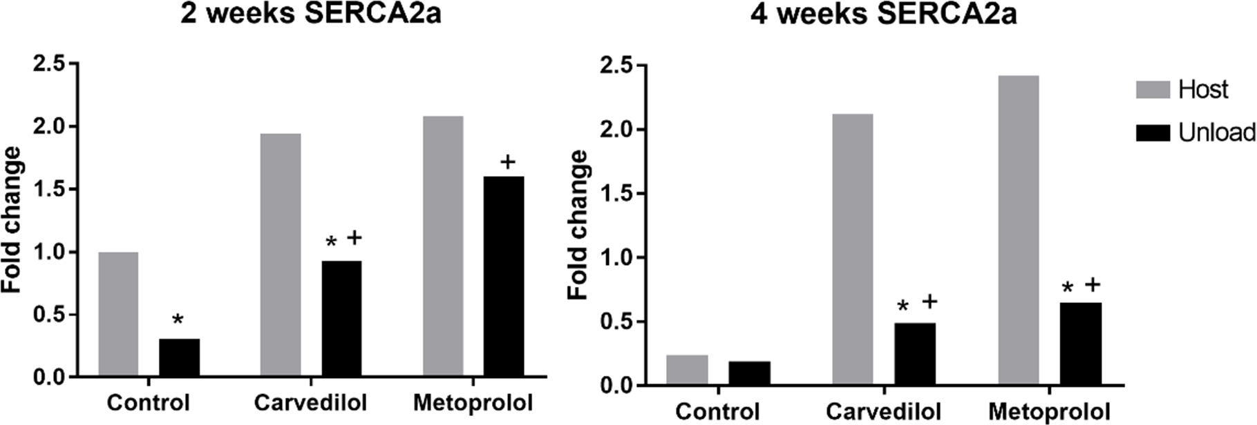
As shown in Figure 3, in the control and carvedilol groups, the mRNA expression of SERCA2a in the unloaded hearts decreased significantly compared with that in the host hearts after 2 weeks. This decrease was not observed in the metoprolol group. In unloaded hearts, SERCA2a mRNA expression in the carvedilol and metoprolol groups increased significantly compared with that in the control group. At 4 weeks after transplantation, in the carvedilol and metoprolol groups, SERCA2a mRNA expression in the unloaded hearts decreased compared with that in the host hearts. In unloaded hearts, SERCA2a mRNA expression in the carvedilol and metoprolol groups increased significantly compared with that in the control group.
Measurement of SERCA2a mRNA expression fold changes by RT-qPCR. All fold changes represent differences relative to the host heart of the control group 2 weeks after transplantation. (*Fold change >2 or <0.5 versus host hearts in the same treatment group; +Fold change >2 or <0.5 versus unloaded hearts in the control group). RT-qPCR = reverse-transcription quantitative real-time polymerase chain reaction.

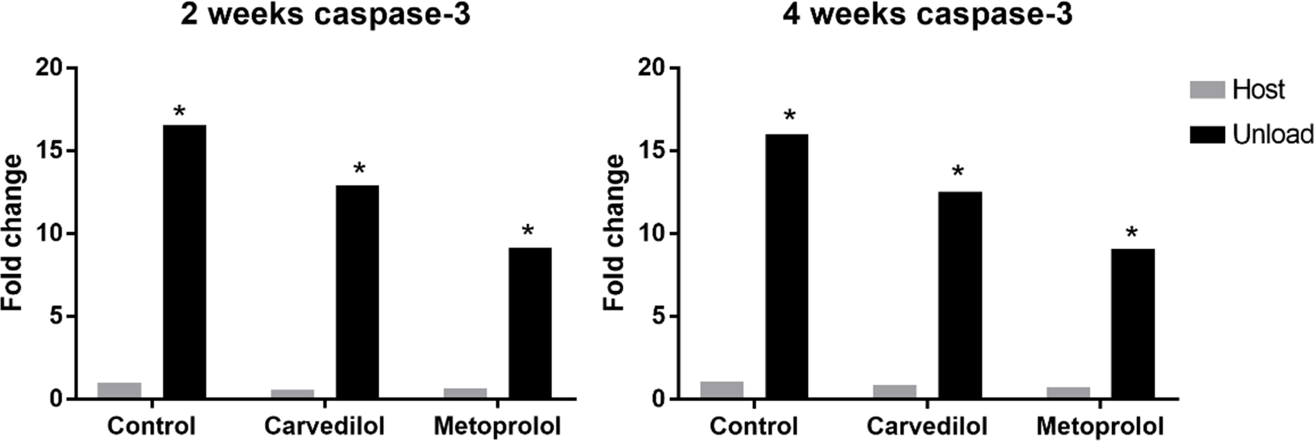
As shown in Figure 4, the unloaded hearts in all groups exhibited significant increases in the mRNA expression of caspase-3 after 2 and 4 weeks following transplantation. However, no significant decreases were observed in the unloaded hearts of the carvedilol and metoprolol groups compared with those of the control group. No significant differences in β1-adrenoreceptor mRNA expression levels were observed between the host and unloaded hearts either within or among all three groups, after either 2 or 4 weeks. In all groups, β2-adrenoreceptor mRNA expression increased significantly in the unloaded hearts compared with that in the host hearts. In unloaded hearts, the β2-adrenoreceptor mRNA expression levels in the metoprolol group 2 and 4 weeks after transplantation were significantly lower than those in the control group.
Measurement of caspase-3 mRNA expression fold changes using RT-qPCR. All fold changes represent differences relative to the host hearts of the control group 2 weeks after transplantation (*Fold change >2 or <0.5 versus host hearts in the same treatment group). RT-qPCR = reverse-transcription quantitative real-time polymerase chain reaction.

Discussion
To succeed in weaning a patient from a left ventricular assist device, specific pharmacological interventions with left ventricular assist device support could be an important aspect of therapy enabling patients to recover from heart failure. Mechanical support with a left ventricular assist device leads to a decrease in neuroendocrine activation and myocyte hypertrophy. Pharmacological interventions are aimed at preventing such pathological hypertrophy and remodelling and at normalising cellular metabolic function. In clinical settings, angiotensin-converting enzyme inhibitors, β-blockers, angiotensin II antagonists, and aldosterone antagonists could be helpful in reversing cardiac remodelling in patients using a left ventricular assist device. Reference Birks, George and Hedger12 This study focused on comparing the effects of the β-blockers carvedilol and metoprolol on unloaded rat hearts.
Cardiac unloading with a left ventricular assist device produces several changes in the myocardium. Loss of cardiomyocytes via apoptosis is one such change that causes cardiac remodelling. Brinks et al. showed that the mechanical unloading of normal rat hearts increases apoptosis, which is aggravated 60 days after transplantation and is related to the loss of myocardial mass. Reference Brinks, Giraud and Segiser5 Razeghi et al reported that myocardial atrophy is determined by a change in the balance of protein degradation. At the molecular level, atrophic remodelling of the normal heart is characterised by the simultaneous activation of proteolysis and protein synthesis by the ubiquitin proteasome proteolytic pathway and mammalian target of rapamycin activation, respectively. Reference Razeghi, Sharma and Ying13
A significant decrease in myocyte diameter in unloaded hearts compared with that in host hearts was observed in the present study. Although left ventricular weights could not be measured in this study, the decrease in myocyte diameter suggested atrophy in the unloaded hearts. However, neither carvedilol nor metoprolol reduced this effect, and no time-based differences were observed after transplantation. This study demonstrated that complete mechanical unloading was an important factor in myocardial atrophy induction. Atrophy is a physiological response to a variety of stimuli, but this process can be reversed by reloading. An experimental study showed that unloading for 1 week caused a significant decrease in the myocyte cross-sectional area and that subsequent reloading for 1 week caused the myocyte cross-sectional area to return to normal. Reference Thompson, Marino, Uboh, Kent and Cooper14 Schena et al also demonstrated that myocardial atrophy was more pronounced in the non-working heart than in the working heart. Reference Schena, Kurimoto and Fukada3
Ventricular fibrosis is thought to involve a process that organises the ventricular remodelling substrate by interrupting intercellular electrophysiological coupling. In the present study, the mechanical unloading of normal rat hearts led to the progression of myocardial fibrosis; this was more prominent in the unloaded hearts of the control group after 4 weeks following transplantation than after 2 weeks. The results of this study are consistent with previous reports that mechanical unloading increases myocardial fibrosis. Reference Oriyanhan, Tsuneyoshi, Nishina, Matsuoka, Ikeda and Komeda15
In the present study, the degree of myocardial fibrosis in unloaded hearts was significantly lower in the metoprolol group than in the control group after 2 and 4 weeks. Consistent with our data, it has been reported that metoprolol treatment normalises myocardial oxygen consumption, decreases myocardial damage, augments cardiomyocyte survival, and improves cardiac function. Reference Sun, Wang and Zhang16 Serpi et al also demonstrated in a dilated cardiomyopathy model in rats that metoprolol treatment helps to reverse remodelling by reducing apoptosis and fibrosis. Metoprolol was found to reduce myocyte hypertrophy and collagen deposition in a rat model of renovascular hypertension-induced cardiac hypertrophy. Reference Serpi, Tolonen and Tenhunen17 Although the latter study was performed in a rat model of obstructive sleep apnea, the authors reported that ventricular fibrosis is markedly suppressed by metoprolol and that it can be decreased by upregulating the profibrotic growth cytokine, transforming growth factor β1. The expression of p38 mitogen-activated protein kinases is regarded as a significant factor in apoptosis and fibrosis in cardiovascular disease. The antifibrotic effect of metoprolol is partially influenced by p38 mitogen-activated protein kinases. Reference Li, Yan and Zhao18
To assess the degree of apoptosis, terminal deoxynucleotidyl-transferase-mediated dUTP nick end-labelling positive cells and caspase-3 mRNA expression levels were analysed. Terminal deoxynucleotidyl-transferase-mediated dUTP nick end-labelling staining is known to detect apoptosis by labelling the DNA-free 3′–OH ends of DNA fragments. Compared with the host hearts, the unloaded hearts exhibited a significant increase in the proportion of terminal deoxynucleotidyl-transferase-mediated dUTP nick end-labelling positive cells. However, in unloaded hearts, the proportion of terminal deoxynucleotidyl-transferase-mediated dUTP nick end-labelling stained cells in the metoprolol group tended to decrease, while no significant differences were observed between the treatment and control groups. In addition, no significant differences in caspase-3 levels were observed in the treatment groups, although decreased caspase-3 expression was observed in the metoprolol group compared with that in the control group.
Other studies have reported significant increases in the proportion of terminal deoxynucleotidyl-transferase-mediated dUTP nick end-labelling positive cells in transplanted hearts compared that with native hearts. Reference Schena, Kurimoto and Fukada3,Reference Tsuneyoshi, Oriyanhan and Kanemitsu4 Tsuneyoshi et al. also reported that caspase-3 expression increased in unloaded hearts and confirmed that clenbuterol was helpful for decreasing the caspase-3 activity. Reference Tsuneyoshi, Oriyanhan and Kanemitsu4 They suggested that β2-adrenoreceptor stimulation might help protect cardiomyocytes from apoptosis. Ahmet et al suggested that the effect of a β2-adrenoreceptor agonist might be regulated through β2-adrenoreceptor–inhibitory G-protein coupling. Inhibitory G blocks the action of stimulatory G, which is considered to be central to β1-adrenoreceptor-stimulated apoptosis. Reference Ahmet, Krawczyk, Heller, Moon, Lakatta and Talan19
SERCA2a is thought to function in intracellular Ca2+ handling. Therefore, downregulation of SERCA2a reflects reduced sarcoplasmic reticulum Ca2+ uptake capacity, disturbed excitation–contraction coupling processes, and impaired contractile function. Reference Terracciano, Hardy, Birks, Khaghani, Banner and Yacoub20 Downregulation of SERCA2a is also observed in hypertrophied or failing hearts despite the opposite hemodynamic changes in unloaded and hypertrophied hearts. Reference Tsuneyoshi, Oriyanhan and Kanemitsu4 Tsuneyoshi et al reported that SERCA2a expression tends to increase in the unloaded–infarcted group compared with that in the infarcted-only group. Reference Tsuneyoshi, Oriyanhan and Kanemitsu10 In the current study, decreased SERCA2a levels were observed in the unloaded hearts compared with those in the host hearts after 2 weeks in the control and carvedilol groups, but no significant differences were observed between the host and unloaded hearts in the metoprolol group. These findings suggest that metoprolol was superior to carvedilol for preventing SERCA2a downregulation, although carvedilol treatment increased SERCA2a levels. Similarly, Zou et al reported that metoprolol improves SERCA2a expression in a rabbit heart failure model. Reference Zou, Liu and Jiang21
The clinical success of carvedilol, a nonselective β1- and β2-adrenoreceptor blocker, is well established. It is known that carvedilol reduces the mortality of patients with heart failure more effectively than metoprolol in clinical trials. Reference Poole-Wilson, Swedberg and Cleland22 However, a meta-analysis of human studies concluded that carvedilol is not superior to metoprolol for the treatment of myocardial infarction. Reference Li, Chen and Gao9 Sun et al. also reported that carvedilol induced a greater increase in SERCA2a expression than metoprolol in a rat model of heart failure. Reference Sun, Hu, Wang, Hu and Zhou23 However, no study has directly compared the two drugs in the unloaded heart.
Interestingly, the present study showed that SERCA2a expression in unloaded hearts after 4 weeks following transplantation was significantly decreased compared with that after 2 weeks. This was consistent with other studies that reported that SERCA2a expression increases in the early period of left ventricular assist device use Reference Heerdt, Holmes and Cai2 and peaks by 20 days. Another study in a rat model of heart failure reported that SERCA2a levels peak and normalise after 4 weeks of unloading, Reference Oriyanhan, Tsuneyoshi, Nishina, Matsuoka, Ikeda and Komeda15 and Xydas et al also demonstrated that cardiac recovery in patients with a left ventricular assist device peaks by day 60. Reference Xydas, Rosen and Ng24 The optimal duration for mechanical unloading is therefore still debatable.
The present study analysed β-adrenoreceptor density by measuring the expression of β1- and β2-adrenoreceptor mRNA. There were no significant differences in β1-adrenoreceptor expression levels among the three groups. Unexpectedly, significant upregulation of the β2-adrenoreceptor was observed in the unloaded heart compared with that in the host heart, and significant downregulation of the β2-adrenoreceptor was observed in the metoprolol group compared with that in the control group. Metoprolol is known to upregulate the myocardial β1-adrenoreceptor and to reverse the uncoupling of the myocardial β2-adrenoreceptor that occurs in heart failure. β2-adrenoreceptor signalling is important for the recovery of unloaded hearts. Reference Brouri, Findji and Mediani25
Wang et al found that chronic partial unloading in failing rat hearts increases β1-adrenoreceptor and β2-adrenoreceptor densities. Reference Wang, Tsukashita and Nishina26 In the latter study, overexpression of β2-adrenoreceptor at even higher levels was shown to lead to the development of cardiomyopathy and early death. Although the β2-adrenoreceptor is easier to downregulate than the β1-adrenoreceptor Reference Brouri, Findji and Mediani25 and some drugs might cause the destruction of β2-adrenoreceptors, the downregulation of β2-adrenoreceptor in the metoprolol group is a controversial result. If the overexpression of β2-adrenoreceptor-induced cardiac toxicity in the unloaded heart in the control group in this study, downregulation of the β2-adrenoreceptor in the metoprolol group may have contributed to functional recovery.
Ahmet et al. studied adrenoreceptors in a rat model of heart failure by using a β1-adrenoreceptor blocker and a β2-adrenoreceptor agonist. They reported that the combination of β2-adrenoreceptor stimulation with a β1-adrenoreceptor blockade increases the therapeutic effectiveness of the β1-adrenoreceptor blockade. Reference Ahmet, Krawczyk and Zhu8 Tsuneyoshi et al. also reported that the β2-adrenoreceptor agonist clenbuterol improves cardiac function and β-adrenergic responsiveness, but that it also decreases β2-adrenoreceptor levels. Reference Tsuneyoshi, Oriyanhan and Kanemitsu4
In a recent prospective study, Birks et al suggested that the pharmacological management of severe heart failure with the use of left ventricular assist devices should comprise two stages. The first stage includes high-dose carvedilol treatment. The second stage includes use of the β1-blocker bisoprolol, followed by clenbuterol treatment when the left ventricular end-diastolic dimension has regressed maximally. Reference Birks, George and Hedger12 Birks et al reported good outcomes for the reversal of heart failure through the combination of mechanical and pharmacological therapy. Although it is known that different β-blockers may help cardiac recovery in the mechanically unloaded heart, the results of this study provide new information about how the effects of the two drugs carvedilol and metoprolol compare.
This study had some limitations. First, it was performed on a small number of rats. Although we performed all procedures within 40 minute, the heterotopic transplantation requires arrest of the donor heart, so ischaemia could have had an influence in the unloaded rat hearts. Moreover, we performed heterotopic transplantation in normal rather than in failing hearts but did not perform functional studies of the myocardium. In addition, partial unloading models are known to improve myocardial function, whereas our transplantation model was completely unloaded. The parameters were measured for only 4 weeks, and further extended studies are needed. To further evaluate the practical effect of β-blockers, we plan to create a dilated cardiomyopathy heart model in rats, transplant the failing heart, and observe the anatomical changes.
In conclusion, it was found that treatment with metoprolol was superior to that with carvedilol for preserving cardiac muscle, preventing fibrosis, and improving calcium handling; however, metoprolol did not influence apoptosis. Further studies are needed to determine the optimal pharmacological interventions for patients with severe heart failure who have left ventricular assist devices.
Acknowledgements
The authors declare that no acknowledgments exist.
Financial support
This study was supported by the Research Institute for Convergence of Biomedical Science and Technology (30-2016-016), Pusan National University Yangsan Hospital.
Conflicts of interest
The authors declare that no conflict of interest exist.
Ethical standards
The authors assert that all procedures contributing to this work comply with the ethical standards of the relevant national guides on the care and use of laboratory animals and has been approved by the institutional committee (Pusan National University Yangsan Hospital).