Surgery was accepted as the consuetudinary treatment for ventricular septal defect closure, which is not the case today.Reference Scully, Morales and Zafar 1 The last 10–20 years have witnessed the invention of new devices, and percutaneous ventricular septal defect closure is being used more commonly with satisfactory results.Reference Lock, Block, McKay, Baim and Keane 2 – Reference Butera, Carminati and Chessa 5 Percutaneous ventricular septal defect closure with the Amplatzer Duct Occluder-II device (St. Jude Medical, St. Paul, Minnesota, United States of America) has been undertaken in the study centre since April, 2011. We have published our initial results previously.Reference Narin, Baykan and Pamukcu 6 We have concluded that the Amplatzer Duct Occluder-II device can be safely and effectively used for ventricular septal defect closures. Our experience, however, was limited to 20 patients and 3.5 years. In the current study, the patient population has increased to 49, and the follow-up period to 5.5 years. In this paper, we aim to share the mid-term results of these patients.
Material and methods
Patients
Percutaneous ventricular septal defect closure of 49 patients was performed using the Amplatzer Duct Occluder-II device in our institution between April, 2011 and October, 2016. A haemodynamically significant left-to-right shunt was detected by clinical examination and echocardiography of the patients. Similar to our previous study, the inclusion criteria were as follows: ventricular septal defect diameter <6 mm, as the waist diameter of the Amplatzer Duct Occluder-II is 6 mm; prominent left-to-right shunt, demonstrated using either telecardiography – revealing cardiothoracic index ⩾0.5 and pulmonary venous congestion – or using echocardiography for left atrial and ventricular enlargement, or by catheterisation (Qp/Qs>1.5). The local research Ethics Committee approved the study. All parents were informed about the procedure and its complications, and written consent was obtained from each of them before the procedure.
Procedure
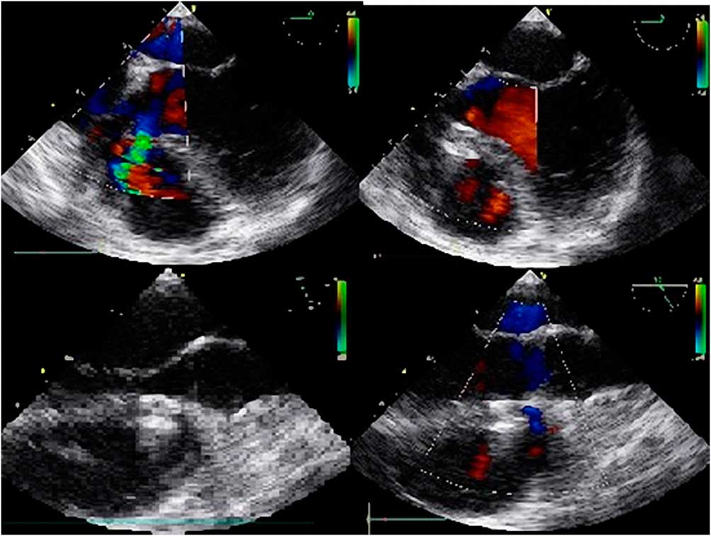
The procedure was performed under general anaesthesia. Intravenous prophylactic antibiotics were administered before the procedure. Heparinisation dosage was 100 IU/kg during the procedure. Additional dosages were added to maintain activating clotting time >200 seconds. Patients were monitored using an electrocardiogram throughout the procedure. Both femoral venous and arterial routes were accessed. The anatomy, location, and size of the defect were defined using a left ventriculogram in left oblique view. The procedure was performed under fluoroscopy with the guidance of trans-thoracic and/or trans-oesophageal echocardiography (Fig 1). The appropriate device size was chosen to be at least 0.5 to 1 mm larger than the defect size as measured at the largest diastolic phase by colour Doppler trans-oesophageal and/or trans-thoracic echocardiography or by ventriculography. The largest device was accepted in case of a discrepancy between two measurements. A partly cut pigtail or a Judkins right catheter sized 4 or 5 Fr was used to pass through the ventricular septal defect. A hydrophilic guidewire was passed across the defect into the right ventricle and then into the pulmonary artery or superior vena cava, where it was snared and withdrawn from the femoral vein, thus establishing an arterio-venous loop. Subsequently, a long sheath (4–5 Fr) was advanced to the left ventricle via the arterio-venous circuit. An occluder was advanced into the delivery sheath, and then positioned on the defect. The device was released if its position was appropriate, as observed by fluoroscopy and echocardiography. The presence of residual shunt was checked using trans-thoracic and/or trans-oesophageal echocardiography and left ventriculography. Cardiac rhythm was monitored in all patients during the first 24 hours after the procedure. All patients were discharged within 2–3 days after the procedure with acetylsalicylic acid (3–5 mg/kg/daily), which was administered for 6 months in all patients. Physical examinations, electrocardiography, and trans-thoracic echocardiography were performed at 1, 3, 6, and 12 months after the procedure, and yearly thereafter, and 24-hour Holter monitoring was performed 3 months after the procedure.
Trans-thoracic echocardiography views of percutaneous ventricular septal defect and atrial septal defect closure of a patient.

Statistical analysis
Data are expressed as frequency or percentage for nominal variables, as median and range for categorical variables, and as mean±SD for continuous variables.
Results
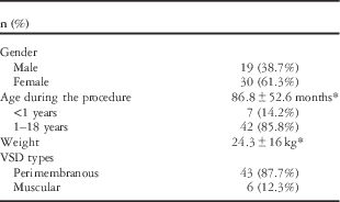
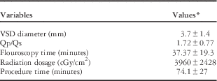
In our institution, during the mentioned period, 49 percutaneous ventricular septal defect closures were performed using the Amplatzer Duct Occluder-II device. The mean age of the patients was 86.8±52.6 months. The youngest patient was 4 months old and the oldest patient was 18 years old. In all, seven patients were <1 year old. The mean weight of the patients during the procedure was 24.3±16 kg, with a minimum body weight of 5 kg and a maximum of 76 kg. The mean defect diameter was 3.7±1.4 mm. Of the patients enrolled in the study, 30 were male and 19 were female. Demographic data of the patients are summarised in Table 1. Associated heart defects were found only in 10 patients: eight of them had an atrial septal defect, one had patent ductus arteriosus, and one had mild aortic valvular stenosis. Atrial septal defect of three patients and patent ductus arteriosus were closed percutaneously in the same session as the closure of the ventricular septal defect.
Demographic features of patients.

VSD=ventricular septal defect
* Mean values expressed as mean±SD
The Amplatzer Duct Occluder-II Additional Size device was used in two patients. One of them was operated upon for ventricular septal defect and had a residual 2-mm-sized defect. This defect was closed with a 4×2-sized Amplatzer Duct Occluder-II Additional Size; the other defect, of size 2.5 mm, was occluded with a device of size 4×6.
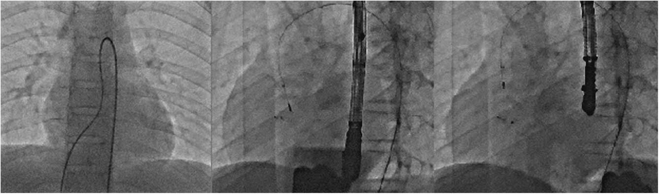
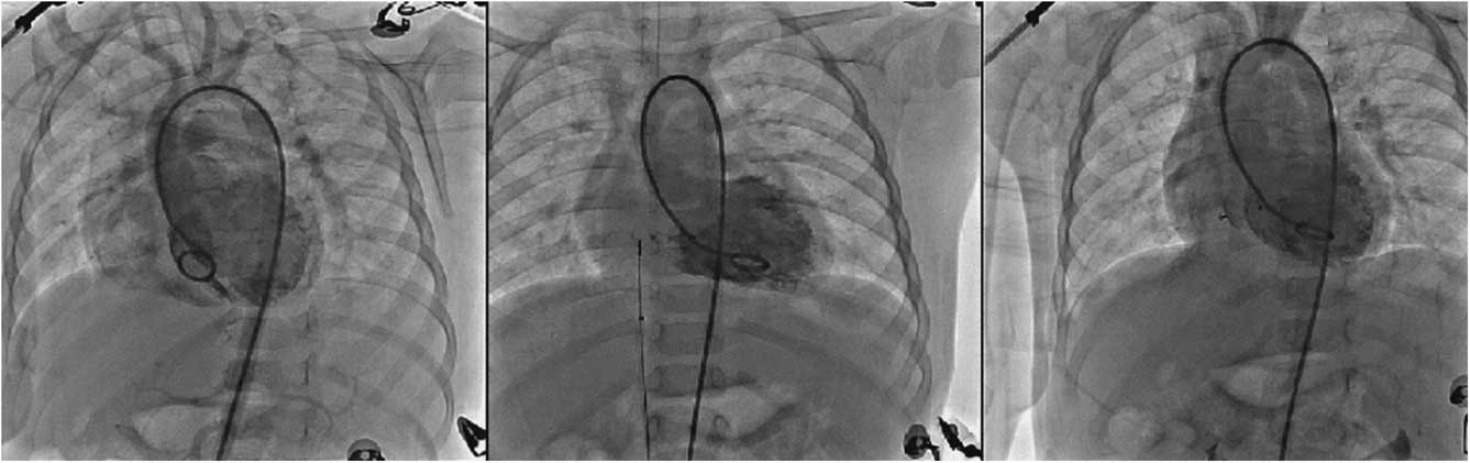
Mean fluoroscopy and total procedure times were 37±19.3 and 74.1±27 minutes, respectively (Table 2). All of the perimembranous defects were aneursymatic and mostly tunnel shaped. In perimembranous aneursymatic defects, the distance of the defect to the aortic valve ranged between 2 and 8 mm. The mean Qp/Qs value was 1.72±0.77 (Table 2). The arterial route was used as the site of closure in only five patients (Fig 2); the venous route was more frequently employed, in 44 patients (Fig 3). The complete closure rate of the ventricular septal defect immediately after the procedure was 85%, as documented by angiography, with seven patients having a residual shunt. The residual shunt persisted in three of these seven patients. Therefore, the overall complete closure rate in all cases was 93.7% on follow-up. The left ventricle and left atrium decreased to normal size during the follow-up with trans-thoracic echocardiography. Cardiac monitoring was carried out during the procedure, and the electrocardiogram was examined after the 1st day and at each follow-up. We observed various degrees of heart block in two patients during the procedure from manoeuvres of the catheter or the guidewire, but they usually did not persist: both cases recovered to sinus rhythm. There were no major arrhythmias such as left bundle branch block, PR prolongation, or complete heart block just after implantation. A single patient was, however, admitted with dyspnoea 6 months after the procedure. Electrocardiography revealed complete atrioventricular block, and the trans-thoracic echocardiogram revealed a 20-mm-sized pericardial effusion, which was the only major complication that we observed. Essentially, there was no problem in the 1st and 3rd follow-up months. Pericardiocentesis was performed, and 4 weeks of anti-inflammatory treatment was given. Despite the treatment, complete atrioventricular block persisted, and a permanent pacemaker was implanted. The patient was last seen during her first follow-up year, and was doing well with the pacemaker. We did not observe any major complications like death, vascular complications, or device embolisation. Aortic regurgitation was already present in two patients before closure, which neither increased nor disappeared after device implantation. There was, however, no new-onset aortic regurgitation in any patient just after the defect closure or during follow-up. The longest follow-up interval was 66 months. The mean follow-up period was 17±13.7 months.
Percutaneous ventricular septal defect closure of a patient from the arterial site.

Percutaneous ventricular septal defect closure of a patient from the venous site.

Angiographic data of patients.

VSD=ventricular septal defect
* These are the mean values, expressed as mean±standard deviation
Discussion
Percutaneous ventricular septal defect closure has been performed using the Amplatzer Duct Occluder-II device in our institution since April, 2011. We believe that this method is safe and effective. We have published our initial results previously,Reference Narin, Baykan and Pamukcu 6 and now wish to share the mid-term results.
In our previous study, we reported that Amplatzer Duct Occluder-II is much softer, has low-profile retention discs and is very safe in treating aneursymatic ventricular septal defects, even when they are close to the aortic valve. We have reported 20 cases, and two of them were <1 year old during the procedure. We did not face any complication during a 2-year follow-up of these infants.
It is important to note that some complications such as heart block may occur later on as a delayed complication. Therefore, we persistently followed up these infants at regular intervals. During this time, the number patients whose ventricular septal defect was closed using Amplatzer Duct Occluder-II increased to 49, and the follow-up period was extended to 66 months.
The closure of the perimembranous ventricular septal defect has certain risks because of its critical location close to the conduction system and the aortic valve.Reference Fischer, Apostolopoulou and Rammos 7 In previous studies, the risk for complete atrioventricular block after surgical closure was reported as 0.7%,Reference Scully, Morales and Zafar 1 , Reference Andersen, Leval and Tsang 8 and as <1% after percutaneous closure.Reference Saurav, Kaushik and Mahesh Alla 9 The incidence of this complication varies across studies: Fu et al reported complete atrioventricular block in 1/35 patientsReference Fu, Bass and Amin 10 ; Butera et al identified 2 patients in the early period and another 4 in the late term.Reference Butera, Carminati and Chessa 11 They used muscular and perimembranous ventricular septal defect occluder devices in their study. There were various factors causing an atrioventricular block, such as the location of the defect, device type, device size, etc. Butera et alReference Butera, Carminati and Chessa 11 suggested that age was an important risk factor and reported that being <6 years old increased the risk. Yang et al defined other risk factors such as having a longer aortic rim width, mismatch between the device size and defect, and shorter distance between the septal tricuspid leaflet and the lower rim of the ventricular septal defect.Reference Yang, Kong and Sheng 12
Finding an ideal device for children is important in order to minimise such complications. The Amplatzer Duct Occluder-II device for ventricular septal defect closure is not widely used and is still off-label. Vijayalakshmi et alReference Vijayalakshmi, Narasimhan and Singh 13 reported non-ductal shunt lesion closure using Amplatzer Duct Occluder-II. They postulated that Amplatzer Duct Occluder-II usage in ventricular septal defect closure decreased rates of complications such as complete heart block. No patient was reported as having a complete heart block in that study; however, the follow-up period was 20 months at most. We faced only one serious complication in this study: complete atrioventricular block 6 months after implantation. Therefore, the complication rate in the current study was 2%. This complication was thought to be related to atypical myocarditis owing to pericardial effusion, but there was no problem in the patient’s 1st and 3rd follow-up months. An A-V block may not cause pericardial effusion by itself. Pericardiocentesis was performed. In the follow-up of the patient, complete atrioventricular block persisted despite anti-inflammatory treatment; therefore, a permanent pacemaker was implanted. Myocarditis seems to be as harmless as the common cold, but the consequences may be serious, such as complete atrioventricular block, congestive heart failure, cardiogenic shock, and even death.Reference Freedman, Haladyn and Floh 14 – Reference Ichikawa, Sumitomo and Komori 16
Another serious risk to be aware of during follow-up is aortic regurgitation. It was already present in two of the study patients before closure. Fortunately, we did not face any new-onset aortic regurgitation in our case series, neither in the early period nor during the follow-up. As emphasised in our previous study, Amplatzer Duct Occluder-II can adapt to different shapes and fit into the defect without disturbing the aortic or the tricuspid valve. Kanaan et alReference Kanaan, Ewert and Berger 17 reported no increase in aortic or tricuspid valve regurgitation after the procedure. During the 66 months of follow-up in our study, we did not face any new-onset aortic or tricuspid insufficiency.
Residual shunts observed immediately after device implantation disappeared during a median follow-up period of 38 months in the study by Kanaan et al.Reference Kanaan, Ewert and Berger 17 In the current study, the total closure rate immediately after device implantation was 85%. A residual shunt, which was detected in seven patients, persisted only in three patients. Complete closure occurred in all cases (93.7%) on follow-up. Our findings were similar to those of Kanaan et al.Reference Kanaan, Ewert and Berger 17
With experience, we were able to include patients of a much younger age. We have commenced percutaneous ventricular septal defect closure of patients under 1 year of age. The data on ventricular septal defect closure for patients under 1 year of age are limited in the literature. Kanaan et alReference Kanaan, Ewert and Berger 17 reported ventricular septal defect closure in 31 patients with Amplatzer Duct Occluder-II over an 8-year period. It is an important study because it has the largest series of ventricular septal defects closed with the Amplatzer Duct Occluder-II device, and also because it involves ventricular septal defect closure of patients aged <1 year. The age range in the above study was too wide, ranging between 3 months and 55 years. They closed the ventricular septal defect in five patients aged <1 year, similar to our study (seven patients). Their median follow-up period was 38 months. As a major complication, device embolisation was seen in one patient and was recaptured. They did not encounter complete heart block, only minor arrhythmias. In this study, they concluded that Amplatzer Duct Occluder-II may be the ideal device for the closure of defects of moderate size (2–5 mm), especially in infants and small children, because of its better profile and trackability. Their complication rate was sufficiently low to be within acceptable limits. The study also reported that as the number of studies increase, Amplatzer Duct Occluder-II may not be off-label, and that it may be approved for ventricular septal defect closure in the future.
We believe that the current study contributes important data to the literature in this respect. The main strengths of our study are as follows: our patient population was the largest series that underwent closure with Amplatzer Duct Occluder-II (49 patients), and seven of these patients were <1 year old, which was similar to the study by Kanaan et al.Reference Kanaan, Ewert and Berger 17 Our follow-up period was 66 months, which is a relatively good time interval, but a longer interval should give more reliable results. When the complications are taken into consideration during the 66-month follow-up period, we can say that the Amplatzer Duct Occluder-II device is a safe and effective choice in selected cases for ventricular septal defect closure, even in infants aged <1 year.
Acknowledgements
None.
Financial Support
This research received no specific grant from any funding agency, commercial or not-for-profit sectors.
Conflicts of Interest
None.
Ethical Standards
The authors assert that all procedures contributing to this work comply with the ethical standards of the relevant national guidelines on human experimentation and with the Helsinki Declaration of 1975, as revised in 2008, and has been approved by the local research ethics committee.