Introduction
High ammonia (NH3) concentrations are postulated to exist in the subsurface of Titan, a moon of Saturn. Current modelling suggests that a primordial subsurface Titan ocean may have had concentrations of ammonia up to 15 wt%. However, today, because of the shrinkage of the ocean, Titan might host a subsurface water body with concentrations as high as 33 wt% (Fortes Reference Fortes2000). Although the connection between the surface and subsurface of Titan has not been demonstrated, the possibility of ammonia–water cryovolcanism has been suggested (Mitri et al. Reference Mitri, Showman, Lunine and Lopes2008), raising the question of the fate of any organics or terrestrial micro-organisms on spacecraft that come into contact with high ammonia solutions. Can terrestrial micro-organisms survive or even proliferate in these extraterrestrial environments of the outer solar system? The answer to this question will be informative for persons responsible for planetary protection regulations for missions to these cold worlds.
Ammonia is known to be toxic to micro-organisms (Deal et al. Reference Deal, Souza and Mack1975). There are three major mechanisms by which it can exert a toxic effect. First, un-ionized ammonium (NH3) is easily transported across the cell membrane and accumulates inside the cell as ammonium ions, where it interferes with the proton gradient across the cell wall by removing free H+. This causes a collapse of the proton motive force driving energy acquisition. Secondly, the reaction with water (NH3+H2O>NH4++OH−) is a redox reaction, potentially interfering with cellular redox reactions. Ammonia, a weak base, also causes an increase in pH. Deleterious effects on cell survival could potentially be caused by an increase in pH.
Free NH3 at concentrations of 6–12 mm (0.011–0.022%) have been suggested as the highest concentrations to be tolerable by a range of micro-organisms, the highest of these levels reported in methanogens (Mc Carty & McKinney Reference Mc Carty and McKinney1961; Braun et al. Reference Braun, Huber and Meyrath1981; De Baere et al. Reference De Baere, Devocht, van Assche and Verstrate1984; Koster & Lettinga Reference Koster and Lettinga1984; Angelidaki & Ahring Reference Angelidaki and Ahring1993). Much of the existing work has been conducted in sludges and digestion reactors where high concentrations of free NH3 accumulate. Siegel et al. reported evidence for the capability of a mutant strain of Penicillium notatum to take up a radiolabelled precursor of DNA, RNA and protein in a glycerol–NH3 medium at −40°C and apparent localization of labelled DNA precursor in the nucleoid region (Siegel et al. Reference Siegel, Nathan and Roberts1968), although these results have not been corroborated. In other studies survival of micro-organisms including a strain of Penicillium brevicompactum was reported on plants exposed to atmospheres with a high NH3 concentration up to 95% (Siegel & Giumarro Reference Siegel and Giumarro1965).
Although actively growing cells are affected by low concentrations of ammonia, there is little data on its effects on inactive organisms or those possessing resting states. The effects of ammonium ions on spore germination have been studied previously and were suggested to enhance Bacillus spore germination at sub-molar concentrations (Wolf & Thorley Reference Wolf and Thorley1957; Preston & Douthit Reference Preston and Douthit1984). Although ammonium is widely reported to be cytotoxic, experiments investigating the effects of ammonium on bacteria (Corynebacterium glutamicum, Escherichia coli and Bacillus subtilis) showed that sodium ions could elicit the same retardation of growth as ammonium when present at concentrations of 750 mm (1.5% v/v) or more, suggesting that ionic or osmotic effects caused growth inhibition, not the presence of ammonium per se (Müller et al. Reference Müller, Walter, Wirtz and Burkovski2006).
The purpose of this study was to ascertain whether organisms on spacecraft would remain intact after exposure to high liquid-ammonia concentrations (up to 35% v/v) in concert with low temperatures, and whether transient exposure to ammonia would compromise their ability to grow when re-exposed to favourable growth conditions.
Experimental procedures
Culturability of soil microbes exposed to NH3
Most micro-organisms within spacecraft assembly clean rooms are either human-associated or of environmental origin. Spore-forming micro-organisms such as Bacillus spp. are common contaminants, as well as other organisms that are resistant to desiccation or have environmentally robust resting states (La Duc et al. Reference La Duc, Osman, Vaishampayan, Piceno, Andersen, Spry and Venkateswaran2009).
To sample a large diversity of potential micro-organisms of interest, subsurface soil (circa 2–6 cm depth) was collected from an unvegetated site adjacent to a lawn at The Open University in Milton Keynes, UK. It was mixed before dividing into two portions. One portion was subjected to ‘activation’ by heating in a water bath at 70°C for 22 minutes, before being left at room temperature (RT) for 26 hours. The ‘unactivated’ soil was likewise incubated at RT. To 10 g soil in 50 ml tubes, 40 ml of a solution containing 5, 17.5 or 35% (v/v) NH3 was added. Ammonia solutions were prepared from a 35% (v/v) stock solution (0.88 S.G., Fisher Scientific, Leicestershire, UK) and diluted as required in distilled water. Controls contained soil and 40 ml distilled water only. Tubes were shaken thoroughly by hand after NH3 addition and were incubated immediately at −80°C or RT. Each treatment had four replicates. In addition to NH3 treatments, additional treatments without NH3 but with pHs amended to correspond as closely as possible to those of NH3 solutions of 5% (pH 11.1), 17.5% (pH 12.1) and 35% (13.1) were prepared by amending distilled water with NaOH prior to addition to soil.
Tubes were sampled (following mixing by hand) after incubation for 1, 12 and 48 weeks, except for RT treatments, which were not sampled at 48 weeks. At each sampling −80°C treatments were thawed at RT prior to sample removal and returned to −80°C as quickly as possible. Samples were serially diluted in distilled water and plated in triplicate on nutrient agar (Oxoid, Basingstoke, UK). Plates were incubated aerobically or anaerobically at RT for 9 days before recording colony forming units (cfu). Regression analyses and analysis of variance (ANOVA) were used to determine whether activation treatment, exposure time and NH3 significantly influenced culturability.
Determination of NH3 by distillation
To determine whether the concentration of NH3 had decreased during the soil-exposure experiment, the concentration in the liquid phase of treatments was determined at 48 weeks. Samples were diluted in dH2O and 0.5 ml added to a solution containing 5 ml boric acid (1% w/v) and a mixed indicator, before distillation in a Vapodest 30. The distilled sample was titrated in duplicate against 0.2 N H2SO4 using a Titronic 96 (Schott). Blanks were also titrated, in addition to the NH3 stock solution. Total N per ml of sample was calculated as

where T=ml of standard acid to titrate sample, B=ml of standard acid to titrate blank, N=normality of standard acid, 14=equivalent weight, Vp=volume distilled, W=weight of sample.
Identification of bacteria isolated following NH3 exposure
Selected bacterial isolates (labels preceded with LKS) were identified by partial sequencing of the amplified 16S rRNA genes (Mclab, USA). Amplicons were generated by colony PCR with primers pA and pH (Bruce et al. Reference Bruce, Hiorns, Hobman, Osborn, Strike and Ritchie1992) where each 50 μl reaction mixture contained 0.2 μm each primer, 200 μm each dNTP (New England Biolabs), 2.5 U Taq DNA polymerase, 2 mm MgCl2 and 1×PCR buffer (200 mm Tris/HCl (pH 8.4) and 500 mm KCl) (Invitrogen Corporation, Paisley, UK). Amplifications were performed in a G-Storm GS1 thermal cycler (GRI Ltd, Essex, UK), with an initial denaturation at 94°C for 5 minutes, followed by 35 cycles of 94 °C for 1 minute, 55°C for 40 seconds and 72°C for 40 seconds, with a final extension for 10 minutes at 72°C. Amplicons were sequenced using primer pH and/or pA. Isolate sequences were searched against the RDP (Ribosomal Database Project) (Cole et al. Reference Cole, Wang, Cardenas, Fish, Chai, Farris, Kulam-Syed-Mohideen, McGarrell, Marsh and Garrity2009) to determine their phylogenetic affiliations. A neighbor-joining phylogenetic tree was constructed from partial 16S rRNA gene sequences of isolates and their closest database relatives, aligned in BioEdit (Hall Reference Hall1999) over E. coli nucleotide positions 112–673. Trees were constructed in MEGA4 (Tamura et al. Reference Tamura, Dudley, Nei and Kumar2007) using the Jukes–Cantor (Jukes et al. Reference Jukes, Cantor and Munro1969) algorithm. Isolate sequences have been deposited in GenBank under accession numbers JF502702–JF502828.
Exposure of spores to NH3 at −80°C
B. subtilis DSM10, which was used as a defined control, and soil isolate LKS-9 (JF502810) were grown on nutrient sporulation medium (NSM), consisting of nutrient agar with 1 mm MgCl2·6H2O. Biomass from 5-month-old plate cultures, grown and maintained at RT, was incubated at −80°C in water containing NH3 at concentrations of 0, 5 and 35% (v/v). All treatments were performed in triplicate. Prior to the addition of biomass, tubes (15 ml) containing NH3 and/or water were cooled on ice. Inocula were prepared in chilled sterile distilled water and added rapidly to prevent spore germination prior to inoculation. Immediately after inoculation, tubes were mixed vigorously to ensure homogeneous suspensions and contents aliquoted into smaller (1.7 ml) tubes to prevent repeated freeze–thaw cycles during future samplings. These tubes were incubated immediately at −80°C. The initial number of cfu was determined by plating immediately on nutrient agar, in triplicate, following serial dilution. Plates were incubated at RT for 6 days prior to counting. Subsequent samplings were performed 44 and 77 days.
Scanning electron microscopy (SEM)
In addition to determinations of cfu, spores of B. subtilis DSM10 and LKS-9 exposed to NH3 in water (as above) or nutrient broth at −80°C, were observed at intervals by SEM. Samples of 1 ml volume were filtered onto 0.2 μm pore size black polycarbonate filters (Fisher Scientific, Leicestershire, UK). Filters were gold coated prior to observation in secondary electron mode with a field emission gun scanning electron microscope (FEGSEM) (Zeiss Supra VP), using an accelerating voltage of 8 kV and a 14 mm working distance.
Results
Culturability of soil microbes exposed to NH3
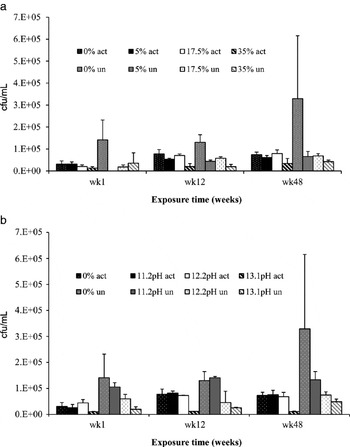
The effects of NH3 on the culturability of soil microbes was assessed by incubating soil, both at RT and −80°C, in the presence of up to 35% (v/v) NH3. Microbial culturability, represented by cfu at 1, 12 and 48 weeks, is shown in Fig. 1. On some plates accurate determination of cfu was hampered by the presence of spreading growth forms with obscured the ability to count colonies, accounting for high standard deviations.
Colony forming units (cfu) obtained on nutrient agar following exposure of activated (act) and unactivated (un) soil at −80 °C, to (a) NH3 and (b) alkaline pH conditions over time.

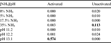
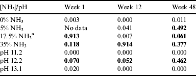
Exposure time significantly affected cfu abundance for both activated and unactivated soils at −80°C (Table 1), as revealed by a one-way ANOVA analysis and confirmed by regression analysis. The activation state of soil was shown to be non-significant in the presence of NH3 at −80°C, with the exception of 17.5% after 12 weeks of exposure (Table 2). Two-way ANOVA analyses revealed a significant interaction of NH3 with time at −80°C (not shown). Significant effects of activation treatment were observed for 11.2 and 13.1 pH treatments, however, significant effects at pH 12.2 were not observed. With some exceptions, differences in culturability between NH3 and the corresponding pH treatment were not significant (not shown).
P-values resulting from a one-way ANOVA analysis to determine the effects of exposure time at −80°C on cfu abundance for activated and unactivated soil at each NH3 concentration and pH. Results in bold indicate those relationships for which regression analysis returned a non-significant P-value (⩾0.05)
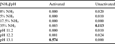
P-values resulting from the one-way ANOVA analysis to determine the effect of activation treatment on soil cfu abundance following exposure at −80°C for 1, 12 and 48 weeks. Results in bold indicate those relationships for which regression analysis returned a non-significant P-value (⩾0.05)
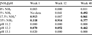
a A high number of fungi on plates in this treatment obscured bacterial counts and may account for this value.
Determination of NH3 by distillation
The % concentration of N in soil treatments did not decrease substantially during the course of the experiment. The average N% in 35% treatments was 0.7752, compared with 0.7952 in the stock solution. The N% of the control tubes was 0.0224.
Identification of bacteria isolated following NH3 exposure
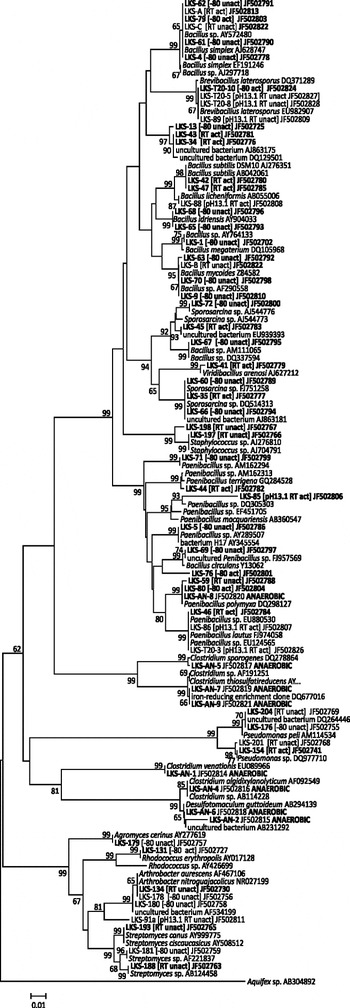
DNA sequencing of bacteria isolated on nutrient agar following soil exposure to NH3 revealed the dominance of Firmicutes, particularly the order Bacillales (Fig. 2). Within this order, bacteria belonging to the genera Bacillus, Sporosarcina, Viridibacillus, Paenibacillus, Staphylococcus and Brevibacillus were identified following exposure to 35% NH3. Also identified, however, were isolates belonging to the phyla Actinobacteria (Arthrobacter and Streptomyces) and Proteobacteria (Pseudomonas), albeit at a much lower frequency. The phylogenetic placement of these isolates relative to their closest matches within the RDP is shown in Fig. 2.
Neighbor-joining phylogenetic tree of partial 16S rRNA gene sequences from soil bacteria isolated after exposure to NH3, and their closest database representatives. Trees were constructed as described in methods, using 1000 bootstrap replications (values shown at nodes). Scale bar represents 1% sequence divergence. Isolates from soil exposed to 17.5 or 35% (v/v) NH3 are indicted in bold. Temperature and soil activation treatment are indicated in parenthesis. Sequences from isolates cultured at RT under anaerobic conditions are also indicated. Additional sequenced isolates belonging to the genera Bacillus, Paenibacillus and Sporosarcina, and identical or almost identical to those shown have been excluded to avoid repetition.

Exposure of spores to NH3 at −80°C
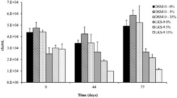
The survival of spores of B. subtilis DSM10 and soil Bacillus isolate LKS-9 (JF502810) at −80°C, in the presence of up to 35% (v/v) NH3 was observed by culturing on nutrient agar following 44 and 77 days incubation. Counts, relative to those at the time of inoculation are shown in Fig. 3.
Culturability (cfu) of spores of B. subtilis DSM10 and soil isolate LKS-9 (JF502810, a Bacillus sp.) both prior to and following exposure at −80°C to NH3 at concentrations of 0, 5 and 35%.

SEM
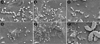
Spores were observed in suspensions prepared in water and stored at −80°C. Spores were also observed in 35% (v/v) NH3 after storage at −80°C (Fig. 4). Preparations containing NH3 and media had mineral rosette crystal structures associated with them (Fig. 4(e)). The composition of a typical crystal, as determined by Energy Dispersive Spectroscopy (EDS) was 57.6% C, 22.7% O, 0.3% Na, 6.9% Mg, 11.5% P and 0.9% K.
SEM of spores of isolate LKS-9 (JF502810) (a–c) and DSM-10 (d–f) exposed to 0 or 35% (v/v) NH3. (a, d) Spores in water (0% NH3) at RT. (b, e) Spores at −80°C in water with 0% NH3. (c, f) Spores at −80°C in media with 35% NH3.

Discussion
To test the hypothesis that a diversity of micro-organisms of interest to planetary protection could survive solutions of high ammonia concentrations, samples of soil were exposed to varying concentrations of ammonia at RT and −80°C.
After 3 months of exposure, a large number of organisms were isolated. Most culturable organisms that were identified were from spore-forming genera, suggesting that these resting states conferred resistance to ammonia. The species isolated, however, may also reflect the specific cultivation methods. Spore-formers are also the organisms most likely to survive on spacecraft surfaces. The majority of isolates were from the phylum Firmicutes, particularly the order Bacillales. Isolates of another spore-forming genus, Streptomyces (phylum Actinobacteria) were also obtained. Following exposure to high ammonia concentrations, soil incubated under anaerobic growth conditions yielded a prevalence of organisms phylogenetically affiliated to the genus Clostridium.
However, non-spore-forming bacteria were also obtained. The survival of the organisms closely affiliated with the non-spore-forming genus Arthrobacter, is consistent with the known environmental tolerances of this genus. They are known to be capable of inhabiting dry, oligotrophic environments (Cacciari & Lippi Reference Cacciari and Lippi1986; Reddy et al. Reference Reddy, Prakash, Matsumoto, Stackebrandt and Shivaji2002). They have been isolated from the surface of rocks (Gounot, Reference Gounot1967; Heyrman et al. Reference Heyrman, Verbeeeren, Schumann, Swings and De Vos2005) and are known to sequester metals from rocks (Brantley et al. Reference Brantley, Liermann, Bau and Wu2001). They are especially known for their capacity to inhabit cold polar environments (Nelson & Parkinson Reference Nelson and Parkinson1978a, Reference Nelson and Parkinsonb; Vishnivetskaya et al. Reference Vishnivetskaya, Petrova, Urbance, Ponder, Moyer, Gilichinsky and Tiedje2006; Cockell et al. Reference Cockell, Osinski, Banerjee, Howard, Gilmour and Watson2010), which is relevant to the context here (i.e. the planetary protection of cold outer solar system bodies). However, this is the first time that tolerance to high ammonia concentrations has been reported. Species affiliated with Pseudomonas, a non-spore-forming Proteobacteria, were recovered. Some representatives of pseudomonads are well characterized with respect to organic solvent tolerance (Huertas et al. Reference Huertas, Duque, Marqués and Ramos1998), although tolerance to high ammonia concentrations is not reported. Identification of organisms was not exhaustive and although every effort was made to sample multiple representatives of the dominant morphologies, other genera may have been overlooked.
The death of vegetative organisms and organisms without robust resting states might be attributed to the factors discussed earlier involving ammonia toxicity. At the extremely high ammonia concentrations examined in this study the solutions are very alkaline. Before the addition of soil, the NH3 solutions at 5, 17.5 and 35% (v/v) had pHs of approximately 11.2, 12.2 and 13.1. The alkaline pH alone was capable of causing significant reductions in the viable population of organisms, suggesting that this factor alone is an important determinant in ammonia toxicity at the concentrations studied here. Although direct cytotoxic effects of ammonia cannot be ruled out, there was no statistically significant difference between effects of NH3 and basic conditions on cfu. The time of exposure to NH3 and basic conditions did have a statistically significant effect on the number of cfu, showing that these conditions were deleterious over time.
The purpose of heat treating the soil prior to use was to germinate at least some spores present within the soil, thus leaving a greater proportion of spores in the ‘unactivated’ soil (Ghosh et al. Reference Ghosh, Zhang, Li and Setlow2009). We hypothesized that spore-formers would be more resistant to NH3 treatment and therefore the unactivated soil would yield higher counts of culturable micro-organisms. Our results, however, suggest that this prior heat treatment had no significant effect on cfu abundances in NH3 treatments −80°C. As the original soil community was of unknown composition, we cannot be sure that the heat treatment had the desired effect, due to different requirements among micro-organisms for germination and sporulation.
Although spores did survive high ammonia concentrations, the use of spores of an isolate from this experiment (LKS-9 (JF502810)) showed that over time the viability of spores was reduced as ammonia concentrations were increased. However, the percentage loss of viability of spores of this species at 77 days was similar to 44 days, suggesting that an early loss of viability results in a population in which subsequent loss of viability might be reduced.
An intriguing observation was a greater germination rate in DSM10 after 77 days compared with 44 days. This data may be consistent with the observation that two of the unactivated treatments (5 and 17.5%) and the 17.5% activated, had higher relative cfu after storage at −80°C at 12 (84 days) weeks compared with week 1, suggesting that long-term exposure to low temperatures improved spore germination.
Our results show that many spore-forming and non-spore forming organisms with environmentally tolerant vegetative states (the very organisms found in spacecraft assembly facilities) can survive after exposure to many months of high ammonia concentrations in combination with low (−80°C) temperatures and subsequently grow. Concomitantly, these results show the potential survival of organisms and their component organics under high ammonia concentrations. Thus, exposure to high extraterrestrial ammonia concentrations, although generally cytotoxic, could not be reliably expected to kill all organisms on a spacecraft or destroy its organic components.
Finally, our data yield new insights into organisms potentially capable of surviving high ammonia concentrations in bioreactors and sludge processing plants, where ammonia concentrations are generally very high (Angelidaki & Ahring Reference Angelidaki and Ahring1993). We note that many of the organisms we cultured (Bacillus and Clostridium) belong to genera with pathogenic taxa and thus very high ammonia concentrations generated in many anaerobic facilities, although considered highly cytotoxic, will not kill these organisms.
Acknowledgements
We thank the European Space Agency for funding (CCN4 to contract 4200019751) for this planetary protection study, and Gerhard Kminek and Amedea Perfumo (European Space Agency) for helpful discussions. We also thank Diane Johnson (PSSRI, Open University, Milton Keynes, UK) and Gordon Imlach (Materials Engineering, Open University, Milton Keynes, UK) for assistance with SEM analyses.