Bovine mastitis is one of the most expensive diseases impacting on dairy industry, both in terms of animal welfare and reduced milk yield and quality (Contreras and Rodríguez, Reference Contreras and Rodríguez2011). The disease results from the inflammatory response of the mammary gland caused by metabolic and physiological changes, traumas, or more frequently environmental or contagious pathogenic microorganisms (Oviedo-Boyso et al., Reference Oviedo-Boyso, Valdez-Alarcón, Cajero-Juárez, Ochoa-Zarzosa, López-Meza, Bravo-Patiño and Baizabal-Aguirre2007). Streptococcus agalactiae is a major obligate pathogen of bovine mammary gland, which is transmitted contagiously within the herd, i.e. from one cow to another during milking (Keefe, Reference Keefe1997). Unlike other important mastitis-causing pathogens, which are generally associated with acute infections, S. agalactiae causes chronic subclinical infections (Keefe, Reference Keefe1997). The herd level prevalence is quite high, especially in countries with emerging dairy industries (Duarte et al., Reference Duarte, Miranda, Bellei, Brito and Teixeira2004). Current control strategies involve hygiene practices on the farm and antibiotic administration. However, treatments using antibiotics are coming under increasing public scrutiny due to the possible development of resistant pathogens (Wilson et al., Reference Wilson, Gonzalez, Case, Garrison and Gröhn1999). Therefore, there is a need to develop new control strategies through selection of cows with better ability to resist udder disease. To achieve this goal our in-depth understanding of the events that occur during infection must be improved.
The response to intramammary infection (IMI) differs substantially between Gram-negative bacteria, such as Escherichia coli and Gram-positive bacteria such as Streptococcus uberis and Staphylococcus aureus (Petzl et al., Reference Petzl, Zerbe, Günther, Yang, Seyfert, Nürnberg and Schuberth2008; Bannernam, Reference Bannerman2009; Günther et al., Reference Günther, Esch, Poschadel, Petzl, Zerbe, Mitterhuemer, Blum and Seyfert2011). These studies demonstrate the importance of dissecting the immune response of the mammary gland to different etiological agents, using transcriptomic profiling to understand the patho-physiology of mastitis and to develop appropriate control strategies. Nevertheless, despite the great significance of S. agalactiae as a causative agent, only a few studies using microarray technique have analyzed the changes in expression of cytokines and chemokines from milk samples during mastitis infection by this pathogen (Fonseca et al., Reference Fonseca, Antunes, Paiva, Lange, Guimarães and Martins2011, Reference Fonseca, Cardoso, Higa, Giachetto, Brandão, Brito, Ferreira, Guimarães and Martins2015). Furthermore, the majority of studies used infection models that measured the response relatively late after experimental inoculation (Bannernam et al., Reference Bannerman, Paape, Lee, Zhao, Hope and Rainard2004a, Reference Bannerman, Paape, Goff, Kimura, Lippolis and Hope2004b; Petzl et al., Reference Petzl, Zerbe, Günther, Yang, Seyfert, Nürnberg and Schuberth2008) or during acute naturally acquired clinical mastitis (Fonseca et al., Reference Fonseca, Antunes, Paiva, Lange, Guimarães and Martins2011) when significant necrotic damage may have been evident. However, little is known about the network of genes that orchestrate local mammary tissue responses at the earliest stages of infection by S. agalactiae.
An ethical and practical issue about the use of in vivo models represents an important limitation in animal studies and the ex-vivo or extracorporeal model approach is a potentially useful alternative. This isolated perfused bovine udder approach has been reported to be suitable in studies of mammary gland metabolism (Hardwick and Linzell, Reference Hardwick and Linzell1960) and pharmacokinetics of substances after intramammary administration (Kietzmann et al., Reference Kietzmann, Löscher, Arens, Maass and Lubach1993, Reference Kietzmann, Niedorf and Gossellin2010) and recently to evaluate gene expression (Pinto et al., Reference Pinto, Fonseca, Brandão, Gern, Guimarães, Carvalho, Brito, Viccini and Martins2017). However, this method has not been widely used to evaluate the transcriptional response of mammary gland against pathogens. In particular, the use of isolated perfused bovine udder could be highly advantageous to investigate the early stage response to mastitis infection. It does not require the live animal to be challenged, and may complement in vitro studies.
High-throughput technologies, such as RNA-Sequencing (RNA-Seq) are valuable tools to address complex biological questions, such as the host and bacterial pathogen responses, by simultaneously measuring the expression of the whole transcriptome within a tissue (Richards et al., Reference Richards, Choi, Pavinski Bitar, Gurjar and Stanhope2013). Capturing subtle transcriptomic changes can provide identification of candidate genes that can be used as markers for resistance to mastitis in dairy cattle breeding programs. Therefore, we applied the RNA-Seq technique aiming to evaluate the transcriptional profile of the isolated perfused bovine udder in response to challenge with S. agalactiae and to identify the most affected gene networks and pathways in mammary alveolar tissue 3 h post-inoculation.
Materials and methods
All animal care and handling procedures followed regulations approved by the animal research ethics committee of Embrapa Gado de Leite (license no. 11/2011).
Experimental animal
Six Girolando dairy cows scheduled for culling and without history of udder diseases were pre-selected from the herd of Campo Experimental José Henrique Bruschi of Embrapa Gado de Leite (Coronel Pacheco, Minas Gerais, Brazil). Before slaughter, the animals were submitted to clinical examination of the udder and the California Mastitis test after milking. At seven and three days prior slaughter, as well as on the day of slaughter, milk samples were collected and submitted to microbiological tests, performed according to NMC (1987) and only the cows that presented negative result to S. agalactiae were selected.
Isolated perfused bovine udder, inoculation, and collection of the samples
Within 10–15 min post slaughter, four udders were collected, cooled at 8 °C and blood clots in the vessels of the glands were cleared using heparinized Tyrode solution (137 mm NaCl; 2.70 mm KCl; 1.36 mm CaCl2·2H2O; 0.50 mm MgCl2·6H2O; 0.36 mm NaH2PO4·H2O; 11.90 mm NaHCO3 and 5.50 mm glucose). The extracorporeal udder system was designed as described by Kietzmann et al. (Reference Kietzmann, Löscher, Arens, Maass and Lubach1993) and Ehinger and Kietzmann (Reference Ehinger and Kietzmann2000), using temperature of the perfusion fluid at 37 °C. Methodological details are in the online Supplementary File. The viability of the perfused udders was controlled using biochemical parameters such as lactate dehydrogenase (LDH)-activity, glucose consumption and lactate production in the perfusate (Ehinger and Kietzmann, Reference Ehinger and Kietzmann2000). Perfusate was sampled at 0 and 3 h post-inoculation (hpi) with S. agalactiae and indicated that the perfused bovine udders were viable throughout the experiment as reported by our research group before (Pinto et al., Reference Pinto, Fonseca, Brandão, Gern, Guimarães, Carvalho, Brito, Viccini and Martins2017).
The left fore and rear quarters (IQ-inoculated quarters) were inoculated with the same strain of S. agalactiae used by Fonseca et al. (Reference Fonseca, Cardoso, Higa, Giachetto, Brandão, Brito, Ferreira, Guimarães and Martins2015). To prepare the inoculum, the culture was resuspended in 1× PBS (2.7 mm KCl; 2 mm K3PO4; 137 mm NaCl; 10 mm NaH2PO4) to a final concentration of 100 CFU/ml determined by spectrophotometer. The bacterial suspension was infused into a final volume of 10 ml. The right fore and rear quarters were infused with sterile 1× PBS (NIQ-non-inoculated quarters) and used as control.
Alveolar tissue samples were collected aseptically by biopsy from all four quarters (two IQ and two NIQ) before inoculation (hour 0) and 3 h later using a Tru-Cut needle and stored in RNAlater® (Ambion, Austin, TX, USA) following the manufacturer's instructions at −20 °C until RNA extractions were carried out. The experimental unit was considered to be the individual quarter. Therefore, a total of 16 tissue samples were obtained for the RNA extraction, sequencing and Real-Time PCR analyses.
RNA sequencing
From total RNA, we used the TruSeq RNA sample preparation kit (Illumina, San Diego, CA, USA) for library preparation following the manufacturer's instructions. Libraries were multiplex, 8 libraries per lane, and paired-end sequenced with a HiSeq 2000 analyzer (Illumina Inc.). Sequences fragments were mapped to the annotated bovine reference genome UMD 3.1 using TopHat algorithm according to protocol described by Trapnell et al. (Reference Trapnell, Roberts, Goff, Pertea, Kim, Kelley, Pimentel, Salzberg, Rinn and Pachter2012), following the default TopHat settings. Quality control analysis was performance using FastQC tool (version 0.65) available on Galaxy platform (https://usegalaxy.org/). In short for the assembly procedure, the individual assembly of the transcripts for each sample was performed using the Cufflinks program (Galaxy tool version 2.2.1.0) (Trapnell et al., Reference Trapnell, Williams, Pertea, Mortazavi, Kwan, van Baren, Salzberg, Wold and Pachter2010).
Expression values were calculated by counting for each gene how many aligned reads overlap its exons using Htseq-count (Anders et al., Reference Anders, Pyl and Huber2014). The significance of gene expression changes across time for a total of four contrasts (0 h IQ × 3 h IQ; 0 h IQ × 0 h NIQ; 3 h IQ × 3 h NIQ; 0 h NIQ × 3 h NIQ) were assessed using edgeR package (Galaxy tool version 0.0.2) (Robinson et al., Reference Robinson, Mccarthy and Smyth2010).
Functional enrichment analyses
The list of differentially expressed genes (DEGs) and regulatory elements was used as target gene list to compare with a background gene list formed by all genes expressed in the mammary tissue. The target vs. background list comparison was performance by up-loading these list into DAVID, used to perform functional enrichment (Huang et al., Reference Huang, Sherman and Lempicki2009). With DAVID genes were annotated in terms of their known function, gene ontology terms, and pathways. The output of DAVID analyses are overrepresented pathways or ontologies associated with target gene list. Significant results after Benjamini–Hochberg correction for multiple testing are reported. To identify regulatory elements in our data set, we used the AnimalTFDB bovine database (http://bioinfo.life.hust.edu.cn/AnimalTFDB/download_index?tr=Bos_taurus; accessed 10 October 2017), which includes classification and annotation of genome wide TF, transcription cofactors, and chromatin remodeling factors.
Real-time PCR and data analyses
To further investigate the immune response induced by S. agalactiae, we selected five genes for validation by real-time PCR because they perform important functions in the immune response (Bax—BCL2 associated X, apoptosis regulator; Bcl2—B-Cell CLL/Lymphoma 2; TLR-2—toll like receptor 2; TLR-4—toll like receptor 4; and IL8—interleukin 8). Beside these five genes, we evaluated the expression of three other genes not present on the list of DEGs by real-time qPCR based on their important role in inflammatory response to mastitis; (IL1β—interleukin β; IL10—interleukin 10; and Casp1- caspase 1). Methodological details are in the Supplementary File.
Results
RNAseq analysis of mammary alveolar tissue challenged with Streptococcus agalactiae
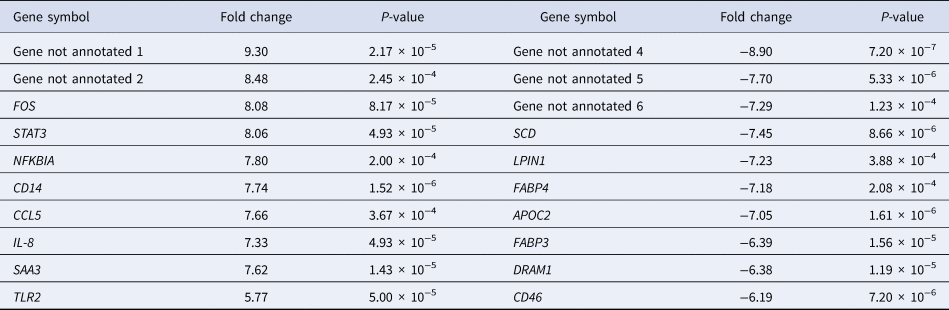
The number of sequences expressed in the mammary alveolar tissue was 27 404 of which 21 691 transcripts corresponded to annotated genes and 5821 as expressed sequences not yet described. Of note, the number of genes deemed expressed and its splice variants would vary with the use of newer and increasingly annotated versions of the bovine reference genome, which should be available in the future (FAANG Consortium et al., Reference Leif, Archibald, Bottema, Brauning, Burgess, Burt, Casas, Cheng, Clarke, Couldrey, Dalrymple, Elsik, Foissac, Giuffra, Groenen, Hayes, Huang, Khatib, Kijas, Kim, Lunney, McCarthy, McEwan, Moore, Nanduri, Notredame, Palti, Plastow, Reecy, Rohrer, Sarropoulou, Schmidt, Silverstein, Tellam, Tixier-Boichard, Tosser-Klopp, Tuggle, Vilkki, White, Zhao and Zhou2015). Just over 6% of expressed genes were differentially expressed in the IQ vs. NIQ 3 h after inoculation with S. agalactiae. Of the 1756 DEGs, 952 were up-regulated and 804 were down-regulated between IQ and NIQ (online Supplementary Table S3). Table 1 shows the top 10 genes up-regulated (9.30 to 5.77-fold). These genes play roles in immune response during infection including CD14 (CD14 molecule), CCL5 (chemokine (C–C motif) ligand 5), TLR2 (toll like receptor 2), IL-8 (interleukin-8 (IL-8)), SAA3 (serum amyloid A3), as well as in transcriptional regulation such as FOS (Fos proto-oncogene, AP-1 transcription factor subunit), STAT3 signal (transducer and activator of transcription 3) and NFKBIA (NFKB inhibitor alpha). The most down-regulated genes (−8.90 to −6.19 fold) 3 h after IMI are also shown in Table 1. Most of these are engaged in lipid metabolism (Stearoyl-CoA desaturase—SCD, Lipin 1—LPIN1, Apolipoprotein C-II—APOC2, Fatty acid binding protein 3—FABP3 and Fatty acid binding protein 4—FABP4). We did not detect differences in the 0 h IQ × 3 h IQ, 0 h IQ × 0 h NIQ, 0 h NIQ × 3 h NIQ contrasts.
List of top 10 up- and down-regulated genes in mammary alveolar tissue 3 h post- inoculation with Streptococcus agalactiae from inoculated quarters compared to non-inoculated quarters

Fos proto-oncogene, AP-1 transcription factor subunit (FOS), signal transducer and activator of transcription 3 (STAT3), NFKB inhibitor alpha (NFKBIA), CD14 molecule (CD14), chemokine (C-C motif) ligand 5 (CCL5), interleukin-8 (IL-8), serum amyloid A3 (SAA3), toll like receptor 2 (TLR2), stearoyl-CoA desaturase (SCD), lipin 1 (LPIN1), fatty acid binding protein 4 (FABP4), Apolipoprotein C-II (APOC2), fatty acid binding protein 3 (FABP3), DNA damage regulated autophagy modulator 1 (DRAM1), CD46 molecule (CD46).
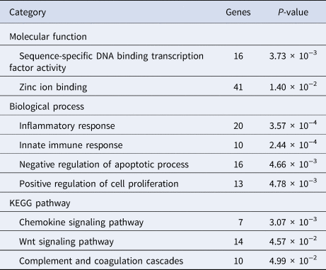
The GO results for molecular function indicated a marked effect on DNA binding transcription factor activity (16 genes) and genes encoding cofactor zinc binding (41 genes; Table 2). The biological process most enriched identified by DAVID (P < 0.01) revealed overrepresentations of genes acting in inflammatory response, innate immune response, negative regulation of apoptosis process and cell proliferation (Table 2). GO analysis revealed that 28 genes including CD14, CD46 (CD46 molecule), ALOX5 (arachidonate 5-lipoxygenase), CCL2 (chemokine (C-C motif) ligand 2), CCL5, PTX3 (pentraxin 3) and SAA3 were involved in inflammatory response and innate immune response (online Supplementary Table S2).
Description of the significant molecular function, biological processes and pathways among the 1756 DEGs between inoculated and non-inoculated quarters at 3 h post-inoculation with Streptococcus agalactiae, identified by GO enrichment analyses using DAVID 6.8

DEGs, differentially expressed genes.
Pathway enrichment analysis with DAVID identified 31 DEGs participated in three KEGG pathways. Among them, 14 genes were enriched in the Wnt signaling pathway and 11 genes encoding complement and coagulation cascade pathway (Supplemental file 2). Chemokine signaling pathway was also overrepresented which included two top up-regulated genes such as CCL5, IL-8 among seven genes belonging in this group (Table 2).
Validation of differentially expressed genes by real-time PCR
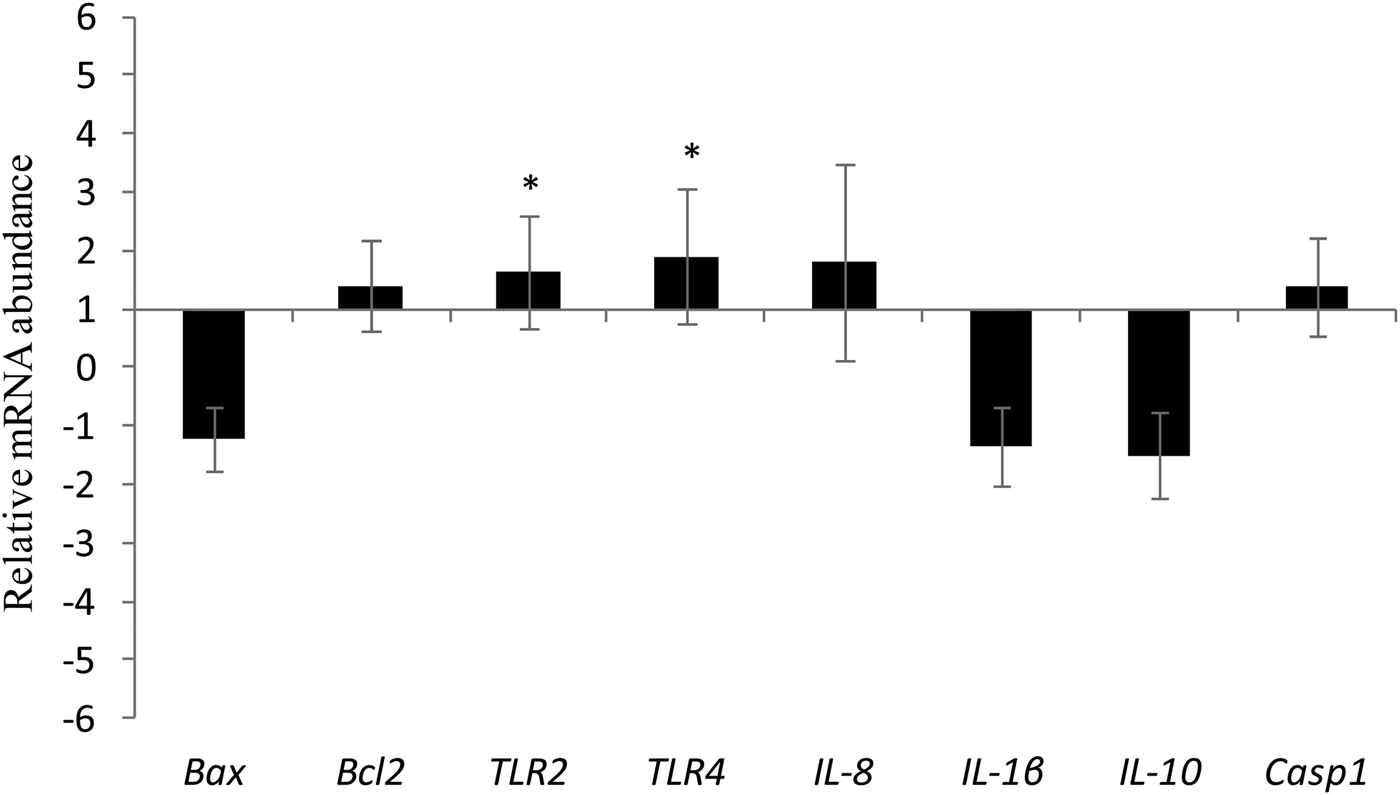
Both the RNAseq and real-time PCR analysis revealed variations in the expression of TLR2 (Table 1 and Fig. 1) and TLR4 (online Supplementary Table S2 and Fig. 1). There was an increase in the mRNA abundance of TLR2 and TLR4 at 3 hpi with S. agalactiae relative to 0 h.
Relative mRNA abundance of Bax, Bcl2, TLR2, TLR4, IL8, IL1β, IL10, Casp1 genes 3 h post-inoculation with the Streptococcus agalactiae strain compared to 0 h (before inoculation) from inoculated quarter (*P < 0.05).

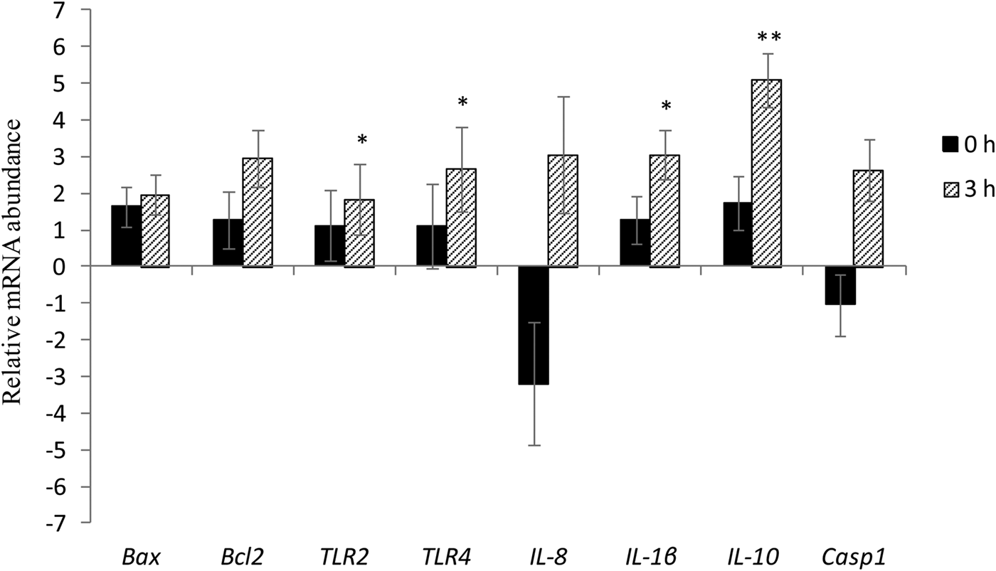
As defined by the RNAseq analysis, all immune-associated genes tested in real-time PCR confirmed no differences in gene expression before inoculation of S. agalactiae (0 h) between IQ and NIQ. However, at 3 hpi we observed increase mRNA abundance of Bcl2 (P = 0.046), TLR2 (P = 0.037), TLR4 (P = 0.049), IL-1β (P = 0.010) and IL-10 (P = 0.008) in IQ compared to NIQ (Fig. 2). IL-8 mRNA abundance was numerically greater in IQ, but this difference was not significant.
Relative mRNA abundance of Bax, Bcl2, TLR2, TLR4, IL8, IL1β, IL10, Casp1 genes at time 0 h and 3 h post-inoculation between inoculated quarter and non-inoculated quarter (*P < 0.05; **P < 0.01).

Discussion
The innate immune response is one of the first lines of host defense to infection as it has the capacity to immediately recognize and respond to earliest signs of infection (Bannerman, Reference Bannerman2009). So far, the majority of experiments in vivo and in vitro have studied pathogen-specific induction of innate immune response after challenge by E. coli (Bannerman et al., Reference Bannerman, Paape, Lee, Zhao, Hope and Rainard2004a; Günther et al., Reference Günther, Liu, Esch, Schuberth and Seyfert2010, Reference Günther, Esch, Poschadel, Petzl, Zerbe, Mitterhuemer, Blum and Seyfert2011), S. aureus (Petzl et al., Reference Petzl, Zerbe, Günther, Yang, Seyfert, Nürnberg and Schuberth2008) and S. uberis (Bannerman et al., Reference Bannerman, Paape, Goff, Kimura, Lippolis and Hope2004b; Swanson et al., Reference Swanson, Stelwagen, Dobson, Henderson, Davis, Farr and Singh2009). In contrast, the transcriptional profile of bovine mammary tissue after challenge with S. agalactiae is poorly characterized. Only a few studies have investigated the changes in gene expression in milk samples after IMI with S. agalactiae (Fonseca et al., Reference Fonseca, Antunes, Paiva, Lange, Guimarães and Martins2011, Reference Fonseca, Cardoso, Higa, Giachetto, Brandão, Brito, Ferreira, Guimarães and Martins2015).
Transcriptome analysis clearly indicated that the mammary alveolar tissue 3 h after inoculation with S. agalactiae experienced a wide transcriptional response which encompassed more than 1700 genes, providing new data that complements the in vivo studies during the first 3 h post-inoculation. As expected, no differences in expression were observed between IQ and NIQ before inoculation of S. agalactiae. This confirmed that the experimental units (udder quarters) were under the same conditions at the beginning of the experiment.
Transcriptional profiling studies have demonstrated that cells implicated in immune defense (including epithelial cells) respond to bacterial stimuli with transcriptional activation program, which are interpreted as generic ‘alarm signal’ for infection. Variations on this common transcriptional theme result from pathogen-specific response (Jenner and Yong, Reference Jenner and Young2005). Of the 10 genes highly up-regulated, three are implicated in sequence-specific DNA binding transcription factors; NFKBIA, FOS and STAT3. The up-regulation of these transcription factors may be underlying the transcriptional changes of the genes involved in innate immune response and inflammatory response. In agreement with our results, Lutzow et al. (Reference Lutzow, Donaldson, Gray, Vuocolo, Pearson, Reverter, Byrne, Sheehy, Windon and Tellam2008) and Kosciuczuk et al. (Reference Kosciuczuk, Lisowski, Jarczak, Majewska, Rzewuska, Zwierzchowski and Bagnicka2017) reported that NFKBIA and FOS are activated in Staphylococcus -induced mastitis whereas Moyes et al. (Reference Moyes, Drackley, Morin, Bionaz, Rodriguez-Zas, Everts, Lewin and Loor2009) noted the same effect when the infection was caused by S. uberis. The signal transducer and activator of transcription 3 (STAT3) is known not only as a mediator of acute-phase response but also in pro-inflammatory pathways (Ahyi et al., Reference Ahyi, Quinton, Jones, Ferrari, Pepper-Cunningham, Mella, Remick and Mizgerd2013) and likely to underpin the observed increase in expression of genes encoding the acute-phase protein serum amyloid A3 (SAA3) in S. agalactiae-inoculated quarters. In vivo studies conducted by Swanson et al. (Reference Swanson, Stelwagen, Dobson, Henderson, Davis, Farr and Singh2009) also showed the induction expression of STAT3 and SAA3 genes by S. uberis, a Gram-positive bacterium (as is S. agalactiae). Our results indicate that these transcription factors have a critical role in the immune response in the udder to S. agalactiae at the earliest stage of IMI. Serum amyloid proteins have immunological properties and the SAA3 isoform (M-SAA3) has been reported as being specific to infected quarters compared to healthy quarters (Molenaar et al., Reference Molenaar, Harris, Rajan, Pearson, Callaghan, Sommer, Farr, Oden, Miles, Petrova, Good, Singh, McLaren, Prosser, Kim, Wieliczko, Dines, Johannessen, Grigor, Davis and Stelwagen2009). It is possible to suggest that the acute-phase protein SAA3 plays an important role in the local inflammatory response with antibacterial activity during S. agalactiae infection.
Perceiving the presence of a pathogen is the first and compulsory step in the innate immune response against an invading pathogen. In our study, a number of genes implicated in innate immune response category, and in particular with TLR signaling have been identified (Table 1 and Supplementary Table S2). This observation confirmed results from Fonseca et al. (Reference Fonseca, Cardoso, Higa, Giachetto, Brandão, Brito, Ferreira, Guimarães and Martins2015) who observed increased expression of both TLR2 and TLR4 in milk samples after experimental in vivo infection with S. agalactiae. These findings indicate that TLR2, TLR4 and CD14 receptors are regulated jointly at the beginning of the innate immune response in mammary alveolar tissue.
The activation of toll-like receptor signaling results in expression of several pro-inflammatory cytokines (IL1β—interleukin 1 beta, IL-6—Interleukin 6), chemokines (IL-8) and others mediators involved in the immune response, cell proliferation and apoptosis (Ibeagha-Awemu et al., Reference Ibeagha-Awemu, Lee, Ibeagha, Bannerman, Paape and X2008). Chemokine signaling pathway was one of the major pathways affected by IMI challenge with S. agalactiae. Three genes belonging to this pathway were up-regulated; IL-8, CCL5 and CCL2 (3.22-fold; Supplementary Table S2). IL-8 is a potent chemokine capable of initiating an inflammatory response and plays a primary role in the recruitment of neutrophils into the udder (Persson et al., Reference Persson, Colditz, Lun and Ostensson2003). In our present study, the IL-8 gene was found up-regulated by RNAseq analysis whereas by real time PCR IL-8 there was a numerical increase at 3 h post-inoculation. According to previous studies, IMI caused by Gram-negative bacteria provokes a much stronger increase in IL-8 expression and secretion than Gram-positive pathogens, which tend to induce a weaker modulation and more delayed response (Bannernam et al., Reference Bannerman, Paape, Goff, Kimura, Lippolis and Hope2004b, Reference Bannerman2009; Günther et al., Reference Günther, Esch, Poschadel, Petzl, Zerbe, Mitterhuemer, Blum and Seyfert2011). Thus, further investigation will be require to verify the likely differential expression of IL-8 in response to IMI by S. agalactiae within 4 or 6 h post-infection. CCL2 and CCL5 are essential chemokines for recruitment of leukocytes which comprise the second line of cellular immune defense (Schukken et al., Reference Schukken, Günther, Fitzpatrick, Fontaine, Goetze, Holst, Leigh, Petzl, Schuberth, Sipka, Smith, Quesnell, Watts, Yancey, Zerbe, Gurjar, Zadoks and Seyfert2011). Our results showed the importance of these chemokines in the earliest stages of the response to IMI infection by S. agalactiae. These results are in accordance with findings of Fonseca et al. (Reference Fonseca, Cardoso, Higa, Giachetto, Brandão, Brito, Ferreira, Guimarães and Martins2015) who showed that CCL2 was up-regulated in milk samples after inoculation with the same strain in the udders. Similarly to our results, only a few studies demonstrated that the chemokine CCL5 (also known as RANTES) is strongly expressed in the infected udder as well as in inflammation stimulated MEC (Günther et al., Reference Günther, Koczan, Yang, Nürnberg, Repsilber, Schuberth, Park, Maqbool, Molenaar and Seyfert2009, Reference Günther, Esch, Poschadel, Petzl, Zerbe, Mitterhuemer, Blum and Seyfert2011).
IL-1β is known as a key pro-inflammatory cytokine that regulates not only the expression of several immune response genes including others cytokines (e.g. IL-8) and acute phase proteins, but also genes involved in cell proliferation and apoptosis (Lahouassa et al., Reference Lahouassa, Moussay, Rainard and Riollet2007). Because IL-1β is a key component of the innate immune response, we expected induction of this cytokine in the first hours post-inoculation. Although we observed no changes in IL-1β expression by RNAseq technique, real-time PCR analysis showed that this gene had increased at 3 hpi in IQ compared to NIQ. This result confirms those found in the literature, as IL-1 is critical in inflammation of the mammary gland of cows caused by Staphylococci (Lutzow et al., Reference Lutzow, Donaldson, Gray, Vuocolo, Pearson, Reverter, Byrne, Sheehy, Windon and Tellam2008; Kosciuczuk et al., Reference Kosciuczuk, Lisowski, Jarczak, Majewska, Rzewuska, Zwierzchowski and Bagnicka2017). The reasons why IL1β was not identified in the RNA-Seq analysis whereas it showed differential expression when measured by Real time PCR are not clear, but may relate to the differing sensitivities of the two technologies. Real-time PCR is considered to be a highly sequence-specific method for expression analysis (Costa et al., Reference Costa, Giménez-Capitán, Karachaliou and Rosell2013).
Because S. agalactiae mastitis is usually characterized by a moderate inflammatory response of the udder, we investigated by real time-PCR if IL-10, one of the major anti-inflammatory cytokines, might participate in the modulation of the inflammation in the early stages of response to this pathogen. Despite a mild up-regulation of IL1β (3.05-fold) during IMI, the observed 5.10 fold up-regulation of IL-10 probably hampered inflammatory activity. In agreement with our findings, previous in vivo experiments have found an increase in milk concentrations of IL-10 after IMI with E. coli and S. uberis (Bannerman et al., Reference Bannerman, Paape, Lee, Zhao, Hope and Rainard2004a, Reference Bannerman, Paape, Goff, Kimura, Lippolis and Hope2004b) while S. aureus infection fails to induce this response in the host (Bannerman et al., Reference Bannerman, Paape, Lee, Zhao, Hope and Rainard2004a). In addition, it is worth pointing out that in those cows with the greatest persistent concentrations of S. aureus or S. uberis in milk, induction of IL-10 is absent or delayed (Bannerman, Reference Bannerman2009). Thus it is reasonable to suggest that earlier induction of IL-10 could be beneficial to the ability of the cows to limit bacterial growth and eradicate the pathogen without an exacerbated inflammatory response that can damage the epithelial lining of the mammary gland and lead to reduction in milk production.
The induction of apoptosis by bacterial pathogens is a well-established cellular process (Weinrauch and Zychlinsky, Reference Weinrauch and Zychlinsky1999). However, only a few studies have reported direct evidence for involvement of apoptosis during mastitis (Long et al., Reference Long, Capuco, Wood, Sonstegard, Tomita, Paape and Zhao2001). We found that several genes belonging to the apoptotic process were down-regulated at 3 h post-inoculation with S. agalactiae. DRAM1 (DNA damage regulated autophagy modulator 1) was the most down-regulated followed by other apoptotic related genes such as Bax (−2.95-fold), Casp6 (−2.15-fold), Casp1 (−3.24-fold). Guan et al. (Reference Guan, Zhang, Sun, Qi, Wu and Qin2015) reported that DRAM1 regulates apoptosis through increasing BAX levels. Given that the apoptosis process was among the enriched biological processes in our study, we investigated the expression of Casp1, Bax and Bcl2 genes belonging to this group by real-time PCR. Only Bcl2 gene was confirmed as being differentially expressed 3 h post-inoculation. Taken together, these results strongly support the idea that in our isolated perfused model there was no evidence for induction of apoptosis at the early stages of response to S. agalactiae- mastitis because no differences were found in the expression of pro-apoptotic genes (Bax and Casp1) for any contrast when measured by real-time PCR. In addition, the 2.95-fold increase in anti-apoptotic Bcl2 expression 3 h post-inoculation between IQ and NIQ corroborates this hypothesis.
Our data revealed that the Wnt signaling pathway was affected by S. agalactiae-inoculation. We observed that 14 differentially expressed genes were associated with this pathway. Wnt signal transduction has been implicated in inflammation and innate immunity and homeostasis via Toll-like receptor and NF-κB pathways (Sun et al., Reference Sun, Hobert, Duan, Rao, He, Chang and Madara2005). Recently, Noyes et al. (Reference Noyes, Brass, Obara, Anderson, Archibald, Bradley, Fisher, Freeman, Gibson, Gicheru, Hall, Hanotte, Hulme, McKeever, Murray, Oh, Tate, Smith, Tapio, Wambugu, Williams, Agaba and Kemp2011) identified potential candidate genes in the Wnt signaling pathway in response to Trypanosoma congolense infection in bovine liver, kidney and lymph nodes. Our findings revealed a role of the Wnt signaling pathway during the early immune response to S. agalactiae.
The complement and coagulation cascades pathway was among the three most affected pathways. Similarly to our results, a previous study has also found that complement and coagulation cascade pathway was the major affected pathway in mammary tissue naturally infected with S. aureus (Huang et al. Reference Huang, Luo, Zhang, Wang, Ju, Qi, Zhang, Wang, Li, Li, Yin, Xu, Moisá, Loor and Zhong2014). The observed up-regulation of C1QC (complement C1q C chain) was similar to results from Moyes et al. (Reference Moyes, Drackley, Morin, Bionaz, Rodriguez-Zas, Everts, Lewin and Loor2009) who observed 1.37 fold—increase in S. uberis-infected mammary quarter. The component C1q is the first step in the initiation of the classical pathway of complement cascade which plays an important role in innate immunity (Rainard, Reference Rainard2003). CD46 plays a primary function in the host's response to pathogens by regulating complement activation which is a critical step in the early control of infection (Meiffren et al., Reference Meiffren, Joubert, Grégoire, Codogno, Rabourdin-Combe and Faure2010). Previous reports showed that pathogen receptor CD46 on the cell surface can trigger cell autophagy to control infection by Streptococcus (Cattaneo, Reference Cattaneo2004; Joubert et al., Reference Joubert, Meiffren, Grégoire, Pontini, Richetta, Flacher, Azocar, Vidalain, Vidal, Lotteau, Codogno, Rabourdin-Combe and Faure2009). Moreover, Wang et al. (Reference Wang, Zhong, Gao, Ju and Huang2014) recently identified a new variant splice designated CD46 transcript variant (CD46-TV) that was associated with resistance to mastitis caused by Streptococcus in dairy cows. In our study, CD46 gene was one of the most down-regulated indicating the relevant role of this pathogen receptor in the risk of mastitis caused by S. agalactiae.
Another interest finding of the RNA-Seq analysis was that several genes involved with lipid metabolism such as, SCD, LPIN1, APOC2, FABP3 and FABP4 were down-regulated in mammary quarters inoculated with S. agalactiae. As in the present study, Swanson et al. (Reference Swanson, Stelwagen, Dobson, Henderson, Davis, Farr and Singh2009) also observed down-regulation of genes related with lipid metabolism (e.g. LPIN1, APOC2, FABP3) in mammary tissue after IMI with S. uberis. This is further supported by Günther et al. (Reference Günther, Koczan, Yang, Nürnberg, Repsilber, Schuberth, Park, Maqbool, Molenaar and Seyfert2009) who observed marked decreases in mRNA expression of factors associated with lipid metabolism in bovine MEC infected by E. coli. The products of these genes are known to be involved in milk fat synthesis (Bionaz and Loor, Reference Bionaz and Loor2008). Our findings highlighted the potential association between activation of inflammatory immune response and suppression lipid metabolism in S. agalactiae-inoculated mammary quarters. Taken together, the observed responses in inoculated mammary alveolar tissue likely reflect transcriptional changes in bovine MEC. Because the experiment has used an isolated perfused udder model, it is reasonable to suggest that the predominant response would be from the resident macrophages which serve both sentinel as well as effector functions of immune defense in healthy udder (Strandberg et al., Reference Strandberg, Gray, Vuocolo, Donaldson, Broadway and Tellam2005).
In conclusion, our study demonstrated that the first 3 h after S. agalactiae- inoculation elicited a strong transcriptomic response leading to an overall up-regulation of genes involved in innate immune and inflammatory response and marked inhibition of lipid synthesis-related genes. The results provide comprehensive information about the early response factors associated with innate immune response in S. agalactiae-inoculated mammary quarter. We showed that TLR2, TLR4, CD14 and IL-1β genes were highly up-regulated in inoculated-quarters which could be crucial stimulators of innate immunity. It is important to note that no apoptosis induction had occurred, either in non-inoculated or inoculated quarters at 3 h after the experimental inoculation, which validates the model to evaluate the early response to other mastitis-causing pathogens
Supplementary material
The supplementary material for this article can be found at https://doi.org/10.1017/S0022029919000451
Acknowledgements
The authors are grateful to the Conselho Nacional de Desenvolvimento Científico e Tecnológico (CNPq/Project number 473414/2011-2) and Fundação de Amparo à Pesquisa do Estado de Minas Gerais (FAPEMIG/ project number APQ-00095-15) for project funding and scholarship (CNPq—WELLER, FONSECA, VICCINI, MUNARI, SILVA, and MARTINS—CNPq, CAPES—PINTO, and SBARDELLA).
Disclaimer
The authors declare that the research was conducted in the absence of any commercial or financial relationships that could be construed as a potential conflict of interest.