





Introduction
Human schistosomiasis is a parasitic Neglected Tropical Disease (NTD) predominantly affecting sub-Saharan Africa (Li et al. Reference Li, Qin, Bergquist, Qian, Xia, Lv, Xiao, Utzinger and Zhou2021; Rausche et al. Reference Rausche, Rakotoarivelo, Rakotozandrindrainy, Rakotomalala, Ratefiarisoa, Rasamoelina, Kutz, Jaeger, Hoeppner, Lorenz, May, Puradiredja and Fusco2023), with Schistosoma mansoni and S. haematobium being the most prevalent species. S. mansoni primarily causes intestinal pathology, while S. haematobium affects the urinary tract and can lead to severe complications, including infertility (Ally et al. Reference Ally, Kanoi, Ochola, Nyanjom, Shiluli, Misinzo and Gitaka2024). The primary strategy for schistosomiasis control is Mass Drug Administration (MDA) with praziquantel as a chemoprophylactic measure. However, as with other NTDs, obtaining accurate estimates of disease burden and programme coverage remains a significant challenge (Rasoamanamihaja et al. Reference Rasoamanamihaja, Rakotoarivelo, Edosoa, Rasamoelina, Montresor, Marchese and Fusco2023). Globally, approximately 250 million people are affected, with over 200,000 deaths annually in sub-Saharan Africa, and more than 90% of those requiring preventive treatment residing in Africa (Ally et al. Reference Ally, Kanoi, Ochola, Nyanjom, Shiluli, Misinzo and Gitaka2024). Among the most affected countries, Madagascar ranks fifth globally in terms of schistosomiasis burden. According to 2015 data, 106 of its 114 districts were classified as endemic. Of these, 40.7% (46/114) reported a prevalence exceeding 50%, while 37.2% (42/114) had rates between 10% and 50%. Additionally, 16.8% (19/114) of districts report a prevalence below 10%, and only 5.3% (6/114) are considered non-endemic. Regarding its geographical distribution, S. haematobium, responsible for urogenital schistosomiasis, is primarily found in the western and northern regions of the island. In contrast, the intestinal form, caused by S. mansoni, is more prevalent in the eastern, southern and central highland regions of the country (Deka, Reference Deka2022; ESPEN, 2023; Rasoamanamihaja et al. Reference Rasoamanamihaja, Rakotoarivelo, Edosoa, Rasamoelina, Montresor, Marchese and Fusco2023). Despite ongoing efforts to control the disease, the implementation of diagnostic and treatment programmes is hindered by limited financial resources, poor coordination and logistical challenges at the local level. As a result, several districts requiring MDA programmes still do not receive the necessary coverage (Rasoamanamihaja et al. Reference Rasoamanamihaja, Rakotoarivelo, Edosoa, Rasamoelina, Montresor, Marchese and Fusco2023; Rausche et al. Reference Rausche, Rakotoarivelo, Rakotozandrindrainy, Rakotomalala, Ratefiarisoa, Rasamoelina, Kutz, Jaeger, Hoeppner, Lorenz, May, Puradiredja and Fusco2023).
Diagnosis plays a crucial role in measuring the impact of interventions, as it determines the levels of disease reduction, infection rates, and parasite transmission. In endemic regions, establishing structured control programmes is essential for an effective approach to disease management (Ally et al. Reference Ally, Kanoi, Ochola, Nyanjom, Shiluli, Misinzo and Gitaka2024). The WHO recommends the use of the Kato-Katz (KK) method in routine schistosomiasis control programmes, as it enables the mapping and monitoring of S. mansoni infections in community-based interventions (WHO, 2022; Loyo et al. Reference Loyo, Gomes, Pieri, Oliveira, Nascimento and Barbosa2024). This method is a simple direct parasitological diagnostic technique that facilitates the visualization and quantification of viable eggs in the faeces of infected individuals (Loyo et al. Reference Loyo, Gomes, Pieri, Oliveira, Nascimento and Barbosa2024). However, its sensitivity is limited, particularly in individuals with low parasite loads or in post-MDA populations (Cai et al. Reference Cai, Weerakoon, Mu, Olveda, Ross, Olveda and McManus2019).
Several Polymerase Chain Reaction (PCR)-based assays have been developed for the molecular diagnosis of schistosomiasis, proving to be valuable tools for the simultaneous detection and identification of multiple Schistosoma species (Weerakoon et al. Reference Weerakoon, Gordon and McManus2018; Schols et al. Reference Schols, Carolus, Hammoud, Mulero, Mudavanhu and Huyse2019). Although these methods offer high sensitivity and specificity, their application in field studies and resource-limited settings presents significant challenges due to the requirement for specialized equipment, trained personnel and technically complex, costly procedures (Chieng et al. Reference Chieng, Okoyo, Simiyu, Gichuki, Mwatele, Kepha, Njenga and Mburu2021; Blin et al. Reference Blin, Senghor, Boissier, Mulero, Rey and Portela2023; Ally et al. Reference Ally, Kanoi, Ochola, Nyanjom, Shiluli, Misinzo and Gitaka2024). Given these limitations, the development of accessible, rapid, user-friendly and highly sensitive and specific diagnostic techniques is essential. In this context, a loop-mediated isothermal amplification (LAMP) method has emerged as a promising alternative, as it enables nucleic acid amplification under isothermal conditions and provides visual results without the need for sophisticated or expensive equipment (Gandasegui et al. Reference Gandasegui, Fernández-Soto, Hernández-goenaga, López-Abán, Vicente and Muro2016; Fernández-Soto et al. Reference Fernández-Soto, Avendaño, Sala-vizcaíno, Crego-Vicente, Febrer-Sendra, García-bernalt Diego, Oleaga, López-Abán, Vicente, Patarroyo and Muro2020; García-bernalt Diego et al. Reference García-bernalt Diego, Fernández-Soto, Febrer-Sendra, Crego-Vicente and Muro2021a). Due to these characteristics, LAMP technology meets the criteria for an efficient diagnostic assay suitable for resource-limited settings and holds great potential as a Point-of-Care (POC) diagnostic method (García-bernalt Diego et al. Reference García-bernalt Diego, Fernández-Soto, Febrer-Sendra, Crego-Vicente and Muro2021a).
In recent years, multiple LAMP assays have been developed as an alternative to PCR for the diagnosis of various parasitic NTDs, with schistosomiasis being one of the conditions in which this technology has advanced the most. Specifically, LAMP assays have been designed and validated for the detection of the three main species responsible for human schistosomiasis (S. haematobium, S. japonicum and S. mansoni), using distinct molecular targets specific to each species (Fernández-Soto et al. Reference Fernández-Soto, Gandasegui Arahuetes, Sánchez Hernández, López Abán, Vicente Santiago and Muro2014; Gandasegui et al. Reference Gandasegui, Fernández-Soto, Carranza-rodríguez, Pérez-arellano, Vicente, López-Abán and Muro2015, Reference Gandasegui, Fernández-Soto, Hernández-goenaga, López-Abán, Vicente and Muro2016; Crego-Vicente et al. Reference Crego-Vicente, Fernández-Soto, Febrer-Sendra, García-bernalt Diego, Boissier, Angora, Oleaga and Muro2021; García-bernalt Diego et al. Reference García-bernalt Diego, Fernández-Soto, Febrer-Sendra, Crego-Vicente and Muro2021a). In addition, a LAMP assay for the simultaneous detection of multiple Schistosoma species (namely, Schisto-LAMP) has been previously developed and evaluated, including S. mansoni, S. haematobium, S. intercalatum and S. bovis, all of which are present in Africa (Fernández-Soto et al. Reference Fernández-Soto, Avendaño, Sala-vizcaíno, Crego-Vicente, Febrer-Sendra, García-bernalt Diego, Oleaga, López-Abán, Vicente, Patarroyo and Muro2020; Crego-Vicente et al. Reference Crego-Vicente, Fernández-Soto, Febrer-Sendra, García-bernalt Diego, Boissier, Angora, Oleaga and Muro2021); furthermore, this assay has also been applied for the identification of hybrid schistosome DNA (García-bernalt Diego et al. Reference García-bernalt Diego, Fernández-Soto, Febrer-Sendra, Crego-Vicente and Muro2021a).
The aim of this study was to evaluate the diagnostic performance of the Schisto-LAMP assay for the detection of Schistosoma spp. in faecal samples, comparing it with the KK method and a commercial qPCR assay, in order to determine its utility as an easy-to-use molecular method for potential application in resource-limited primary healthcare POC settings.
Materials and methods
Ethics statement
The study was approved by the Human Research Ethics Committee of the Universitat de València (Ref. H1506340075678) on October 5, 2017, and was conducted in accordance with the fundamental principles of the Declaration of Helsinki, the Council of Europe Convention on Human Rights and Biomedicine, and the UNESCO Declaration. Signed informed consent was obtained from all subjects involved in the study or their parents/legal guardians.
Population and setting
The study was conducted using stool samples collected from schoolchildren aged 5–15 years attending Sekoly Willka School, located in the village of Soavinarivo, within the Ankazobe district of the Analamanga region, Madagascar.
Sample collection
In August 2018, the educational centre was visited with the support of the local Non-Governmental Organization (NGO) FAMI (Fikambanana ho an’ny Ankizy MIvoatra), which manages the Sekoly Willka School, and a total of 102 faecal samples were collected. Parents or legal guardians of the participants were informed about the study procedures, emphasizing that participation was entirely voluntary. Those interested in participating were required to complete and sign an informed consent form. Each legal guardian was provided with an individual sample collection kit, which included a plastic screw-capped container labelled with a unique identification code, a spatula, and a sheet of magazine paper. Instructions were given to ensure that defecation occurred on the magazine paper to prevent contamination with soil. Using the spatula, participants were instructed to collect three portions from different areas of the stool sample, with a total weight of approximately 5 g, and deposit them into the container. Samples were collected the following day and were processed and stored in a classroom temporarily adapted as a laboratory.
Parasitological analysis
A single stool sample was collected from each participant. In the field, within a classroom of the Sekoly Willka School that had been set up as a provisional field laboratory, a single KK smear was prepared per sample using fresh faeces. The KK smears were analysed on site for the identification and quantification of Schistosoma eggs. Parasite burden, expressed as eggs per gram of faeces (epg), was calculated by multiplying the number of eggs observed in the thick smears by 24, based on the use of a 41.7 mg template. The intensity of schistosomiasis infection was classified according to epg values as light (1–99 epg), moderate (100–399 epg), and heavy (≥ 400 epg) (Spencer et al. Reference Spencer, Penney, Russell, Howe, Linder, Rakotomampianina, Nandimbiniaina, Squire, Stothard, Bustinduy and Rahetilahy2017). The remaining faecal material was immediately preserved in sterile tubes containing 70% ethanol and, together with the KK slides, sent to the Parasitology Laboratory of the Faculty of Pharmacy of the University of Valencia (Spain) for analysis and subsequent DNA extraction for molecular diagnosis. Upon arrival in Spain, the KK slides were re-examined to confirm the schistosomiasis diagnoses. The re-reading performed in Spain fully confirmed the initial field results with 100% concordance (no discrepancies were found); therefore, the field reading dataset was utilized as the final definitive dataset.
DNA extraction from stool samples
For total DNA extraction, approximately 5 g of each stool sample were first concentrated using the Midi Parasep® Solvent Free (SF) filter device (Apacor Ltd, Wokingham, United Kingdom). Subsequently, DNA was extracted from 200 mg of the concentrate using the commercial QIAamp DNA Stool Mini Kit (Qiagen, Hilden, Germany), following the manufacturer’s instructions. The extracted DNA was purified, eluted in 100 μL of PCR-grade water, and stored at −20°C until further molecular analysis.
qPCR assay for schistosoma spp. detection
For the molecular diagnosis of schistosomiasis, the commercial quantitative PCR (qPCR) LightMix® Modular Assay for Schistosoma spp. was used (manufactured by TIB Molbiol, distributed by Roche Diagnostics, Mannheim, Germany), designed to amplify a specific 78-base pair (bp) fragment of the ITS-2 ribosomal RNA gene. qPCR reactions were carried out in a final volume of 20 µL, consisting of 15 µL of reaction mix (10.5 μL ultrapure PCR-grade water, 0.5 μL primers and hydrolysis probe, 4.0 μL Roche Master) and 5 µL of DNA template. Each reaction included positive and negative controls (DNase/RNase-free water), both supplied with the kit. The reactions were conducted using the CFX96™ thermocycler (Bio-Rad, Marnes-la-Coquette, France), and amplification curves were analysed with the CFX Manager IVD 1.6 software. Samples were considered positive if the cycling threshold (Ct) was less than 37 cycles according to the manufacturer’s instructions.
LAMP assay for schistosoma spp. detection
The detection of Schistosoma spp. DNA was also performed using the colorimetric Schisto-LAMP assay, following the protocol previously described by Fernández-Soto et al. (Fernández-Soto et al. Reference Fernández-Soto, Gandasegui Arahuetes, Sánchez Hernández, López Abán, Vicente Santiago and Muro2014, Reference Fernández-Soto, Avendaño, Sala-vizcaíno, Crego-Vicente, Febrer-Sendra, García-bernalt Diego, Oleaga, López-Abán, Vicente, Patarroyo and Muro2020; Crego-Vicente et al. Reference Crego-Vicente, Fernández-Soto, Febrer-Sendra, García-bernalt Diego, Boissier, Angora, Oleaga and Muro2021). The reactions were carried out using previously described conditions, with the exception of the final reaction volume, which was adjusted from 25 μL to 15 μL. Briefly, LAMP reaction mixtures (15 μL) contained 40 pmol each of FIP and BIP primers, 5 pmol each of F3 and B3 primers, 0.4 μM of each LF primer (Table 1), 1.4 mM of each dNTP (Bioron, Germany), 1x Isothermal Amplification Buffer – 20 mM Tris-HCl (pH 8.8), 50 mM KCl, 10 mM (NH4)2SO4, 2 mM MgSO4, 0.1% Tween20 (New England Biolabs Ltd., Ipswich, MA, USA) – supplemented with 6 mM MgSO4 and 8 U of Bst 2.0 WarmStart DNA polymerase (New England Biolabs Ltd., Ipswich, MA, USA) with 2 μL of template. Schisto-LAMP reactions were performed in 0.5 mL tubes and incubated in a heat block at 65 °C for 60 min and then at 80 °C for 5–10 min to stop the reaction. A portable, compact, lightweight, and cost-effective dry block heater from Thermo Fisher Scientific was used, designed to facilitate transport and enable the application of the LAMP method under field conditions. This device was selected to simulate a POC environment and assess its feasibility in resource-limited settings.
Primer sets used for Schisto-LAMP assay for Schistosoma spp. DNA detection

Table 1 Long description
Schisto-LAMP primer information for detecting Schistosoma species DNA is provided, listing primer name, nucleotide sequence in the five-prime to three-prime direction, and length in base pairs. Five primers are included: F3, B3, FIP, BIP, and LF. Lengths range from 18 base pairs for F3 to 43 base pairs for BIP, with B3 at 19, LF at 22, and FIP at 39. The inner primers are longer than the outer and loop primers, with BIP the longest and F3 the shortest. A literature citation is given for the assay in the row containing F3, referencing Fernández-Soto and colleagues 2020 and Crego-Vicente and colleagues 2021.
F3, forward outer primer; B3, backward outer primer; FIP, forward inner primer (comprising F1c and F2 sequences); BIP, backward inner primer (comprising B1c and B2 sequences); LF, loop forward primer; bp, base pairs.
Schisto-LAMP amplification results were visually inspected by adding 1 μL of SYBR Green I fluorescent dye (Invitrogen, Waltham, MA, USA) diluted 1:10 at a 10 000x concentration to each reaction tube after amplification. Positive Schisto-LAMP reactions exhibited green fluorescence, whereas negative reactions appeared orange. All assays included positive (DNA from S. mansoni) and negative controls, which comprised a no-template control (purified water) and a human faecal sample previously confirmed negative. Tubes were carefully opened before adding the dye to prevent potential cross-contamination with amplified products.
Statistical analysis
Data were analysed using frequency distributions. To evaluate the diagnostic accuracy of the KK method, qPCR, and Schisto-LAMP assays, sensitivity and negative predictive value (NPV) were calculated using a composite reference standard (CRS). This CRS was defined such that a sample was considered positive if it tested positive in at least one of the three diagnostic methods used. Confidence intervals (CI) were set at 95%. Comparisons between diagnostic methods (KK vs. Schisto-LAMP, KK vs. qPCR, as well as Schisto-LAMP vs. qPCR) were performed using McNemar’s test, with a P-value < 0.05 considered statistically significant. Additionally, the Kappa index (κ) was used to evaluate the level of concordance between diagnostic tests. All statistical analyses were conducted using IBM SPSS Statistics version 29.0.2 for Windows.
Results
Diagnosis of schistosomiasis by Kato–Katz, Schisto-LAMP and qPCR
The overall prevalence detected using the three diagnostic methods for schistosomiasis was 75.5% (77/102). The highest proportion of Schistosoma spp. infections was detected by qPCR, with 69.6% of cases testing positive. The Schisto-LAMP method exhibited a prevalence rate of 61.8%; the KK identified a S. mansoni infection rate of 39.2%.
Performance of qPCR and schisto-LAMP according to infection intensity by KK
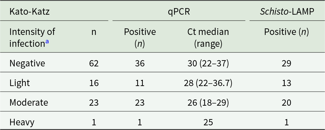
The parasitological dataset obtained from the initial field readings was fully validated by the reference laboratory re-examination in Spain, showing perfect agreement in all cases. According to the KK classification (Table 2), moderate-intensity infections were the most frequent (23/40). qPCR detected all cases (median Ct: 26), whereas Schisto-LAMP detected 20/23. Light-intensity infections accounted for 16/40 cases; qPCR detected 11/16 (median Ct: 28) and Schisto-LAMP 13/16. Discrepancies were observed (Supplementary Table S1): five KK-positive samples were detected only by Schisto-LAMP (nos. 707, 726, 744, 790 and 795), whereas two samples positive by KK and qPCR were negative by Schisto-LAMP (nos. 709 and 757).
Diagnostic performance of qPCR and Schisto-LAMP stratified by infection intensity according to Kato-Katz

Table 2 Long description
Diagnostic results are stratified by Kato-Katz infection intensity groups (negative, light, moderate, heavy) and report sample counts plus positives for qPCR and Schisto-LAMP, with qPCR cycle threshold medians and ranges. Among Kato-Katz negative samples, 36 of 62 were qPCR positive with a median Ct of 30 (range 22 to 37), while 29 of 62 were Schisto-LAMP positive. In light infection, 11 of 16 were qPCR positive with median Ct 28 (range 22 to 36.7), and 13 of 16 were Schisto-LAMP positive. In moderate infection, qPCR detected all 23 of 23 with a lower median Ct of 26 (range 18 to 29), whereas Schisto-LAMP detected 20 of 23. In heavy infection, both tests were positive in the single case, with qPCR Ct 25. Across intensity groups, qPCR shows increasing positivity and decreasing Ct values as intensity increases, consistent with higher parasite burden. Schisto-LAMP is relatively high in light infection but does not increase in moderate infection compared with qPCR. The heavy category has only one sample, so conclusions for that group are limited.
a Categorized according to WHO criteria (WHO, 2013).
Heavy-intensity infection was least frequent (Table 2), with a single case detected by all three methods. qPCR identified more positive cases among KK-negative samples than Schisto-LAMP.
Agreement between diagnostic techniques used for schistosoma spp
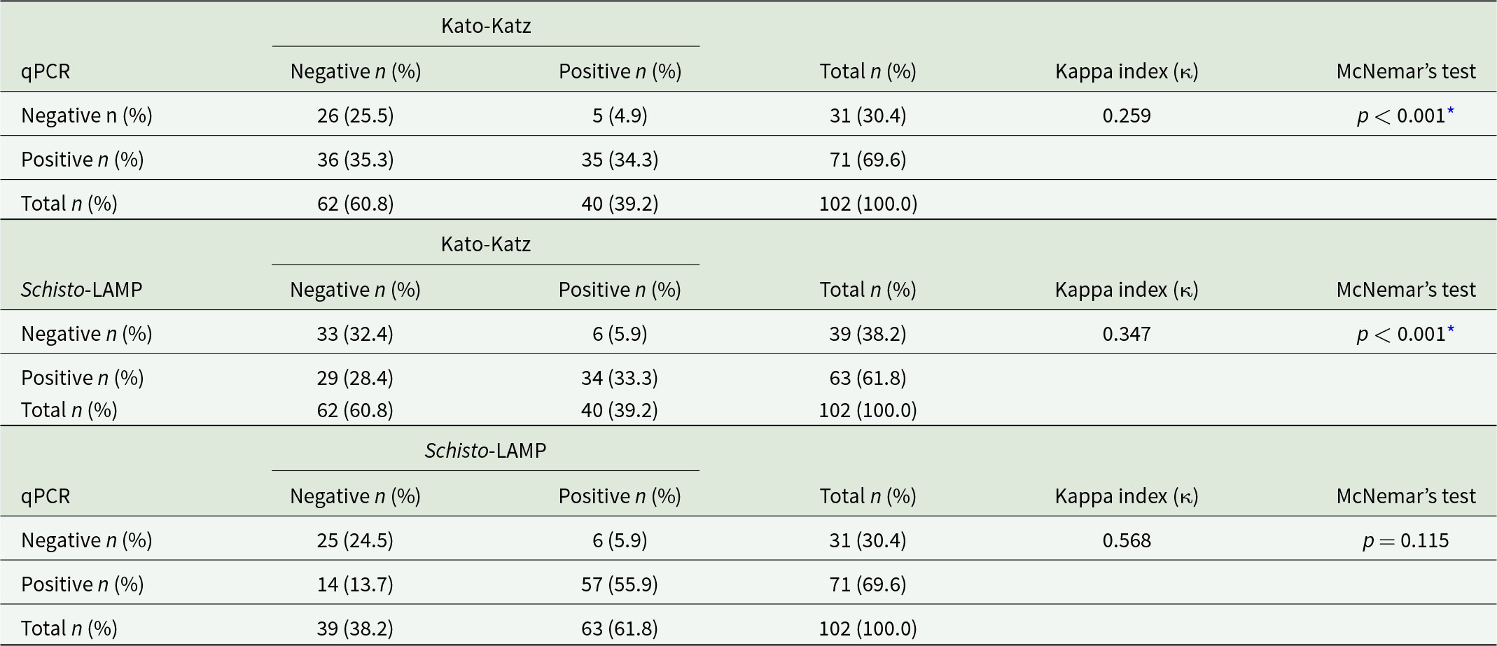
Low agreement was observed between KK and qPCR (κ = 0.259), and between KK and Schisto-LAMP (κ = 0.347) (Table 3), with significant differences in both comparisons. In contrast, moderate agreement was found between Schisto-LAMP and qPCR (κ = 0.568), with no significant differences. Agreement between Schisto-LAMP and qPCR was observed in 55.9% of positive cases and 24.5% of negative cases.
Concordance between Kato-Katz, qPCR and Schisto-LAMP results

Table 3 Long description
Three two-by-two cross-tabulations compare test results across 102 samples for Kato-Katz, qPCR, and Schisto-LAMP, reporting counts and prevalence percentages plus agreement and paired-difference tests. qPCR versus Kato-Katz: 35 samples are positive on both, 26 negative on both, 36 are qPCR positive but Kato-Katz negative, and 5 are qPCR negative but Kato-Katz positive; agreement is low and the paired difference is statistically significant. Schisto-LAMP versus Kato-Katz: 34 are positive on both, 33 negative on both, 29 are Schisto-LAMP positive but Kato-Katz negative, and 6 are Schisto-LAMP negative but Kato-Katz positive; agreement is low to fair and the paired difference is statistically significant. qPCR versus Schisto-LAMP: 57 are positive on both and 25 negative on both, with 14 qPCR positive but Schisto-LAMP negative and 6 qPCR negative but Schisto-LAMP positive; agreement is moderate and the paired difference is not statistically significant. Overall totals show Kato-Katz identifies 40 positives, while qPCR identifies 71 positives and Schisto-LAMP identifies 63 positives, indicating more positives by the molecular methods than by Kato-Katz. Agreement values should be interpreted alongside the differing positive rates, which can affect concordance measures.
n: number of cases; %: prevalence on the total samples (N = 102);
* significant association (p < 0.05).
Comparative analysis of the diagnostic efficacy of the techniques used
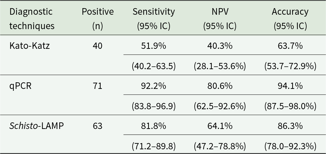
Diagnostic performance was assessed using a CRS (N = 77), considering a sample positive if detected by at least one method. Most positives were detected by multiple techniques, with only a few samples positive exclusively by qPCR (n = 8) or Schisto-LAMP (n = 1).
qPCR showed the highest diagnostic performance (sensitivity 92.2%, NPV 80.6%, accuracy 94.1%) (Table 4), followed by Schisto-LAMP (sensitivity 81.8%, NPV 64.1%, accuracy 86.3%), while KK performed the lowest.
Diagnostic performance of Kato-Katz, qPCR and Schisto-LAMP using a composite reference standard (N = 77)

Table 4 Long description
Diagnostic performance is compared across Kato-Katz, qPCR, and Schisto-LAMP against a composite reference standard in 77 samples. qPCR reports 71 positives, sensitivity 92.2% with 95% confidence interval 83.8 to 96.9, negative predictive value 80.6% with interval 62.5 to 92.6, and accuracy 94.1% with interval 87.5 to 98.0. Schisto-LAMP reports 63 positives, sensitivity 81.8% with interval 71.2 to 89.8, negative predictive value 64.1% with interval 47.2 to 78.8, and accuracy 86.3% with interval 78.0 to 92.3. Kato-Katz reports 40 positives, sensitivity 51.9% with interval 40.2 to 63.5, negative predictive value 40.3% with interval 28.1 to 53.6, and accuracy 63.7% with interval 53.7 to 72.9. Across all three metrics, qPCR ranks highest, Schisto-LAMP is intermediate, and Kato-Katz is lowest. Confidence intervals indicate uncertainty around each estimate, and the number of positives differs by technique, which can affect direct comparisons.
NPV, negative predictive value.
N, Number of positive samples, irrespective of the technique utilized.
Discussion
In Madagascar, despite the existence of national programmes for schistosomiasis control, remote rural areas such as Soavinarivo remain underserved with no implementation of these interventions to date. This lack of coverage contributes to the persistently high prevalence of infection. As a result, schistosomiasis continues to pose a significant public health burden, particularly among children. In this study, the overall prevalence of schistosomiasis was 75.5%. Previous investigations conducted in Malagasy children have reported prevalence rates ranging from 35% to as high as 94% (Spencer et al. Reference Spencer, Penney, Russell, Howe, Linder, Rakotomampianina, Nandimbiniaina, Squire, Stothard, Bustinduy and Rahetilahy2017, Reference Spencer, Linder, Penney, Russell, Hyde, Sheehy, Reid, Andriamasy, Raderalazasoa, Rakotomampianina, Nandimbiniaina, Ranaivoson, Andrianiaina, Cruickshank, Bustinduy, Stothard, Edosoa and Rahetilahy2021; Isaiah et al. Reference Isaiah, Sólveig Palmeirim and Steinmann2023). These findings highlight the urgent need for the integration of diagnostic tools that are not only sensitive and affordable, but also easily deployable in endemic settings. The prevalence rates obtained using Schisto-LAMP (61.8%) and qPCR (69.6%), both performed in a well-equipped laboratory in Spain, were highly comparable and significantly higher than those obtained using KK.
This fact can likely be attributed to several limitations inherent to traditional parasitological diagnostics. Firstly, egg-detection methods fail to identify low-intensity or residual infections, in which eggs excretion may be intermittent or nearly absent. Additionally, the heterogeneous distribution of schistosome eggs within stool samples reduces the likelihood of detection, particularly in newly acquired or mild infections (Cavalcanti et al. Reference Cavalcanti, Cunha and Peralta2019). Another critical limitation is the reduced sensitivity of the KK method when only a single thick smear is analysed per individual, as was the case in this study. The small amount of faecal material examined in a single preparation significantly constrains the diagnostic accuracy of the method. The literature recommends performing at least three slides per sample or, preferably, examining multiple stool samples from each individual to achieve optimal sensitivity (Glinz et al. Reference Glinz, Silué, Knopp, Lohourignon, Yao, Steinmann, Rinaldi, Cringoli, N’goran and Utzinger2010).
When evaluating the performance of the molecular methods according to infection intensity as determined by KK, greater variability in detection was observed in light-intensity infections than in moderate-intensity infections, although both molecular methods demonstrated high detection rates across all categories. In addition, qPCR identified a greater number of positive cases among samples classified as KK-negative than Schisto-LAMP, which may reflect an enhanced ability to detect infections with parasite burdens below the detection threshold of conventional microscopy. Our findings are consistent with those reported by Allam et al., who also observed discrepancies between KK and molecular methods in low-intensity infections, with greater agreement in moderate infections (Allam et al. Reference Allam, Kamel, Farag, Raheem, Shehab and Hagras2022). The performance observed for Schisto-LAMP, close to that of qPCR, may be explained by its low limit of detection (LOD), estimated at 32 fg for S. mansoni (Mwangi et al. Reference Mwangi, Agola, Mugambi, Shiraho and Mkoji2018). Although a formal dilution series was not performed to establish the operational limit of detection of the qPCR assay under the specific laboratory conditions of this study, the commercial assay targets the multicopy ITS-2 gene region and has a well-documented analytical sensitivity, consistent with the high diagnostic performance observed. Furthermore, the decrease in Ct values with increasing parasite burden supports the biological plausibility of the qPCR results (Guegan et al. Reference Guegan, Fillaux, Charpentier, Robert-gangneux, Chauvin, Guemas, Boissier, Valentin, Cassaing, Gangneux, Berry and Iriart2019). Overall, these findings highlight the challenges associated with detecting low parasite burden infections and the need for highly sensitive diagnostic tools in endemic settings.
The Kappa index analysis revealed low agreement between KK and the two molecular techniques evaluated. These results confirm the superior diagnostic performance of molecular methods compared to traditional microscopy (Ally et al. Reference Ally, Kanoi, Ochola, Nyanjom, Shiluli, Misinzo and Gitaka2024). In contrast, the Kappa analysis between qPCR and Schisto-LAMP yielded a coefficient of 0.568, indicating moderate agreement, with no statistically significant differences between the two tests. When diagnostic performance was evaluated using a CRS, specificity and positive predictive value (PPV) were intentionally excluded. Since the CRS denominator is defined by a positive result in any of the assays evaluated, specificity and PPV are mathematically fixed at 100% for all tests, rendering them uninformative in this specific comparative framework. Consequently, qPCR demonstrated higher sensitivity, NPV and accuracy. Nevertheless, Schisto-LAMP performance was remarkably similar to qPCR, supporting its potential as an alternative diagnostic tool for the detection of Schistosoma infections (including S. mansoni). In contrast, the KK method showed the lowest diagnostic performance. Our findings align with previous studies, such as Mesquita et al., who reported a sensitivity of 85.7% in snails and Mwangi et al., who reported 97% sensitivity in human stool samples, both achieving 100% specificity (Mwangi et al. Reference Mwangi, Agola, Mugambi, Shiraho and Mkoji2018; Mesquita et al. Reference Mesquita, Neves, Scholte, Carvalho, Fonseca and Caldeira2021).
Taken together, these findings, along with those reported in other studies, support that the LAMP technique enables DNA amplification with high sensitivity and specificity under isothermal conditions. Furthermore, in many settings its performance approaches that of PCR, reinforcing its utility as an alternative diagnostic method, particularly in resource-limited environments where the implementation of more complex techniques may be constrained (Mori and Notomi, Reference Mori and Notomi2009; Fernández-Soto et al. Reference Fernández-Soto, Gandasegui Arahuetes, Sánchez Hernández, López Abán, Vicente Santiago and Muro2014; Lodh et al. Reference Lodh, Mikita, Bosompem, Anyan, Quartey, Otchere and Shiff2017; Avendaño and Patarroyo, Reference Avendaño and Patarroyo2020; García-bernalt Diego et al. Reference García-bernalt Diego, Fernández-Soto and Muro2021b; Ally et al. Reference Ally, Kanoi, Ochola, Nyanjom, Shiluli, Misinzo and Gitaka2024).
Overall, the findings of this study indicate that Schisto-LAMP represents a promising diagnostic tool for the detection of Schistosoma infections, demonstrating performance close to that of qPCR and superior to KK, particularly in low-intensity infection settings. The LAMP assay offers important operational advantages, especially in hard-to-reach and resource-limited environments (García-bernalt Diego et al. Reference García-bernalt Diego, Fernández-Soto, Febrer-Sendra, Crego-Vicente and Muro2021a; Park, Reference Park2022). Unlike qPCR, which requires specialized infrastructure, costly reagents, strict laboratory conditions and highly trained personnel, Schisto-LAMP can be implemented using more affordable, portable and user-friendly equipment, such as a portable electric generator together with a small centrifuge and a compact heating block, facilitating its application in decentralized settings (García-bernalt Diego et al. Reference García-bernalt Diego, Fernández-Soto and Muro2021b; Park, Reference Park2022). Moreover, as it is a colorimetric method, it allows for direct visual interpretation of results (Gandasegui et al. Reference Gandasegui, Fernández-Soto, Muro, Simões Barbosa, Lopes de Melo, Loyo and de Souza Gomes2018a). These attributes position Schisto-LAMP as a viable tool for decentralized schistosomiasis diagnostic strategies and community-level screening, particularly where logistics and human resources are constrained (Fernández-Soto et al. Reference Fernández-Soto, Avendaño, Sala-vizcaíno, Crego-Vicente, Febrer-Sendra, García-bernalt Diego, Oleaga, López-Abán, Vicente, Patarroyo and Muro2020).This potential has been demonstrated in previous studies in which LAMP assays were applied at the POC in endemic areas and subsequently compared with their performance in reference laboratories, yielding comparable results (Qin et al. Reference Qin, Xu, Feng, Lv, Qian, Zhang, Li, Lv, Bergquist, Li and Zhou2018; Gandasegui et al. Reference Gandasegui, Fernández-Soto, Dacal, Rodríguez, Saugar, Yepes, Aznar-ruiz-de-alegría, Espasa, Ninda, Bocanegra, Salvador, Sulleiro, Moreno, Vicente, López-Abán and Muro2018b; Crego-Vicente et al. Reference Crego-Vicente, Febrer-Sendra, Nindia, Pessela, Aixut, Martínez-campreciós, Mediavilla, Silgado, Sulleiro, Treviño, Molina, Muro, Salvador and Fernández-Soto2023).
However, LAMP assays still present relevant limitations, as in most studies, including the present one, they require a prior DNA extraction step. In addition, the risk of contamination due to carryover of amplified products must be considered (Zhou et al. Reference Zhou, Li, Qiu, Feng, Lv, Deng, Bergquist, Xu, Li and Qin2025). Although some studies have explored the direct application of LAMP to biological samples without prior DNA extraction using simplified protocols, these approaches still require further validation and optimization (Gandasegui et al. Reference Gandasegui, Fernández-Soto, Dacal, Rodríguez, Saugar, Yepes, Aznar-ruiz-de-alegría, Espasa, Ninda, Bocanegra, Salvador, Sulleiro, Moreno, Vicente, López-Abán and Muro2018b). Therefore, additional studies aimed at simplifying or eliminating the DNA extraction step are needed to consolidate its applicability at the POC. In this regard, the progressive implementation of such tools could contribute significantly to improving the diagnosis, surveillance and control of schistosomiasis in endemic areas.
This study has several limitations that should be considered when interpreting the findings. First, the use of a single KK thick smear per participant may have reduced the diagnostic sensitivity of this method, particularly in low-intensity infections. Secondly, molecular analyses were conducted under controlled laboratory conditions; therefore, further studies are required to assess their diagnostic performance and operational feasibility in field settings. Finally, this was a retrospective study based on previously collected samples, which may have affected the quality and integrity of the DNA due to storage conditions and repeated freeze–thaw cycles, potentially influencing the performance of the molecular assays. Nevertheless, these limitations are unlikely to have substantially affected the overall conclusions of the study, as all samples were processed and analysed under the same conditions, allowing for a consistent and reliable comparison between diagnostic methods.
Conclusion
Schisto-LAMP demonstrated robust diagnostic performance for the detection of Schistosoma spp., with sensitivity and accuracy superior to those of the KK technique and comparable to those of qPCR. This supports its potential as an alternative diagnostic tool for the detection and surveillance of schistosomiasis in endemic areas. The use of a CRS confirmed the limited ability of the KK method to detect infections, particularly those of low intensity.
Supplementary material
The Supplementary Material for this manuscript can be found at https://doi.org/10.1017/S0031182026102534.
Acknowledgements
The authors are very grateful to the director of the NGO FAMI and the volunteers of the Willka Project, for their unconditional support with the study and collaboration in translation and communication with the community.
Author contributions
G.T., M.T. and M.V.F: Conceptualization, Data curation, Methodology. M.T., M.V.F and P.F.-S: Funding acquisition, Resources. G.T., M.T., M.V.F., D.A.D.R and P.F.-S: Investigation, Methodology. D.A.D.R., M.T., P.F.-S.: Supervision, Validation. G.T.-V., M.G., B.C.-V., M.T. and P.F.-S: Writing – original draft. G.T.-V., M.T., P.F.-S., M.G., M.V.F and D.A.D.R: Writing – review & editing
Financial support
This work was supported by the Development Cooperation Projects of the Vice-Rector for Internationalization and Cooperation, University of Valencia.
Competing interests
The authors declare that they have no competing interests.
Ethical standards
The study was approved by the Human Research Ethics Committee of the Universitat de València (Ref. H1506340075678) on October 5, 2017 and was conducted in accordance with the fundamental principles of the Declaration of Helsinki, the Council of Europe Convention on Human Rights and Biomedicine, and the UNESCO Declaration. Signed informed consent was obtained from all subjects involved in the study or their parents/legal guardians.



 Open access

