Introduction
Very few representatives of anuran monogenetic flukes under the family Polystomatidae are reported from the Indian subcontinent: the genus Polystoma Zeder, 1800 has only one representative and Eupolystoma Kaw, Reference Kaw1950, two. Of the latter genus, E. rajai was described by Kaw (Reference Kaw1950) from Rana sp. in Punch (Jammu and Kashmir, North India) and E. chauhani by Pandey (Reference Pandey1969) from Bufo sp. in Lucknow (the state of Uttar Pradesh). The lone species of Polystoma from India, i.e. P. indicum Diengdoh & Tandon, Reference Diengdoh and Tandon1991, was described from a rhacophorid, Rhacophorus maximus ( = R. nigropalmatus) of Meghalaya and subsequently also reported from other rhacophorid hosts, namely R. bipunctatus ( = R. reinwardtii) and Polypedates leucomystax and from Hyla annectans from Nagaland, a north-eastern state of India bordering Myanmar (Burma) in the east, Assam in the west and Arunachal Pradesh in the north (Tandon et al., Reference Tandon, Imkongwapang and Kar2001).
During a further exploratory study on anuran amphibians for helminth parasite diversity in Nagaland, a few monogenetic flukes were recovered from the urinary bladder of a megophryid frog host, Xenophrys glandulosa Fei, Ye & Huang, 1991. The present communication deals with the description of this fluke, which appears to be new to science and for which it is proposed to establish a new genus under the subfamily Polystomatinae of family Polystomatidae.
Materials and methods
Thirteen specimens of a monogenean parasite were recovered from the urinary bladder of 11 of the 14 X. glandulosa hosts examined. The host specimens were collected during the period early September to early November from Yisemyong site, 15 km east of Mokokchung, Nagaland, situated 26°11′68″N to 26°46′31″N and 94°17′20″E to 94°42′8″E at an altitude of >1000 m above sea-level from a stream covered by thick forest and also from a tributary to the River Dikhu (Jhanji). In the same month two host specimens were also collected from a small stream from Tuensang District situated east of Mokokchung.
The parasite specimens were flattened and fixed in 70% ethyl alcohol and processed for whole-mount preparation using borax carmine or Mayer's carmalum as the stains. Two specimens were also fixed in 4% neutral buffered formalin (NBF) and processed for scanning electron microscopy following a standard protocol (Roy & Tandon, Reference Roy and Tandon1991). The specimens were viewed under a scanning electron microscope (SEM) (model JEOL JSM 35CF). The measurements are all in millimetres, unless otherwise mentioned.
Neoriojatrema mokokchungensis n. g., n. sp
Description
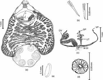
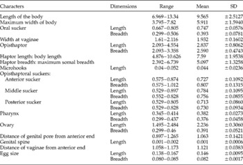
Based on eight mature specimens (figs 1–3). Main body or soma leaf-like, tapering at anterior end, terminating at junction with opisthaptor, 9.565 (6.969–13.34) in length, 5.911 (3.795–7.82) in maximum width across mid-body region. Prohaptor a subterminal oral sucker encircling mouth. Opisthaptor with six prominent suckers, width-wise relatively smaller compared to maximum width of body, lacking hamuli and provided with a pair of needle-like microhooks with cleft base lying between the two most posterior suckers. Pharynx prominent, muscular, bulbous in shape, leading to bifurcate intestine. Intestinal caeca sending out on both lateral and median aspects numerous diverticulations at irregular intervals, which, in turn, give off secondary branches, interspersed with 6–10 smaller unbranched diverticula; diverticula not anastomosing throughout caecal length, not extending into opisthaptoral area. Testis forming insurmountable diffused follicular mass in postovarian intercaecal zone, commencing just behind vitello-vaginal duct; vas deferens arising in region of junction of female genital duct, running anteriad slightly to side of median line; cirrus sac small, tipped with genital crown of 11–12 minute spines; male terminal duct opening into common genital sinus in front of female genital pore; genital sinus opening to exterior close behind intestinal bifurcation. Ovary elongate, retort-shaped with rosette-like lobed proximal end, situated on right of median line; Mehlis' gland complex median, near base of ovary; uterus tubular, emerging from junction of oviduct and vitelline reservoir, proceeding anteriad, containing many (7–49) partly embryonated eggs. Genito-intestinal canal present, given off from same junction as uterus, opening into right caecum. Vaginae opening on either lateral margin of body, on a level with pharynx, between genital pore and oral sucker, at short distance from anterior end. Vitellaria follicular, extensive, commencing from level of pharynx, extending posteriorly up to base of soma.The morphometric measurements of the body and its organs are provided in table 1.
Neoriojatrema mokokchungensis n. g., n. sp.: (a) entire worm, dorsal view; (b) microhooks with cleft base; (c) female genitalia (GP, genital pore; M.Gl, Mehlis'gland; OV, ovary; UT, uterus; VP, vaginal pore); (d) genital crown; (e) egg.

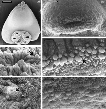
Scanning electron micrographs of Neoriojatrema mokokchungensis n. g., n. sp.: (a) entire worm, ventral view (scale bar = 1 mm); (b) anterior end, front view (scale bar = 100 μm); (c, d) ridge of oral sucker, showing numerous dome-shaped papillae with cilia-like extensions (arrow) (scale bar = 10 μm); (e) genital pore, with ciliate papillae in its vicinity (arrow) (scale bar = 10 μm); (f) single papilla with its cilium (arrow) at higher resolution (scale bar = 5 μm).

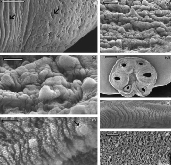
Scanning electron micrographs of tegument of Neoriojatrema mokokchungensis n. g., n. sp.: (a) anterolateral vaginal region (scale bar = 50 μm); (b) ventral body surface (scale bar = 20 μm); (c) same at higher resolution (scale bar = 10 μm); (d) opisthaptor – surface papillae are missing in the region (scale bar = 500 μm); (e) opisthaptoral sucker rim showing fine radial foldings (scale bar = 10 μm); (f) haptoral sucker to show large domed papillae in between the fine beaded texture (arrow) (scale bar = 5 μm); (g) opisthaptor (scale bar = 50 μm).

Morphometric measurements (mm) of Neoriojatrema mokokchungensis n. g., n. sp.

Scanning electron microscopy revealed the fine surface topography of the fluke (figs 2 and 3). The tegument is studded with dense papillae-like outgrowths all over the somal region except the circum-vaginal region. These outgrowths appear to be mounted in between a mesh-like network, showing a sponge-like texture; papillae in the anterior region immediately behind the oral sucker are poked each with a hole, from which a cilium-like extension is seen to emerge. The opisthaptoral surface is clearly devoid of any papillate structures, although the tegument surrounding the suckers is thrown into a pattern of concentric ring-like foldings, which indicate the underlying musculature and limits of each sucker; under higher magnification the rim of these suckers shows many radial foldings and reveals a finely beaded texture that is interrupted by randomly distributed large, domed papillae. Sparsely distributed papillae abound in between the mesh-like texture of the opisthaptoral tegument.
Diagnostic characters
Polystomatidae, Polystomatinae. Opisthaptor lacking large hooks, not broader than forebody, with two needle-like microhooks with cleft base. Pharynx bulbous, of varying shapes. Intestinal caeca highly diverticulate but not anastomosing medially and not uniting posteriorly, not extending into opisthaptoral zone. Ovary retort-shaped, with rosette-like lobed proximal end, located in anterior third of body length; vaginal apertures present. Genital crown with 11–12 spines. Eggs many in utero, 0.138–0.167 × 0.080–0.085 in size.
Type host. Xenophrys glandulosa Fei, Ye & Huang, 1991.
Habitat. Urinary bladder.
Type locality. Mokokchung, 26°11′68″N to 94°17′20″E and 26°46′31″N to 94°42′8″E (Nagaland, India).
Other locality. Tuensang (Nagaland, India) 25°55′45.12″N to 94°33′08″E and 26°02′23.78″N to 94°44′25.82″E (Nagaland, India).
Deposition of specimens. Holotype: W9247/1; Paratype: W9248/1 – deposited in Zoological Survey of India, Kolkata. Other paratypes: NEHU/Z-MA/14.1-14.5 (five numbers), deposited in the helminthological collection of the Department of Zoology, NEHU, Shillong, India.
Etymology. Genus named after the closely related genus in the subfamily; species named after the type locality.
Discussion
In having the following characters – leaf-like or pyriform body; discoid opisthaptor with six suckers; prohaptor in the form of an oral sucker situated terminally or subterminally, followed by a well-developed bulbous pharynx; bifurcate intestine with the caeca extending up to the posterior region of the body; median genital pore, situated near the level of the intestinal bifurcation; presence of a genital crown; reticulate testis; follicular vitellaria and the presence of vaginae – the present form can be undoubtedly relegated to the superfamily Polystomatoidea Price, Reference Price1936. In having a compound six-suckered opisthaptor and testis in the form of a follicular mass, the present form belongs to family Polystomatidae and subfamily Polystomatinae. So far, there are six genera included under the subfamily; these are Polystoma, Polystomoides Ward, 1917, Parapolystoma Ozaki, 1935, Eupolystoma Kaw, Reference Kaw1950, Pseudopolystoma Yamaguti, 1963, and Riojatrema Lamothe-Argumetho, 1964. The present form comes close to Eupolystoma, Pseudopolystoma and Riojatrema, in all of which the opisthaptor lacks the large hooks or hamuli; in the other three genera the opisthaptor is provided with a pair of hamuli. As mentioned earlier, of the various polystomatid genera only two, i.e., Eupolystoma and Polystoma, have so far been reported to occur in anuran Amphibia in India. The present form cannot be relegated to the genus Polystoma, the members of which are characterized by possessing a pair of large opisthaptoral hamuli. It comes close to Eupolystoma in sharing the feature of opisthaptor without large hamuli. Of the two species of Eupolystoma described so far from the Indian subcontinent, E. rajai is characterized by having the opisthaptor armed with 16 microhooks with undivided roots, the intestinal caeca united medially just in front of the haptor, numerous follicular testes, and the genital crown with eight minute hooks; in E. chauhani, six microhooks are present between the last pair of suckers on the opisthaptor, the intestinal caeca lack diverticula and the genital crown has six minute hooks. However, location of the ovary in the anterior one-third of the body in the present form is a distinct character that makes it stand apart from the genera Eupolystoma and Pseudopolystoma; in both these genera the ovary characteristically lies in the posterior part of the body.
The diagnostic characters of the third genus, i.e. Riojatrema, as described by Lamothe-Argumedo (Reference Lamothe-Argumedo1964), include: opisthaptor wider than the forebody; pharynx cylindrical with a constriction in the middle; intestinal diverticula anastomosing prehaptorally and uniting in the region of opisthaptor; and vitellaria follicular and extending into the opisthaptoral region. In all these features the present form differs from Riojatrema.
So far the genus Riojatrema includes only two species: R. bravoae Lamothe-Argumedo, 1964 from Tomodactylus amulae and Bufo simus from Mexico, and R. ecuadorensis Dyer, 1985 from Bufo typhonius from Ecuador. In R. bravoae, characteristically the intestinal caeca possess numerous diverticula forming three to four anastomoses in the median field of the body and unite posteriorly in the opisthaptoral region; the genital crown includes 8–9 spines, the ovary is pyriform, and the vitelline follicles are widely scattered and extend into the opisthaptoral region. The diagnostic characters of R. ecuadorensis include: opisthaptoral disc wider than the main body; pharynx cylindrical, with a constriction in the middle; caecal diverticula anastomosing prehaptorally and caeca uniting medially in the region of the opisthaptor; and vitellaria extensively distributed.
The only characters that the present form shares with known species of the genus Riojatrema are: opisthaptor lacking large hooks and the uterus lying on a level with the anteriorly placed ovary. However, the following characters – opisthaptor with relatively much smaller width than that of the body; pharynx bulbous (and not cylindrical with a constriction), intestinal diverticulation in both lateral and marginal aspects but caeca not uniting or anastomosing prehaptorally; ovary long, with a lobed or rosette-like proximal end, genital crown comprising 11–12 spines, and vitellaria not extending into the opisthaptoral area – completely set it apart from the species under Riojatrema. Besides these differences, the present form was recovered from a different host, a megophryid frog.
A high degree of host specificity and restricted geographical distribution demonstrated among the anuran monogeneans have also been assigned significant importance in species differentiation within the polystomatid genera (Maeder et al., Reference Maeder, Euzet and Combes1970; Combes & Knoepffler, Reference Combes and Knoepffler1977; Dupouy, Reference Dupouy1978; Combes & Channing, Reference Combes and Channing1979; Murith, Reference Murith1981; Kok & Van Wyk, Reference Kok and Van Wyk1986; Kok & Seaman, Reference Kok and Seaman1987; Diengdoh & Tandon, Reference Diengdoh and Tandon1991). However, the extent to which host specificity could be depended upon as a differential character of taxonomic value is controversial (Prudhoe & Bray, Reference Prudhoe and Bray1982). The foregoing differences in generic diagnostic characters, along with several distinct characters (i.e. width of the forebody greater than opisthaptor; pharynx not cylindrical and constricted in the middle but round and bulbous in shape; caeca not extending into the opisthaptoral region and not uniting medially; genital crown with 11–12 spines; two needle-like, cleft-base microhooks between the posterior-most opisthaptoral suckers; ovary retort-shaped with a lobed end; vitellaria not extending into the opisthaptoral region; and a different anuran host) that deviate the present form from the known species of the genus Riojatrema, warrant establishment of a new genus to accommodate it. Thus, a new genus is herewith established and given the name Neoriojatrema, with N. mokokchungensis as its type species.
Neoriojatrema mokokchungensis n. g., n. sp. is the third polystomatid monogenean genus represented in anuran hosts from the Indian subcontinent.
Acknowledgements
The authors are thankful to the Heads of the Department of Zoology at both Nagaland University and North-Eastern Hill University for providing infrastructural support and to the Director, Sophisticated Analytical Instrumentation Facility (SAIF) at NEHU, Shillong for allowing the use of the SEM facility. Support from AICOPTAX Project (Ministry of Environment & Forests, Government of India) to V.T. is gratefully acknowledged. Thanks are due to Drs Bishnupada Roy and Pramod Kumar Prasad for their assistance in SEM work.