Introduction
The main breeding colonies of the Magellanic penguin Spheniscus magellanicus (Forster, 1781) in the south-western Atlantic Ocean (SWA) are found from Cape Horn to latitude 42°S, including the Falkland Islands (Williams, Reference Williams1995). After the breeding and moulting season (September–April), these Magellanic penguins migrate northwards during the austral winter, following the seasonal movements of Argentine anchovy, Engraulis anchoita Hubbs & Marini, 1935 (Clupeiformes, Engraulidae), up to the coast of south-eastern Brazil (Dantas et al., Reference Dantas, Almeida, Maracini, Serra, Chame, Labarthe, Kolesnikovas, Siciliano, Matias, Moura, Campos, Mader and Serafini2013, Stokes et al., Reference Stokes, Boersma, Casenave and García-Borboroglu2014).
Spheniscus magellanicus has been reported to be parasitized by 21 different helminth species, of which nine were recorded from individuals found on the Brazilian coast (Brandão et al., Reference Brandão, Moreira and Luque2014). However, in southern Brazil, the region where the highest mortality of S. magellanicus occurs, the gastrointestinal parasite Contracaecum pelagicum Johnston & Mawson, 1942 (Ascaridida, Anisakidae) was, until now, the only nematode known to parasitize S. magellanicus (Altrão et al., Reference Altrão, Paula, Tavares, Ott and Silva-Souza2017).
Herein, we describe a new species of Diomedenema Johnston & Mawson, 1952, a desmidocercid aproctoid spiruromorph, parasitizing the lungs of S. magellanicus specimens found in southern Brazil during their winter migration.
Materials and methods
Collection of nematodes
Forty (22 females, 11 males and 7 undetermined) young specimens of Magellanic penguins, S. magellanicus (weight 1.63–3.70 (2.35 ± 0.63) kg and total length 47.80–68.40 (56.67 ± 4.89) cm) were collected during June, July and August of 2013 and 2014, from between the beaches of Itapeva, municipality of Torres (29°21′32″S; 49°44′09″W) and Dunas Altas, municipality of Palmares do Sul (30°38′18″S; 50°28′20″W), state of Rio Grande do Sul (RS), Brazil. All individual penguins involved in this research were donated, with 31 having been found dead by the monitoring team of the Centro de Estudos Costeiros Limonológicos e Marinhos, Instituto de Bio-Ciências, Universidade Federal do Rio Grande do Sul, RS, Brazil (CECLIMAR/IB/UFRGS) and nine found moribund by the Setor de Reabilitação de Animais Selvagens e Marinhos, CECLIMAR/IB/UFRGS, the latter having died without being fed or treated with vermifuge. Specimens were labelled, placed individually in plastic bags and transported on ice in isothermal boxes to the laboratory of CECLIMAR/IB/UFRGS for analysis. They were identified in accordance with Williams (Reference Williams1995) and skins and skeletons of representative specimens were deposited in the Ornithological Scientific Collection of the Museu de Ciências Naturais (MUCIN 318, 319342, 448, 547, 549, 550, 561, 808, 903, 904) of CECLIMAR/IB/UFRGS.
The host specimens were necropsied and their lungs inspected for helminths. Eleven specimens (nine females and two males) were parasitized by a nematode species. These nematode specimens were placed in Petri dishes with 0.90% NaCl solution, fixed in AFA (glacial acetic acid, formaldehyde and 70% ethanol in the proportions of 2:3:95), preserved in a solution of 70% ethanol plus 5% glycerin and clarified with Amman's lactophenol. Some of the nematode specimens were then mounted in Canada balsam, as described by Knoff & Gomes (Reference Knoff, Gomes, Molinaro, Caputo and Amendoeira2012), identified taxonomically in accordance with De Ley & Blaxter (Reference De Ley, Blaxter, Cook and Hunt2004) and Anderson & Bain (Reference Anderson, Bain, Anderson, Chabaud and Willmott2009).
The parasite indexes used were prevalence, mean intensity and mean abundance, and followed Bush et al. (Reference Bush, Lafferty, Lotz and Shostak1997). The holotype and paratypes were deposited in the Helminthological Collection of the Instituto Oswaldo Cruz, Fiocruz (CHIOC), Rio de Janeiro, state of Rio de Janeiro, Brazil. Photos of type material (designated as syntypes and paratypes) of Diomedenema diomedeae Johnston & Mawson, 1952, deposited in the Australian Helminthological Collection (AHC) of the South Australian Museum (SAM), North Terrace, Adelaide, Australia, were analysed for taxonomic comparison.
Morphological analysis
Morphological identification used nematode specimens that were mounted provisionally between slides and coverslips, using Amman's lactophenol to clarify the specimens for observation of their structures. After observations, the specimens were preserved in 70° GL ethanol. Some of the nematodes were sectioned at the cephalic end and the sections were clarified in glycerine. The holotype and its allotype were mounted in Canada balsam. Measurements of specimens were made by means of bright-field microscopy using an Olympus BX 41 microscope (Tokyo, Japan) and are presented in millimetres (mm), with the range followed by the mean in parentheses, unless otherwise indicated. Drawings were executed using a drawing tube connected to the microscope.
For topographic description of the cuticular surface, some of the specimens were subjected to analyses by scanning electron microscopy (SEM). For this, samples were fixed in 2.5% glutaraldehyde with 0.1 m sodium cacodylate buffer (pH 7.4), postfixed in 1% osmium tetroxide, dehydrated in an ethanol series, dried using the CO2 critical-point drying method, coated in gold, and examined and photographed using a Tescan Vega-3 scanning electron microscope (Brno-Kohoutovice, Czech Republic), with 15 kV acceleration voltage.
Results
Aproctoidea Yorke & Maplestone, 1926, Desmidorcercidae Cram, 1927, Diomedenema Johnston & Mawson, 1952, Diomedenema tavaresi n. sp.
Description
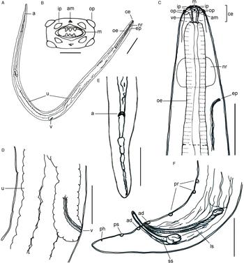
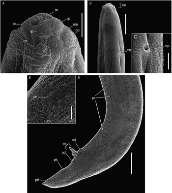
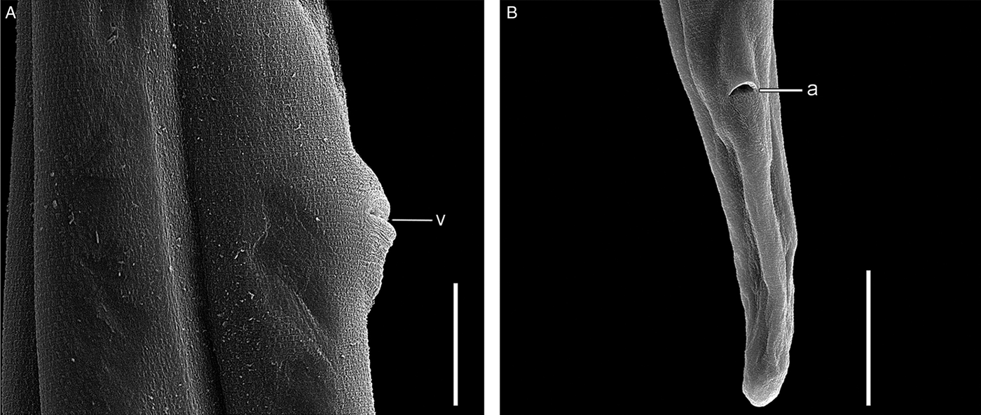
The description (figs 1–3) is based on observations of 30 specimens: 2 whole mounts (1 male and 1 female) and 18 wet mounts (9 males and 9 females) in Amman's lactophenol, for measurements; and 10 (4 males and 6 females) for SEM analysis.
Diomedenema tavaresi n. sp. (A) Female, ventral view entire worm: cephalic end (ce), oesophagus (oe), excretory pore (ep), nerve ring (nr), uteri amphidelphic (u) and vulva (v), lateral view; posterior end bent with anus (a). (B) Cephalic end, apical view: outer papilla (op), inner papilla (ip), amphid (am) and mouth (m). (C) Female, lateral view anterior end: cephalic end (ce), outer papillae (op), inner papillae (ip), amphid (am), mouth (m), vestibule (ve), oesophagus (oe), excretory pore (ep) and nerve ring (nr). (D) Female, lateral view: detail of vulva (v) and part of uterus (u). (E) Female, lateral view posterior end: anus (a), ventral view. (F) Male, posterior end, large spicule (ls) and small spicule (ss); three pairs of precloacal papillae (pr), two pairs of adcloacal papillae (ad), one pair of postcloacal papillae (ps) and one pair phasmids (ph). Scale bars: A = 400 μm; B = 25 μm; C–F = 100 μm.

Diomedenema tavaresi n. sp., SEM. (A) Dorso-lateral view of detail of cephalic end: mouth (m) with two lateral tricuspid teeth on entrance of buccal cavity; inner papillae (ip), amphid (am) and outer papillae (op). (B) Ventro-lateral view of anterior end: cephalic end (ce) and excretory pore (ep). (C) Ventral view of detail of excretory pore (ep). (D) Lateral view of posterior end of male: extroverted spicules (es), three pairs precloacal papillae (pr), two pairs adcloacal (ad) one pair postcloacal (ps), and one pair phasmids (ph). (E) Detail of phasmid (ph). Scale bars: A and C = 10 μm; B and D = 50 μm; E = 4 μm.

Diomedenema tavaresi n. sp., SEM of female. (A) Lateral view of mid-body: detail of vulva (v). (B) Ventral view of posterior end: detail of anus (a). Scale bars: A = 30 μm; B = 50 μm.

General description
Small-sized nematodes with anterior end compressed laterally; eight cephalic papillae in two rings, one inner and one outer (a set of four papillae distributed in two lateral pairs). Two amphids present, each one between the inner and outer pair of papillae. Mouth dorso-ventrally elongated, entrance to buccal cavity with two lateral tricuspid teeth; on lateral mouth is a plaque-like extension flanked by the inner papillae present on lateral mouth border; vestibule present, well developed as a tubular cavity; oesophagus much shorter than half the length of the body, not differentiated externally into two regions, posterior region to nerve ring wider. Nerve ring anterior to excretory pore. Excretory pore ventral. Tail short in males and females, rounded in females and pointed in males, without alae. Vulva post-oesophageal, located ventrally at the mid-body. Uteri amphidelphic. Oviparous; eggs submedium-sized, thick-shelled. Posterior end of female with ventrally positioned anus. Posterior end of male curved ventrally; spicules unequal. Cuticle with small punctations arranged in annuli until the end of the tail, in both sexes.
Males
Body 4.900–5.600 (5.230) (n = 10) long by 0.150–0.175 (0.170) (n = 10) wide; vestibule 0.025 (n = 10) long by 0.013 (n = 10) wide; oesophagus 0.650–0.690 (0.669) (n = 10) long; excretory pore 0.190–0.220 (0.201) (n = 10) from anterior end and nerve ring at 0.125–0.175 (0.149) (n = 10); cloaca 0.130–0.145 (0.136) (n = 10) from tip of pointed tail; large spicule 0.170–0.22 (0.20) (n = 10) long, small spicule 0.135–0.140 (0.137) (n = 10) long; three pairs precloacal papillae, two pairs adcloacal papillae and one pair postcloacal papillae; one pair phasmids posterior to postcloacal pair of papillae.
Females
Body 5.825–6.050 (5.970) (n = 10) long by 0.175 (n = 10) wide; vestibule 0.028–0.035 (0.030) (n = 10) long by 0.013–0.015 (0.0145) (n = 10) wide; oesophagus 0.675–0.725 (0.707) (n = 10) long; excretory pore 0.180–0.220 (0.196) (n = 10) from anterior end, nerve ring at 0.150–0.160 (0.155) (n = 10) and vulva at 3.000–3.075 (3.035) (n = 10); anus, 0.150–0.170 (0.160) (n = 10) from tip of rounded tail. Eggs oval 30–37.5 (36) μm × 17.5–25 (18.2) μm (n = 30).
Taxonomic summary
Type host
Spheniscus magellanicus (Forster, 1781), Aves, Sphenisciformes; Magellanic penguin.
Type locality
Beach of Imbé, Imbé, RS, Brazil (29°91′04″S; 50°08′84″W).
Other locality
Beach of Arroio do Sal, Arroio do Sal, RS, Brazil (29°56′96″S; 49°89′69″W).
Site of infection
Lung.
Specimens collected
Three hundred and nineteen adult nematodes (145 males and 174 females).
Prevalence
Eleven hosts of 40 collected, 27.5%.
Mean intensity
29 ± 35.66.
Mean abundance
7.98 ± 22.30 (0–131).
Range of infection
1–131.
Material deposited
Holotype CHIOC no. 38655a (male), and allotype CHIOC 38655b (female) (whole mounts), other paratypes CHIOC 38506a (males) and CHIOC 38506b (females) (wet materials).
Etymology
The specific name is in honour of Mauricio Tavares MSc (biologist of CECLIMAR/IB/UFRGS) who, for several years, has tirelessly monitored the mortality of marine tetrapods, especially birds, on the beaches of RS, Brazil. He has collected and prepared specimens, including parasites, for scientific collections and for several studies, aiding the development of research and the expansion of knowledge about the animals that live or visit the Brazilian coast.
Material examined
Photos of D. diomedeae AHC 4287 syntype (one male) and AHC 1729 paratypes (two females).
Discussion
The new species is very similar morphologically to D. diomedeae Johnston & Mawson, 1952 found in a yellow-nosed albatross, Diomedea chrysostoma Forster, collected in Brighton, South Australia, in possessing a body with a laterally compressed anterior end; eight large cephalic papillae in two rings, an inner and an outer ring, amphids present; mouth dorsoventrally elongate, buccal cavity with two lateral tricuspid teeth; vestibule present, well developed as a tubular cavity; oesophagus much shorter than half the length of the body, not differentiated externally into two regions, posterior region to nerve ring wider; nerve ring anterior to excretory pore; male and female tail short, without alae; vulva behind oesophagus; uteri amphidelphic; posterior end of male curved ventrally; spicules unequal; and cuticle with small punctations, arranged in annuli until the end of the tail. However, it differs by having a shorter (by more than half) body length in both sexes; having a set of caudal papillae in males with an additional pair of adcloacal papillae (three pairs of precloacal papillae, two pairs of adcloacal papillae and one pair of postcloacal papillae instead of three pairs of precloacal papillae, one pair of adcloacal and one pair of postcloacal papillae); having precloacal papillae with the first two pairs closer to each other than the third pair; the pointed tail of males being longer than the rounded tail of females; and the vulva of females being located at mid-body, instead of at the anterior third of the body (Johnston & Mawson, Reference Johnston and Mawson1952).
The new species was allocated to the genus Diomedenema in its original diagnosis, adding the female characteristic of the vulva located at mid-body, instead of creating a new genus based on this single different feature of the new taxon. In the original description of this genus, the vulva is described as ‘Vulva anterior, post-oesophageal’; however, there was no accompanying drawing showing the location, and in the original description it was described as ‘Vulva anterior, 4.7 mm from head end in worm 14.7 long.’ (Johnston & Mawson, Reference Johnston and Mawson1952). The exact location of the vulva was observed in photos of a female D. diomedeae, AHC 1729 paratype, deposited in SAM, confirming it to be at the end of the anterior third of the body. Yamaguti (Reference Yamaguti1961) cited the location of the vulva as ‘vulva postesophageal’ and Anderson & Bain (Reference Anderson, Bain, Anderson, Chabaud and Willmott2009) cited it as ‘Vulva behind oesophagus’, both of whom based their claims on the observation of Johnston & Mawson (Reference Johnston and Mawson1952). Thus, we felt this to be sufficient for confirming the generic diagnosis of specimens of the present study.
This is the first report of a nematode infecting the lung of a sphenisciforme host, thus extending its occurrence to another marine bird.
Acknowledgements
The authors would like to thank Maurício Tavares MSc (CECLIMAR/IB/UFRGS), who collecting the hosts and kindly donated them to us to proceed with this parasitological research; Angélica Alves de Paula and Carla Suzuki Altrão (students of the Programa de Pós-Graduação em Ciências Biológicas, Universidade Estadual de Londrina (UEL), state of Paraná, Brazil) for their assistance during necropsies and parasite collection; Dr Leslie Chisholm and Dr Lesley Rae Smales Warner (South Australian Museum's Parasitology Collection) for kindly sending the photos of the syntypes and paratypes of D. diomedeae deposited in the Australian Helminthological Collection of the South Australian Museum; and Mr Ricardo Baptista Schmidt (Serviço de Produção e Tratamento de Imagens, IOC, Fiocruz) for processing the figures
Financial support
Fiocruz, UEL, Universidade Federal Rural da Amazônia (UFRA) and UFPA are thanked for their partial financial support.
Conflict of interest
None.
Ethical standards
The authorization for the capture of the hosts and collection of parasites, no. 20185, was obtained by Maurício Tavares MSc from CECLIMAR/IB/UFRGS in the Sistema de Autorização e Informação em Biodiversidade (SISBIO) of the Instituto Chico Mendes de Conservação da Biodiversidade, Ministério do Meio Ambiente, Brazil.