




No CrossRef data available.
Article contents
Natural IgM antibodies in sera from various animals but not the cat kill Toxoplasma gondii by activating the classical complement pathway
Published online by Cambridge University Press: 01 March 2004
Abstract
Sera from swine, rabbit, and dog, that had never been exposed to Toxoplasma gondii, demonstrated significant killing of T. gondii tachyzoites in vitro, while cat serum did not. Swine and rabbit sera contained natural IgM antibody against the tachyzoites, and the classical complement pathway was activated by the binding of natural IgM antibody to the tachyzoites, leading to lysis. Anti-T. gondii antibodies, induced in swine or cat infected with T. gondii, had no killing effect by themselves but killed the tachyzoites in the presence of swine complement. However, the anti-T. gondii antibodies of swine or cat demonstrated a very low killing effect in the presence of cat complement. This suggests that T. gondii tachyzoites have an evasion mechanism to prevent lysis which is specific for cat complement.
Information
- Type
- Research Article
- Information
- Copyright
- 2004 Cambridge University Press
INTRODUCTION
Toxoplasma gondii (T. gondii) is a protozoan parasite with an extremely broad host range. Cats are the definitive host for the parasite, with most warm-blooded animals including birds, wild animals, pets, livestock, and humans acting as intermediate hosts (Innes, 1997).
The role of complement in the host defence mechanism against infection of T. gondii is not well understood. In the case of human serum, in vitro investigations showed that T. gondii tachyzoites are rapidly lysed by the activation of complement through the classical pathway in the presence of specific antibodies (Schreiber & Feldman, 1980; Suzuki & Kobayashi, 1985), although the parasites are resistant to complement in the absence of antibodies (Fuhrman & Joiner, 1989; Feldman, 1956; Strannegard & Lycke, 1966). Sera of several hosts seem to have a lethal effect on the tachyzoites in the absence of specific antibodies (Sabin & Feldman, 1948), but the mechanisms underlying the killing activity have not been elucidated.
Serum contains natural IgM antibodies, which are capable of binding to a particular antigen or a pathogen, even if the host has never been exposed to it (Boes, 2000). Naturally occurring IgM antibodies to T. gondii have been reported in swine (Takahashi & Konishi, 1986) and human sera (Konishi, 1991), and they enhance phagocytic and microbicidal activities of neutrophils (Konishi & Nakao, 1992). However, their involvement in complement activation has not been studied despite the fact that IgM is a much more efficient complement-fixing antibody than IgG because of its pentameric structure (Boes, 2000).
This study demonstrated the in vitro killing activities of sera derived from several hosts to T. gondii tachyzoites. Furthermore, it was shown why serum from cat, a definitive host of T. gondii, showed low killing activity.
MATERIALS AND METHODS
Parasites and cells
Toxoplasma gondii (RH strain) tachyzoites engineered to express green fluorescent protein (GFP) constitutively were used (Xuan et al., manuscript in preparation). Tachyzoites were serially passaged in Vero cell culture every 4 or 5 days. Vero cells and parasites were maintained in Dulbecco's Modified Eagle's Medium (DMEM) (Nissui, Japan) supplemented with 5% heat-inactivated foetal calf serum (FCS), 50 μg/ml streptomycin, and 50 units/ml penicillin. Porcine kidney-derived CPK cells (Komaniwa, Fukusho & Shimizu, 1987) and rabbit kidney-derived RK13 cells (ATCC, CCL-37) were maintained in Eagle's MEM (Nissui, Japan) containing 7·5% FCS.
Sera and antibodies
Blood samples were obtained from 6-month-old specific pathogen-free pigs (Zen-noh Institute of Animal health, Chiba, Japan), and from healthy Japanese White strain rabbits. Blood samples were obtained also from healthy adult cats and dogs kept at the Veterinary Medical Center of the University of Tokyo through the courtesy of Professor G. Tsujimoto, at the Graduate School of Veterinary Medicine, The University of Tokyo. Blood samples were allowed to clot at room temperature for 60 min, centrifuged and the supernatants were aliquoted and stored at −80 °C and thawed just before use to minimize the loss of complement activity. All sera were confirmed to be seronegative for Toxoplasma antibodies by the latex agglutination test with Toxocheck-MT (Eiken Chemical Co., Ltd, Tokyo, Japan). Western blot analysis of all the sera failed to recognize major antigens of T. gondii (data not shown).
IgG-depleted swine serum was prepared by passing normal swine serum through protein A conjugated beads (HiTrap™ Protein A HP; Amersham Biosciences, Tokyo, Japan) column.
Anti-T. gondii sera were obtained from mice infected with the attenuated PLK strain of T. gondii. Sera from pigs and cats that were naturally infected with T. gondii, were also used as anti-T. gondii sera. Antibody titres of these antisera ranged from 1[ratio ]256 to 1[ratio ]2048 using the latex agglutination test. All antisera were stored at −20 °C and lost complement activity during the storage at this temperature.
Goat anti-human C1q antibody was purchased from Sigma (St Louis, MO). Affinity-purified goat anti-swine IgM (μ-specific) antibodies with or without horse-radish peroxidase (HRP) label were purchased from Kirkegaard & Perry Laboratories Inc. (Gaitherburg, MD). Afffinity purified goat anti-rabbit-IgM (μ-specific) antibody was purchased from Bethyl Laboratories, Inc. (Montgomery, TX).
Polyclonal mouse antibodies against CPK and RK13 cells were described previously (Maeda et al. 2002).
Serum neutralization assay
The extent of killing of T. gondii tachyzoites during incubation in serum was determined. Tachyzoites were released from infected Vero cells by rapid extrusion through a 27-gauge needle, and cell debris was subsequently removed by filtration through a polycarbonate filter with a pore size of 5 μm (Millipore, Bedford, MA). The purified tachyzoites were pelleted by centrifugation at 500 g and re-suspended in DMEM containing 1% FCS. In a 100 μl reaction mixture, 104 to 105 purified tachyzoites were incubated at 37 °C for 30 min with various amounts of serum in DMEM. The killing effect of sera did not vary in this range of tachyzoites. After centrifugation at 500 g to remove the supernatant, the pellet was re-suspended in DMEM containing 1% FCS. Diluted tachyzoite samples were applied to confluent monolayers of Vero cells on 48-well plates. After overnight incubation at 37 °C, the infected cell monolayers were overlaid with DMEM containing 1·8% Bacto Agar to prevent formation of secondary plaques. Parasites were allowed to grow for 2–3 days, and the number of fluorescent parasites clusters (plaques) was counted under a fluorescent microscope.
To examine the factors that contributed to serum killing, the conditions of the assay were varied. In some assays, serum was used after chelation with 10 mM EDTA or after heat-inactivation. Heat inactivation to abolish serum complement activity was carried out at 56 °C for 30 min. In order to prevent the classical pathway activation, sera were chelated with 10 mM ethylene glycol-bis-(β-aminoethyl ether)-N,N,N′,N′-tetraacetic acid (EGTA) containing 2 mM MgCl2. Antibodies to swine-IgM, rabbit-IgM, and human C1q were used to inactivate the components attributable to serum killing individually. The concentrations of anti-IgM antibodies were 2·5 μg/μl for swine serum, 5 μg/μl for rabbit serum respectively, and 990 μg of anti-C1q antibody was added to 3·5 μl of both swine and rabbit serum. The antibody to human C1q was shown to cross-react with swine and rabbit C1q by Western blot analysis (data not shown).
Preparation of protein A bead antibody affinity column
Rabbit anti-swine IgM antiserum was prepared by immunizing New Zealand white rabbits with swine IgM purified from swine serum by affinity chromatography using HiTrap IgM purification HP column (Amersham Pharmacia Biotech UK Ltd, UK) and gel-filtration using a Biogel A-5m gel (Bio-Rad Laboratories, Hercules, CA) column. Anti-swine IgM antibody was immobilized on protein A beads according to the method described by Gersten & Marchalonis (1978). Briefly, 2 ml of rabbit anti-swine IgM serum were mixed with a 2 ml suspension of protein A-Sepharose 4B beads (Sigma, St Louis, MO) and incubated at room temperature for 1 h with gentle rocking. The beads were washed twice with 10 ml of 0·2 M sodium borate buffer (pH 9·0) and re-suspended in 10 ml of the same buffer. Di-methylpimelimidate was added to the suspension solid to bring the final concentration to 20 mM, and this was mixed for 30 min at room temperature. The reaction was stopped by washing the beads once in 0·2 M ethanolamine (pH 8·0) and then incubating for 2 h at room temperature in 0·2 M ethanolamine.
Preparation of IgM and IgG-free pig serum
Protein A beads coupled with rabbit anti-swine IgM antibodies as described above was poured into a small column to a bed volume of 1·5 ml and washed extensively with PBS. Then 0·5 ml of swine serum was applied to the top of the column and eluted with PBS. The pass-through protein fraction was collected, aliquoted, stored at −80 °C and thawed immediately before use as a source of IgM and IgG-free swine complement.
Statistical analysis
Results were expressed as the mean±standard deviation of data obtained from 3 or more separate experiments with different samples of sera (Figs 1 and 6). In other experiments the results were expressed as the mean±standard deviation of triplicate data in a single experiment. The same experiments were carried out at least twice and the results were similar. Statistical analyses were performed using a two-tailed Students t-test. * and ** indicate P<0·05 and P<0·01, respectively.
RESULTS
Killing effect of sera from various animals
The viability of T. gondii tachyzoites was assessed by fluorescent plaque assay after incubation in normal sera derived from various animals. The concentrations of the sera were varied from 2·5 to 40%, but the killing effect did not differ significantly at a concentration of more than 10%. Therefore, 20% serum was employed. The killing effect was observed in non-immune sera of swine, dog, and rabbit, while cat sera showed very little killing effect (Fig. 1). The killing effect of rabbit and dog sera appeared to be stronger than that of swine sera but statistical analysis revealed no significant difference among these sera.

Fig. 1. Killing of Toxoplasma gondii by various animal sera. Tachyzoites were incubated with 20% serum at 37 °C for 30 min, and viable tachyzoites were counted as described in the Materials and Methods section. The results were presented as a percentage of the control value. The mean±standard deviation of several experiments with different batches of sera, performed in triplicate, are shown. Sera from 4 pigs, 2 rabbits, 3 dogs and 6 cats were tested. Control: tachyzoites treated with medium alone. *P<0·05; **P<0·01, respectively.
It is known that complement activity is heat-labile and dependent on Ca2+ and Mg2+. Heat-inactivated swine, dog and rabbit sera as well as EDTA-treated sera failed to kill tachyzoites (Fig. 2), indicating that the lethal effect of these sera is complement dependent.

Fig. 2. Complement dependency for the tachyzoites killing effect of pig (A), rabbit (B) and dog serum (C). Tachyzoites were incubated with medium (Control), 20% serum (Serum), heat-inactivated serum (H-Serum), or serum chelated with 10 mM EDTA (EDTA). The experiment was repeated more than twice. **P<0·01.
Analysis of complement activation pathways
To determine the complement activation pathway in these sera, the parasite viability assay was performed with sera treated with EGTA, a selective chelating reagent for Ca2+, or anti-human C1q antibody. It is known that activation of the classical pathway of complement requires the presence of Ca2+ while the alternative pathway does not. Treatment with both EGTA and anti-C1q antibody (Fig. 3) deprived the sera of their killing effect. Therefore, it is conceivable that the tachyzoite killing effect is mediated by an activation of the classical complement pathway.

Fig. 3. Effect of EGTA and anti-C1q antibody on the tachyzoite killing activity of pig (A), rabbit (B) and dog sera (C). Tachyzoites were incubated with medium (Control), 20% serum (Serum), serum treated with 10 mM EGTA and 2 mM Ca2+ (EGTA), or serum with anti-C1q antibody (anti-C1q). The experiments were repeated twice and generated similar results. *P<0·05; **P<0·01, respectively.
Complement-activation mediated by IgM
Although all the sera used above came from animals that have never been exposed to T. gondii, the data suggested the presence of antibodies, which are reactive to the parasites and activate the classical complement pathway. Since most of the natural antibodies found in the non-immune animal were IgM, we carried out the following experiments to determine whether the complement activating antibody in swine and rabbit sera was IgM. It was found that addition of anti-swine IgM antibody to swine serum, and anti-rabbit IgM antibody to rabbit serum negated the killing effect of each serum (Fig. 4A and B). Further addition of anti-T. gondii antibodies, obtained from swine infected with T. gondii, to the mixture of serum and anti-IgM antibody resulted in the recovery of the killing activity, thereby obviating the possibility that complement was consumed by addition of anti-IgM antibody. The anti-T. gondii antibodies obtained from infected swine were of IgG isotype (data not shown). Swine IgM- and IgG-depleted serum (SMGDS) demonstrated very little killing effect while IgG-depleted swine serum demonstrated the same degree of the killing effect as normal swine serum (Fig. 4C). Replenishment of swine IgM fraction to SMGDS restored the killing activity. These results indicate that IgM indigenous to the swine sera is responsible for the activation of the classical complement pathway.

Fig. 4. IgM antibodies are responsible for the killing activity of swine serum and rabbit serum. (A) Tachyzoites were incubated with medium (Control), 20% swine serum (Serum), swine serum+anti-swine IgM antibodies (anti-IgM), swine serum+anti-swine IgM antibodies+anti Toxoplasma gondii antibodies (anti-IgM+antiserum), or anti-T. gondii antibodies (antiserum). (B) Tachyzoites were incubated with medium (Control), 10% rabbit serum (Serum), rabbit serum+anti-rabbit IgM antibodies (anti-IgM), rabbit serum+anti-rabbit IgM antibodies+anti-T. gondii antibodies (anti-IgM+antiserum), or anti-T. gondii antibodies (antiserum). The experiments were performed 3 times. (C) T. gondii tachyzoites were incubated with 20% normal swine serum (Serum), 40% swine serum depleted with IgM and IgG (SMGDS), SMGDS replenished with gel-filtrated swine IgM fraction (SMGDS+IgM), swine serum depleted with IgG (IgG(−)) and gel-filtrated swine IgM fraction (IgM) without complement. All experiments were repeated 3 times. *P<0·05; **P<0·01.
Resistance against cat sera
We attempted to find out the reason why sera from cats were not capable of killing tachyzoites. The following experiments confirmed that sera from cats contained comparable amounts of the components of the complement classical pathway as swine or rabbit sera. CPK cells incubated with mouse anti-CPK antibody were not killed, but when cat serum was added as a source of complement, they were killed effectively as judged by Trypan blue exclusion tests (Fig. 5A). All sera obtained from different cats demonstrated similar killing activity with anti-CPK antibodies, while heat-inactivated sera and EDTA-treated sera did not (Fig. 5A). It was also confirmed that sera from swine and rabbit showed the same degree of killing effects as cat sera on CPK cells in the presence of anti-CPK antibodies (data not shown). Furthermore, the concentrations of the complement components for the classical pathway were compared between cat serum and SMGDS by measuring the killing activities on RK13 cells. When RK13 cells were sensitized with mouse anti-RK13 cell antibody, they were killed in the presence of 1% cat serum as a source of complement while not in the presence of 2% of SMGDS. At higher concentrations of SMGDS, sensitized RK13 cells were killed (data not shown). Therefore it can be concluded that the concentration of the complement components for the classical pathway was higher in cat sera than in SMGDS.

Fig. 5. (A) Verification of complement activity in cat sera. CPK cells sensitized with mouse anti-CPK antibody were incubated with 20% cat serum at 37 °C for 30 min. Trypan blue was used to stain the dead cells. The percentages of viable cells were shown. The mean values of experiments with 6 different cat sera preparations were presented. αCPK: CPK cells sensitized with mouse anti-CPK antibodies. Serum+αCPK: CPK cells sensitized with mouse anti-CPK antibodies were incubated with 20% cat serum. H-serum+αCPK: CPK cells sensitized with mouse anti-CPK antibodies were incubated with 20% heat inactivated cat serum. Serum+αCPK+EDTA: CPK cells sensitized with mouse anti-CPK antibodies were incubated with 20% cat serum that had been chelated with 10 mM EDTA. (B) Effect of cat complement and cat anti-Toxoplasma gondii antibodies on T. gondii. Control: T. gondii tachyzoites were incubated with medium. Cat: T. gondii tachyzoites were incubated with 20% cat serum. Cat+antiserum: T. gondii tachyzoites were incubated with 20% cat serum and 5% cat anti-T. gondii antiserum. The antiserum obtained from the cat infected with T. gondii was heat inactivated and used as a source of cat anti-T. gondii antibodies. Antiserum: T. gondii tachyzoites were incubated with 5% cat anti-T. gondii antiserum.
Anti-T. gondii antibodies were induced in cats infected with T. gondii but these antibodies, without complement, had little killing effect on T. gondii as shown in Fig. 5B. Addition of fresh cat serum as a source of complement enhanced the killing effect only a little. However, when SMGDS was added as a source of complement, a strong killing effect by cat anti-T. gondii antibodies was seen (Fig. 6A). Swine anti-T. gondii antibodies, the titre of which was much higher than that of cat anti-T. gondii antibodies, did not kill T. gondii without complement as shown in Fig. 4. Even at low concentrations of swine anti-T gondii antibodies, a strong killing effect was seen when SMGDS was added as a source of complement (Fig. 6B). However, when cat serum was added as a source of complement, very little killing effect was seen at low concentrations of swine anti-T gondii antibodies. Only when a high concentration of swine anti-T gondii antibodies was added was there some killing effect seen (Fig. 6B). It was also observed that the swine natural IgM containing fraction, which demonstrated a strong killing effect with SMGDS as shown in Fig. 6, did not demonstrate any killing effect with cat serum as the source of complement (data not shown). These results show that cat complement was poorly activated by anti-T. gondii IgG antibodies or IgM natural antibodies on the surface of T. gondii.

Fig. 6. (A) Toxoplasma gondii tachyzoites sensitized by cat anti-T. gondii antibodies were killed more efficiently by swine complement than cat complement. Cat: T. gondii tachyzoites were incubated with 5% cat anti-T. gondii antiserum and 20% cat serum as a source of complement. SMDGS: T. gondii tachyzoites were incubated with 5% cat anti-T. gondii antiserum and 40% SMDGS as a source of complement. Bars represent the ratio of the number of the viable tachyzoites treated with the complement source and cat anti-T. gondii antibodies to the number of viable tachyzoites treated with the complement source alone. (B) T. gondii tachyzoites sensitized by swine anti-T. gondii antibodies were killed more efficiently by swine complement than cat complement. T. gondii tachyzoites were sensitized with 5% swine anti-T. gondii antiserum and incubated with 20% cat serum or 40% SMDGS as sources of complement. The same experiments were carried out using 1/20 and 1/200 diluted swine anti-T. gondii antiserum. Bars represent the ratio of the number of the viable tachyzoites treated with the complement source and swine anti-T. gondii antibodies to the number of viable tachyzoites treated with the complement source alone.
DISCUSSION
In this study, parasite viability was assessed by plaque assay with tachyzoites that express GFP. This rendered the counting process easier in contrast to the normal plaque method. The number of fluorescent plaques was not different from that of normal plaques under light microscopy (data not shown), which proved the accuracy of this method. Moreover, the fluorescent plaques were countable at 3 days post-infection, while the normal plaque assay takes 10 days to visualize the plaques (Roos et al. 1994).
The killing effect of serum on T. gondii has been investigated by several researchers (Schreiber & Feldman, 1980; Suzuki & Kobayashi, 1985; Fuhrman & Joiner, 1989; Feldman, 1956; Strannegard & Lycke, 1966; Sabin & Feldman, 1948). Most of them are dedicated to human serum, despite the fact T. gondii is capable of infecting a wide range of animals. We thought it of interest to examine the killing effect of sera from several species of animals.
The results demonstrated that serum of swine, rabbit, and dog killed T. gondii, while cat serum did only poorly. The relation to species susceptibility is not clear. The mechanisms underlying the killing effect of serum were investigated. It was demonstrated that T. gondii is killed by normal swine and rabbit sera through IgM-dependent classical pathway complement activation. The observation that heat-inactivated serum and serum treated with EDTA failed to kill tachyzoites indicated the participation of complement in the killing event. We found that chelation of sera with EGTA, which is known to inactivate the classical pathway, and treatment of sera with anti-C1q antibody, which is a component of the classical pathway, abrogated the killing activity of sera. These findings strongly suggest that killing is mediated by activation of the classical pathway.
The observation that T. gondii did not activate the alternative pathway is in line with previous reports in which normal human serum was used (Schreiber & Feldman, 1980; Suzuki & Kobayashi, 1985; Fuhrman & Joiner, 1989; Feldman, 1956; Strannegard & Lycke, 1966). T. gondii is similar to two protozoa, Leishmania donovani promastigotes (Pearson & Steigbigel, 1980) and Giardia lamblia (Hill, Burge & Pearson, 1984), in its ability to activate the classical pathway, but differs from other protozoa such as Trichomonas vaginalis (Gillin & Sher, 1981), Entamoeba histolytica (Ortiz, Capin & Capin, 1987; Huldt, Davies & Allison, 1979) and Naegleria fowleri (Holbrook et al. 1980), which are killed by normal human serum when complement is activated via the alternative pathway.
Addition of anti-swine IgM antibody to swine serum and anti-rabbit IgM antibody to rabbit serum negated the killing effect. Furthermore, the killing effect of SMGDS was insignificant compared with that of normal swine serum. Supplementing the IgM-enriched fraction to SMGDS restored the killing activity. These results demonstrated that it is IgM that is responsible for the activation of complement. This is in accordance with the fact that IgM antibodies are strong complement activators due to their multimetric structure (Boes, 2000).
The question arises as to the origin of these parasite-specific antibodies in the normal swine and rabbit sera used in this study. The presence of parasite-reactive antibodies, including IgM, in normal human serum has been reported in Leishmania (Feldman, 1956; Schmunis & Herman, 1970) and Pentatrichomonas hominis (Shaio & Chen, 1989). It is possible that antibodies to T. gondii arise in response to antigens shared with other organisms or materials encountered in the environment. The presence of natural antibodies as a result of normal physiological development is an alternative explanation. A large proportion of IgM antibodies tend to have low affinities but broad specificities due to the lack of somatic mutations in germline V gene segments in which IgM antibodies are encoded (Boes, 2000).
Finally, we analysed the reason why sera from cats, a definitive host of this parasite, were not capable of killing tachyzoites. The Trypan blue exclusion assay showed that the cat sera were capable of killing CPK cells in the presence of anti-CPK antibody, while heat-inactivated sera and EDTA-treated sera were not. Thus, the complement activity of cat sera was validated. The addition of anti-T. gondii antibody to the cat serum did not provide significant killing effect, while the addition of the same antibody to SMGDS conferred significant killing activity. This observation suggests that T. gondii possesses a complement-resistant mechanism specific to cat sera. It has been reported earlier that T. gondii tachyzoites were resistant to non-immune human serum (Feldman, 1956; Strannegard & Lycke, 1966). Fuhrman & Joiner (1989) found that T. gondii bound a limited amount of C3 via the alternative complement pathway and this is an important mechanism for serum resistance in T. gondii (Fuhrman & Joiner, 1989). However, human sera containing antibodies directed against T. gondii were directly lytic for the parasite due to the activation of the classical complement pathway (Schreiber & Feldman, 1980; Suzuki & Koybayashi, 1985). Therefore, the cat appeared to be very unique among the hosts of T. gondii in that IgM and IgG directed to T. gondii failed to activate the cat complement classical pathway. The ineffective killing effect of the serum might allow the parasites to survive easily in the cat body, and this might be related to the fact that cat is the only definitive host of T. gondii.
References
REFERENCES
BOES, M.
(2000).
Role of natural and immune IgM antibodies in immune responses. Molecular Immunology 37, 1141–1149.CrossRefGoogle Scholar
FELDMAN, H. A.
(1956).
The relationship of Toxoplasma antibody activator to the serum-properdin system. Annals of the New York Academy of Sciences 66, 263–267.CrossRefGoogle Scholar
FUHRMAN, S. A. &
JOINER, K. A.
(1989).
Toxoplasma gondii: mechanism of resistance to complement-mediated killing. Journal of Immunology 142, 940–947.Google Scholar
GERSTEN, D. M. &
MARCHALONIS, J. J.
(1978).
A rapid, novel method for the solid-phase derivatization of IgG antibodies for immune-affinity chromatography. Journal of Immunological Methods 24, 305–309.CrossRefGoogle Scholar
GILLIN, F. D. &
SHER, A.
(1981).
Activation of the alternative complement pathway by Trichomonas vaginalis. Infection and Immunity 34, 268–273.Google Scholar
HILL, R. D.,
BURGE, J. J. &
PEARSON, R. D.
(1984).
Susceptibility of Giardia lamblia trophozoites to the lethal effect of human serum. Journal of Immunology 132, 2046–2052.Google Scholar
HOLBROOK, T. W.,
BOACKLE, R. J.,
PARKER, B. W. &
VESELY, J.
(1980).
Activation of the alternative complement pathway by Naegleria fowleri. Infection and Immunity 30, 58–61.Google Scholar
HULDT, G.,
DAVIES, P.,
ALLISON, A. C. &
SCHORLEMMER, H. U.
(1979).
Interactions between Entamoeba histolytica and complement. Nature, London 277, 214–216.CrossRefGoogle Scholar
INNES, A. E.
(1997).
Toxoplasmosis: comparative species susceptibility and host immune response. Comparative Immunology, Microbiology and Infectious Diseases 20, 131–138.CrossRefGoogle Scholar
KOMANIWA, H.,
FUKUSHO, A. &
SHIMIZU, Y.
(1987).
Micro-method for neutralization test of transmissible gastroenteritis virus using kidney cell line CPK cells. Nippon Juigaku Zasshi 49, 141–144.CrossRefGoogle Scholar
KONISHI, E.
(1991).
Naturally occurring immunoglobulin M antibodies to Toxoplasma gondii in Japanese populations. Parasitology 102, 157–162.CrossRefGoogle Scholar
KONISHI, E. &
NAKAO, M.
(1992).
Naturally occurring immunoglobulin M antibodies: enhancement of phagocytic and microbicidal activities of human neutrophils against Toxoplasma gondii. Parasitology 104, 427–432.CrossRefGoogle Scholar
MAEDA, K.,
HAYASHI, S.,
TANIOKA, Y.,
MATSUMOTO, Y. &
OTSUKA, H.
(2002).
Pseudorabies virus is protected from complement attack by cellular factors and glycoprotein C. Virus Research 84, 79–87.CrossRefGoogle Scholar
ORTIZ-ORTIZ, L.,
CAPIN, R.,
CAPIN, N. R.,
SEPULVEDA, B. &
ZAMACONA, G.
(1978).
Activation of the alternative pathway of complement by Entamoeba histolytica. Clinical and Experimental Immunology 34, 10–18.Google Scholar
PEARSON, R. D. &
STEIGBIGEL, R. T.
(1980).
Mechanism of lethal effect of human serum upon Leishmania donovani. Journal of Immunology 125, 2195–2201.Google Scholar
ROOS, S. D.,
DONALD, R. G. K.,
MORRISSETTE, N. S. &
MOULTON, A. L. C.
(1994).
Molecular tools for genetic dissection of the protozoan parasite Toxoplasma gondii. Methods in Cell Biology 45, 27–63.Google Scholar
SABIN, A. B. &
FELDMAN, H. A.
(1948).
Dyes as microchemical indicators of a new immunity phenomenon affecting a protozoan parasite (Toxoplasma). Science 108, 660–663.CrossRefGoogle Scholar
SCHMUNIS, G. A. &
HERMAN, R.
(1970).
Characteristics of so-called natural antibodies in various normal sera against culture forms of Leishmania. Journal of Parasitology 56, 889–896.CrossRefGoogle Scholar
SCHREIBER, R. D. &
FELDMAN, H. A.
(1980).
Identification of the activator system for antibody to Toxoplasma as the classical complement pathway. Journal of Infectious Diseases 141, 366–369.CrossRefGoogle Scholar
SHAIO, M. &
CHEN, J.
(1989).
Immunoglobulin M-dependent classical complement pathway activation in killing of Pentatrichomonas hominis. Infection and Immunity 57, 902–906.Google Scholar
STRANNEGARD, O. &
LYCKE, E.
(1966).
Properdin and the antibody effect on Toxoplasma gondii. Acta Pathologica, Microbiologica et Immunologica Scandinavica 66, 227–238.CrossRefGoogle Scholar
SUZUKI, Y. &
KOBAYASHI, A.
(1985).
Requirement for calcium ions in antibody-dependent complement mediated cytolysis of Toxoplasma gondii. Zentralblatt für Bakteriologie, Mikrobiologie und Hygiene 259, 426–431.CrossRefGoogle Scholar
TAKAHASHI, J. &
KONISHI, E.
(1986).
Quantification of antibodies to Toxoplasma gondii in swine sera by enzyme-linked immunosorbent assay. Journal of Immunoassay 7, 257–272.CrossRefGoogle Scholar

Fig. 1. Killing of Toxoplasma gondii by various animal sera. Tachyzoites were incubated with 20% serum at 37 °C for 30 min, and viable tachyzoites were counted as described in the Materials and Methods section. The results were presented as a percentage of the control value. The mean±standard deviation of several experiments with different batches of sera, performed in triplicate, are shown. Sera from 4 pigs, 2 rabbits, 3 dogs and 6 cats were tested. Control: tachyzoites treated with medium alone. *P<0·05; **P<0·01, respectively.
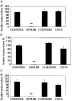
Fig. 2. Complement dependency for the tachyzoites killing effect of pig (A), rabbit (B) and dog serum (C). Tachyzoites were incubated with medium (Control), 20% serum (Serum), heat-inactivated serum (H-Serum), or serum chelated with 10 mM EDTA (EDTA). The experiment was repeated more than twice. **P<0·01.
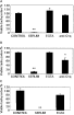
Fig. 3. Effect of EGTA and anti-C1q antibody on the tachyzoite killing activity of pig (A), rabbit (B) and dog sera (C). Tachyzoites were incubated with medium (Control), 20% serum (Serum), serum treated with 10 mM EGTA and 2 mM Ca2+ (EGTA), or serum with anti-C1q antibody (anti-C1q). The experiments were repeated twice and generated similar results. *P<0·05; **P<0·01, respectively.
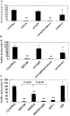
Fig. 4. IgM antibodies are responsible for the killing activity of swine serum and rabbit serum. (A) Tachyzoites were incubated with medium (Control), 20% swine serum (Serum), swine serum+anti-swine IgM antibodies (anti-IgM), swine serum+anti-swine IgM antibodies+anti Toxoplasma gondii antibodies (anti-IgM+antiserum), or anti-T. gondii antibodies (antiserum). (B) Tachyzoites were incubated with medium (Control), 10% rabbit serum (Serum), rabbit serum+anti-rabbit IgM antibodies (anti-IgM), rabbit serum+anti-rabbit IgM antibodies+anti-T. gondii antibodies (anti-IgM+antiserum), or anti-T. gondii antibodies (antiserum). The experiments were performed 3 times. (C) T. gondii tachyzoites were incubated with 20% normal swine serum (Serum), 40% swine serum depleted with IgM and IgG (SMGDS), SMGDS replenished with gel-filtrated swine IgM fraction (SMGDS+IgM), swine serum depleted with IgG (IgG(−)) and gel-filtrated swine IgM fraction (IgM) without complement. All experiments were repeated 3 times. *P<0·05; **P<0·01.

Fig. 5. (A) Verification of complement activity in cat sera. CPK cells sensitized with mouse anti-CPK antibody were incubated with 20% cat serum at 37 °C for 30 min. Trypan blue was used to stain the dead cells. The percentages of viable cells were shown. The mean values of experiments with 6 different cat sera preparations were presented. αCPK: CPK cells sensitized with mouse anti-CPK antibodies. Serum+αCPK: CPK cells sensitized with mouse anti-CPK antibodies were incubated with 20% cat serum. H-serum+αCPK: CPK cells sensitized with mouse anti-CPK antibodies were incubated with 20% heat inactivated cat serum. Serum+αCPK+EDTA: CPK cells sensitized with mouse anti-CPK antibodies were incubated with 20% cat serum that had been chelated with 10 mM EDTA. (B) Effect of cat complement and cat anti-Toxoplasma gondii antibodies on T. gondii. Control: T. gondii tachyzoites were incubated with medium. Cat: T. gondii tachyzoites were incubated with 20% cat serum. Cat+antiserum: T. gondii tachyzoites were incubated with 20% cat serum and 5% cat anti-T. gondii antiserum. The antiserum obtained from the cat infected with T. gondii was heat inactivated and used as a source of cat anti-T. gondii antibodies. Antiserum: T. gondii tachyzoites were incubated with 5% cat anti-T. gondii antiserum.

Fig. 6. (A) Toxoplasma gondii tachyzoites sensitized by cat anti-T. gondii antibodies were killed more efficiently by swine complement than cat complement. Cat: T. gondii tachyzoites were incubated with 5% cat anti-T. gondii antiserum and 20% cat serum as a source of complement. SMDGS: T. gondii tachyzoites were incubated with 5% cat anti-T. gondii antiserum and 40% SMDGS as a source of complement. Bars represent the ratio of the number of the viable tachyzoites treated with the complement source and cat anti-T. gondii antibodies to the number of viable tachyzoites treated with the complement source alone. (B) T. gondii tachyzoites sensitized by swine anti-T. gondii antibodies were killed more efficiently by swine complement than cat complement. T. gondii tachyzoites were sensitized with 5% swine anti-T. gondii antiserum and incubated with 20% cat serum or 40% SMDGS as sources of complement. The same experiments were carried out using 1/20 and 1/200 diluted swine anti-T. gondii antiserum. Bars represent the ratio of the number of the viable tachyzoites treated with the complement source and swine anti-T. gondii antibodies to the number of viable tachyzoites treated with the complement source alone.
 You have
Access
- 26
- Cited by