For patent ductus arteriosus in older adult patients, CT is a widely accepted modality for describing its precise morphology. Reference Thai, Harper and Seneviratne1 However, a highly calcified arterial duct presents with a complicated internal structure and accurate sizing of the duct may be difficult even with CT. Here, we report a case of a calcified arterial duct with such complex morphology that CT sizing was inaccurate. Intravascular ultrasonography proved to be useful in the evaluation of the duct and as a guide for safe device closure.
Case report
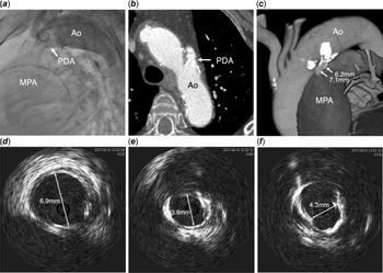
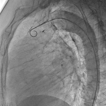
A 73-year-old woman presented with a chief complaint of mild fatigue on exertion. Acceleration of the second heart sound on auscultation was heard, and the patient was diagnosed with patent ductus arteriosus based on transthoracic echocardiography findings. Cardiac catheterisation was performed, and the obtained data were as follows: ratio of pulmonary blood flow to systemic blood flow, 1.75; mean pulmonary artery pressure, 30 mmHg; and ratio of pulmonary artery resistance to systemic artery resistance, 0.10. The patient was referred to our hospital for arterial duct closure. Three-dimensional CT performed at the previous hospital revealed significant calcification around the duct and irregular narrowing of the inner structure (Fig 1b and c). The pulmonary orifice diameter of the duct was 7.1 mm, and the length was 13 mm. Although determining the exact minimum diameter of the duct was difficult, it was measured to be approximately 6.2 mm (Fig 1c). Aortic angiography in the right anterior oblique 20° projection and lateral view was performed prior to treatment. Owing to contrast streaming and the duct overlapping with the ascending aorta, we could not obtain a clear image or measure the duct (Fig 1a). We then performed an intravascular ultrasonographic study of the duct. The duct was crossed with a 0.035-inch Radifocus-type guidewire from the aorta, and a 4-F JR catheter was used to exchange for a 0.018-inch Thruway guidewire (Boston Scientific, Marlborough, MA). The intravascular ultrasonography system (Vision PV 0.018, Volcano Corporation, Rancho Cordova, CA, USA) was mounted on the guidewire and pulled through the duct from the main pulmonary artery to the aorta. An intravascular ultrasonographic image showed encircling calcification of the duct. The pulmonary orifice diameter was measured to be 6.9 mm (Fig 1d), and the narrowest part of the duct, which was located in the middle, was 3.8 mm in diameter (Fig 1e). We chose a 10/8 Amplatzer Duct Occluder I (ADOI, AGA Medical, Golden Valley, MN) because for adult patients we use an Amplatzer Duct Occluder I that is one size larger than that used for children. We placed it successfully using a conventional antegrade approach with the retrograde wire-assisted technique. Reference Hsin, Lin and Hwang2 Aortography performed after device deployment confirmed complete closure (Fig 2).
Aortography, CT, and intravascular ultrasound images of patent ductus arteriosus (PDA). (a) The lateral view of the aortogram shows overlapping of PDA with the ascending aorta and an indistinct outline of the PDA. (b) Transverse plane of a two-dimensional CT image showing the irregular inner structure of the PDA. (c) A three-dimensional CT image showing massive calcification of the PDA. (d–f) Intravascular ultrasonography images of the PDA. (d) The pulmonary orifice diameter was 6.9 mm. (e) The narrowest point showing encircling calcification and a diameter of 3.8 mm. (f) The diameter was 4.3 mm at the aortic end. Ao = aorta; MPA = main pulmonary artery; PDA = patent ductus arteriosus.

Lateral view of an aortogram showing no residual shunt after Amplatzer Duct Occluder I release.

Discussion
The lesson learned from this case is that a highly calcified arterial duct in an adult has a complicated internal structure, and accurate measurement of the duct can be difficult even with CT. Intravascular ultrasonography can be useful in evaluating the anatomy of a highly calcified duct and as a guide for treatment.
A highly calcified adult arterial duct has a complicated internal structure, and accurate sizing of the duct can be difficult even with CT. The arterial duct in adults has specific anatomical features such as cranial shift and large calibre of the aorta, which makes visualisation by angiography difficult. Reference Shafi, Singh, Smith and Rogers3 Therefore, CT is a widely used diagnostic imaging method for patent ductus arteriosus in adults, and the accuracy of measurement of the size of the duct has been reported. Reference Thai, Harper and Seneviratne1 However, in our case, due to extensive and marked calcification, evaluation of detailed anatomy and the narrowest point of the arterial duct, which is crucial for appropriate device size selection, was difficult.
Intravascular ultrasonography is useful for the evaluation of the anatomy of highly calcified duct and as a guide for treatment in such cases. As reported previously, arterial duct measurement by intravascular ultrasonography showed close correspondence with duct size measurement by angiography and CT. Reference Hijazi, Ahmad, Geggel and Marx4,Reference LaBarbera, Storch and Staniloae5 In our case, the narrowest point of the duct, which is usually situated in the pulmonary orifice, was located at the highly calcified middle portion of the duct. Measurement of the minimum diameter of the duct was possible only by intravascular ultrasonography.
As transcatheter arterial duct closure in older adult patients has become a common procedure, opportunities to encounter markedly calcified ducts are expected to increase. Reference Bentham, Thomson and Gibbs6,Reference Gu, Zhang and Sun7 In this patient population, repeated contrast use should be avoided because renal dysfunction is common; therefore, intravascular ultrasonography can be a useful option in cases where arterial duct imaging by angiography or CT is insufficient for accurate size determination.
As regards intracardiac echo which is widely used for arterial duct device closure, it provides thorough monitoring of the procedure, whereas intravascular ultrasound provides solely diameters of the duct. The usefulness of intracardiac echo for highly calcified arterial duct, however, has not yet been reported. In addition, intracardiac echo requires experience and practice to obtain appropriate images and also needs extra venous access to introduce the catheter. Reference Yoshimoto, Yasuto and Inoue8 Therefore, intravascular ultrasound is a more simple and sound method for the measurement of such ducts.
In conclusion, a highly calcified arterial duct in an adult has a complicated internal structure, and accurate measurement of the duct can be difficult even with CT. Intravascular ultrasonography may be useful for the evaluation of the anatomy of such ducts and as a guide for safe and effective transcatheter closure.
Acknowledgements
None.
Financial support
This research received no specific grant from any funding agency, commercial, or not-for-profit sectors.
Conflicts of interest
None.
Ethical standards
The authors assert that all procedures contributing to this work comply with the ethical standards of the relevant national guidelines on human experimentation (Ethical Guidelines for Medical and Health Research Involving Human Subjects) and with the Helsinki Declaration of 1975, as revised in 2008. An informed consent for patient information to be published was obtained by the patient.